
- EN
- DE

Bacteria
Klebsiella granulomatis (Granuloma inguinale, Donovanosis)


- Organ System
- Genitourinary Tract, Skin and Mucosa, Lymphatic System
- Diagnostic Method
- Cytology

- Geographical Region
- Islands,
- Tropical and Subtropical Region
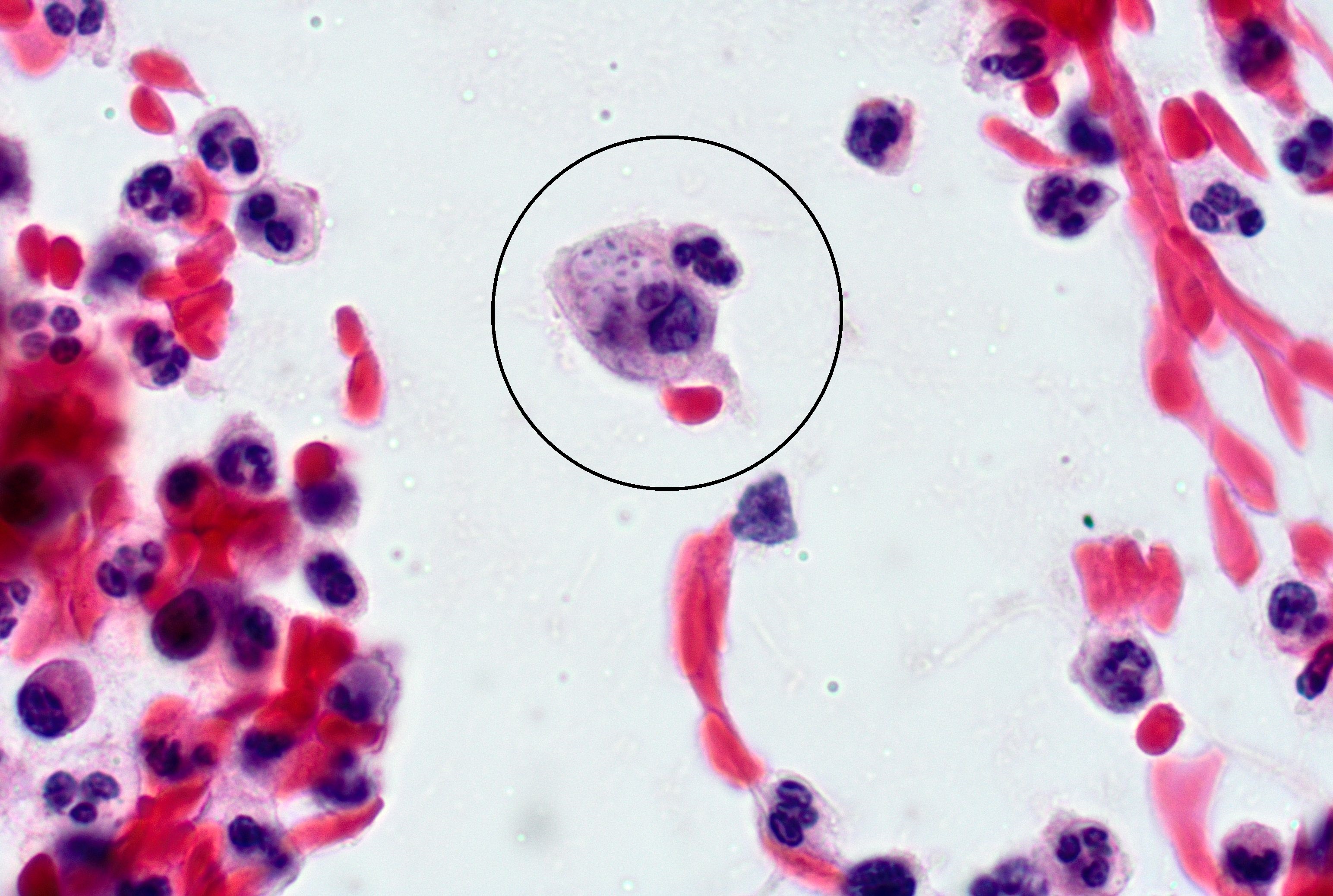 Pap smear, intracellular bacteria, so-called…
Morphology
Cytology:
- Intracellular gram-negative bacterium
- Diagnosis requires visualization of Donovan bodies: dark-staining, encapsulated, pleomorphic rods (1-2 µm) with characteristic bipolar ("safety-pin") appearance due to prominent polar granules
- Organisms located within single or multiple intracytoplasmic vacuoles of large mononuclear cells (histiocytes/macrophages); extracellular organisms may also be present
- Best visualized with Giemsa, Wright, or Leishman stain; also detectable on Papanicolaou-stained smears
- Tissue crush preparation from the lesion base is the preferred specimen
- Very few epithelial cells
- Intakt capillaries may be seen due to ulceration of overlying epithelium
Histology:
- Dense dermal/subepithelial inflammatory infiltrate composed of plasma cells, histiocytes/macrophages, and neutrophils
- Large mononuclear cells with single or multiple intracytoplasmic vacuoles containing Donovan bodies
- Overlying pseudoepitheliomatous hyperplasia is characteristic but not specific, and may histologically mimic squamous cell carcinoma (SCC)
- In advanced cases, tissue destruction and extension beyond the dermis may occur. Transepithelial elimination of the organisms has been reported. (1)
Clinical notes
Sexually transmitted; low infectivity (repeated contact usually required). Painless, slowly progressive, "beefy red" genital ulcers that bleed easily; no lymphadenopathy - distinguishing feature from other genital ulcer diseases. Subcutaneous granulomas (pseudobuboes) may occur. Extragenital spread possible (pelvis, bone, oral). Co-infection with syphilis and HIV common. Without treatment, progressive tissue destruction over months to years; donovanosis may mimic SCC clinically.
Rare in the United States; sporadic cases reported from India, South Africa, and South America. Australia now extremely rare. (2)
Ancillary Testing
Culture difficult (requires HEp-2 cell monolayers); PCR assayAdditional Information
HIV screening is recommended in all patients with genital ulcers.
Differential Diagnosis
References
- Ramdial, P.K., Kharsany, A.B.M., Reddy, R. and Chetty, R. (2000), Transepithelial elimination of cutaneous vulval granuloma inguinale. Journal of Cutaneous Pathology, 27: 493-499. https://doi.org/10.1034/j.1600-0560.2000.027010493.x
- Workowski KA, Bachmann LH, Chan PA, Johnston CM, Muzny CA, Park I, Reno H, Zenilman JM, Bolan GA. Sexually Transmitted Infections Treatment Guidelines, 2021. MMWR Recomm Rep. 2021 Jul 23;70(4):1-187. doi: 10.15585/mmwr.rr7004a1. PMID: 34292926; PMCID: PMC8344968.
Copyright ® 2025 Österreichische Gesellschaft für Zytologie