
- EN
- DE

Virus
Differential Diagnosis of Klebsiella granulomatis (Granuloma inguinale, Donovanosis):
Herpes simplex virus (HSV)

- Organ System
- Genitourinary Tract, Digestive Tract, Skin and Mucosa, Respiratory Tract
- Diagnostic Method
- Cytology, Histology

- Geographical Region
- Dry Areas,
- Islands,
- Tropical and Subtropical Region,
- Polar and Subpolar Zone,
- Temperate Zone
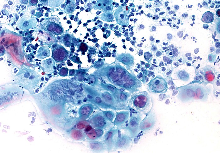
 Cervical smear, superficial and metaplastic…
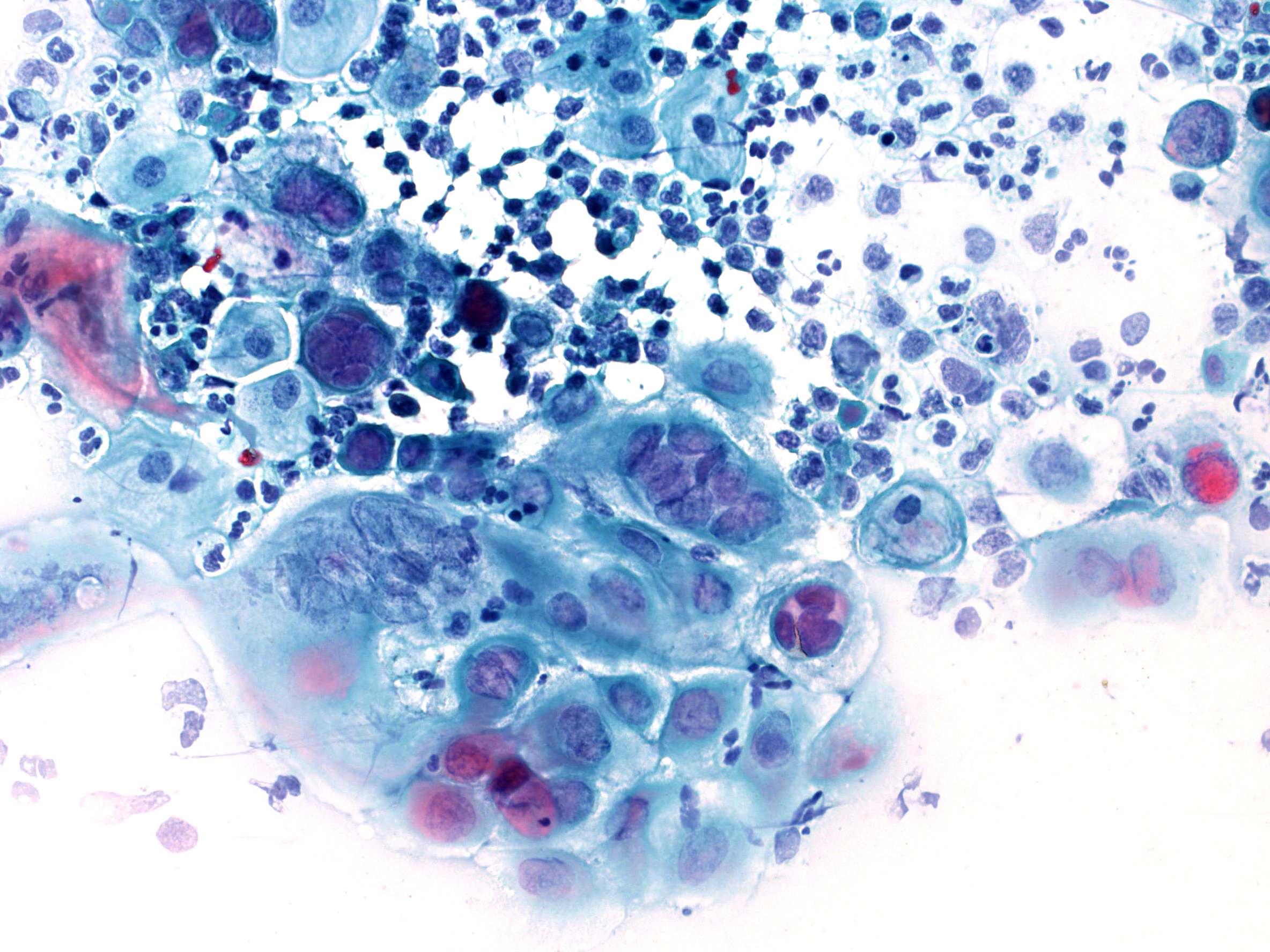
 Bronchial secretion, HSV cytopathic changes…
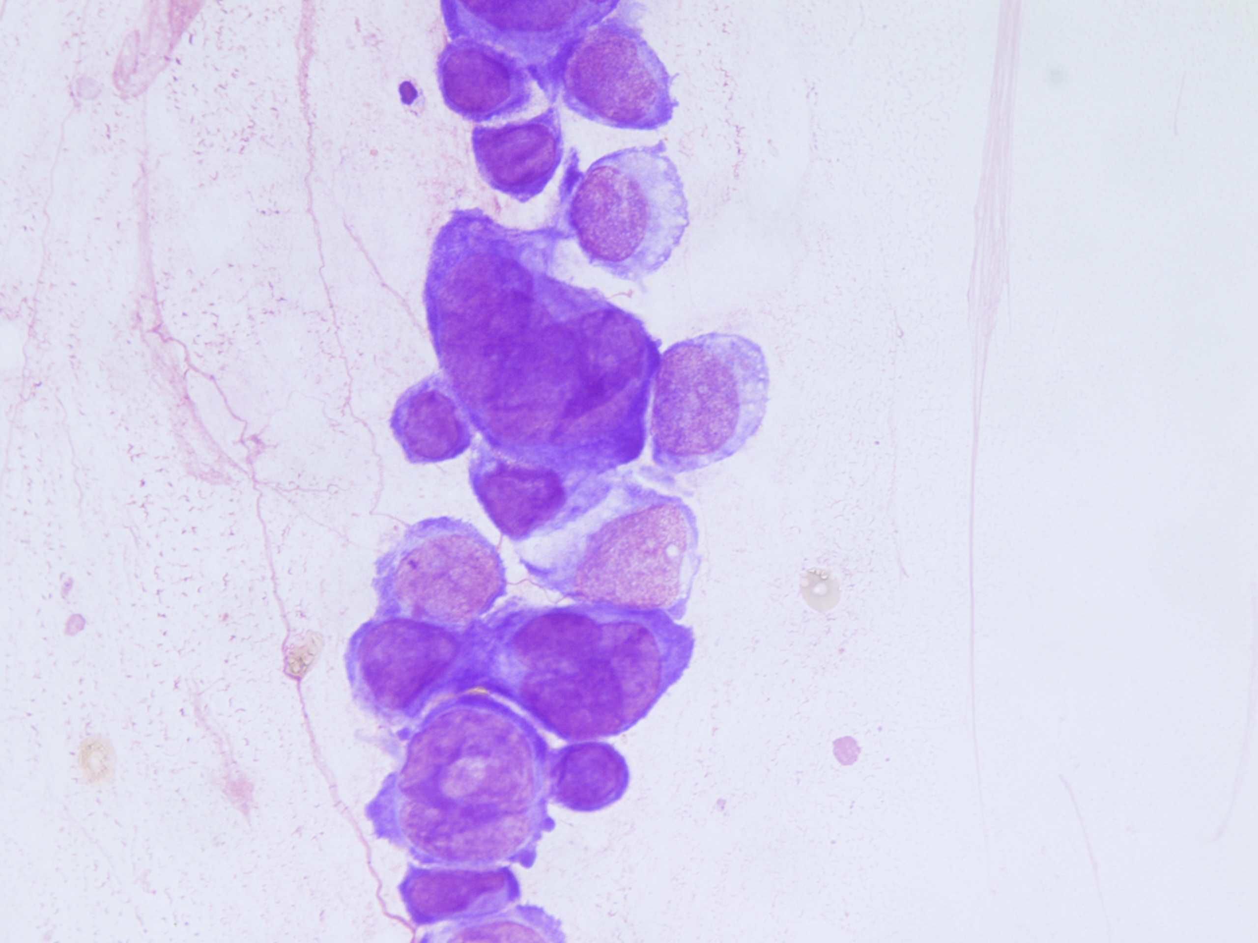
 Esophagus, brush smear, dense cell cluster…
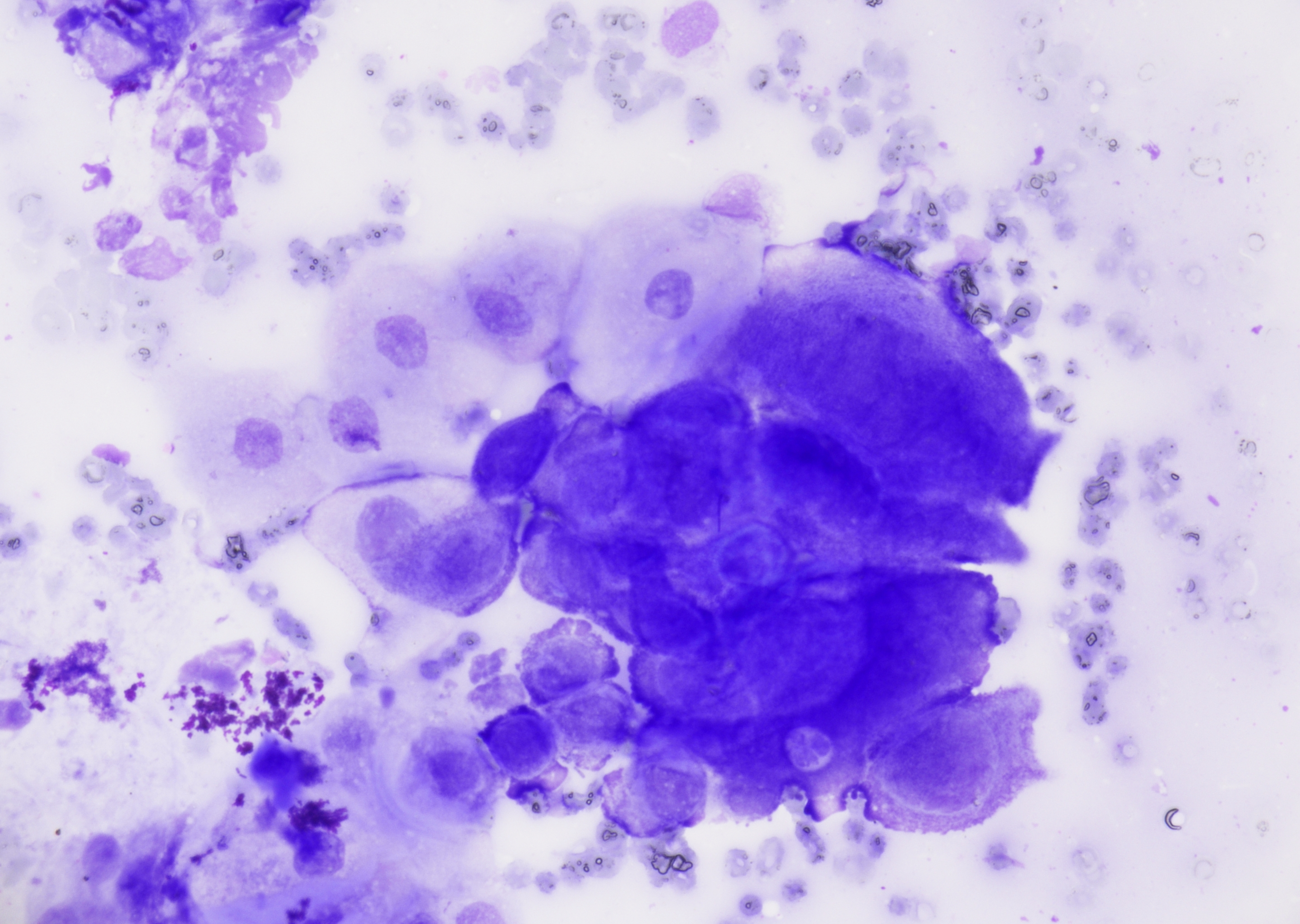
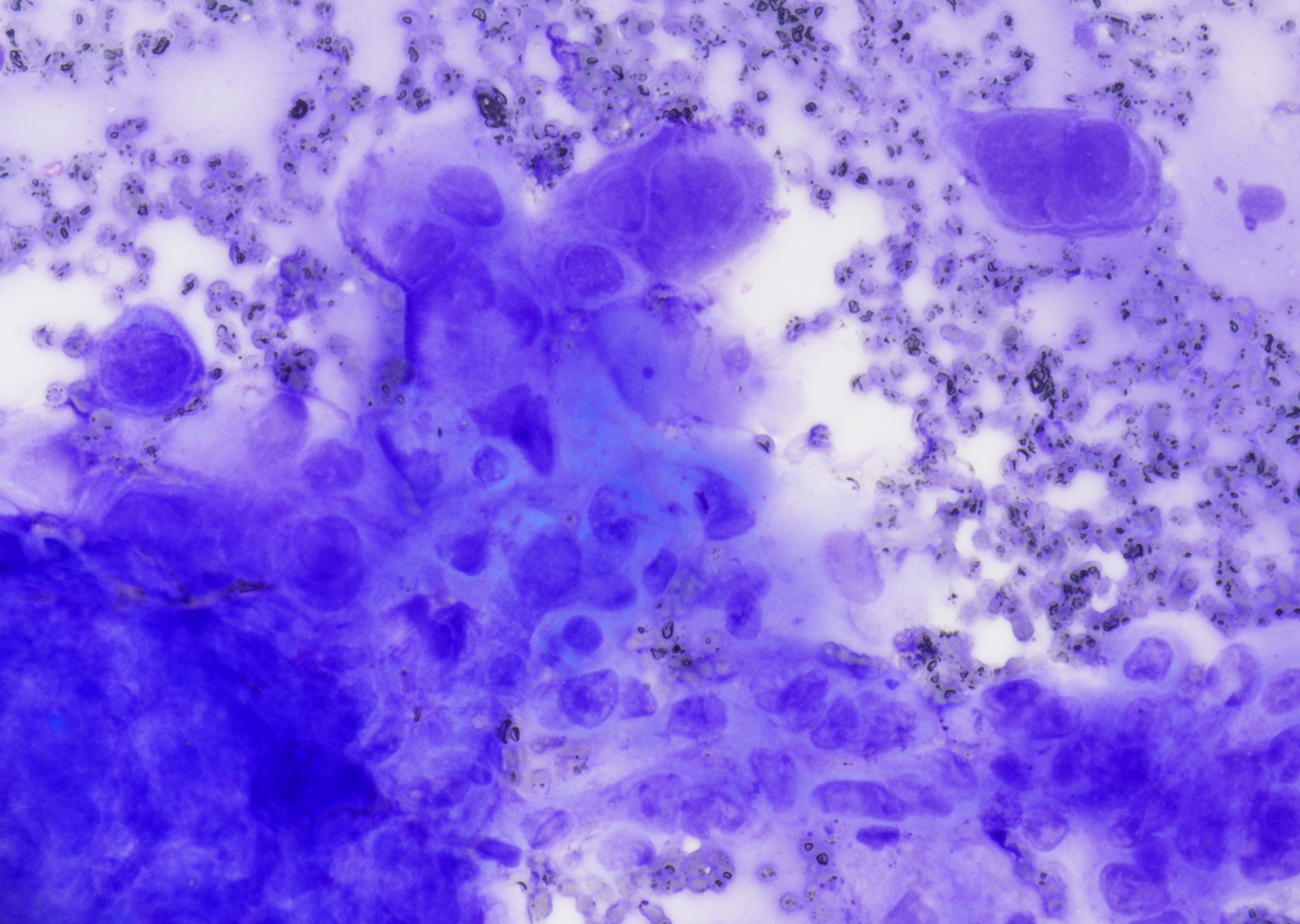 Esophagus, brush smear, multinucleation…
 Voided Urine with multinucleated urothelial…
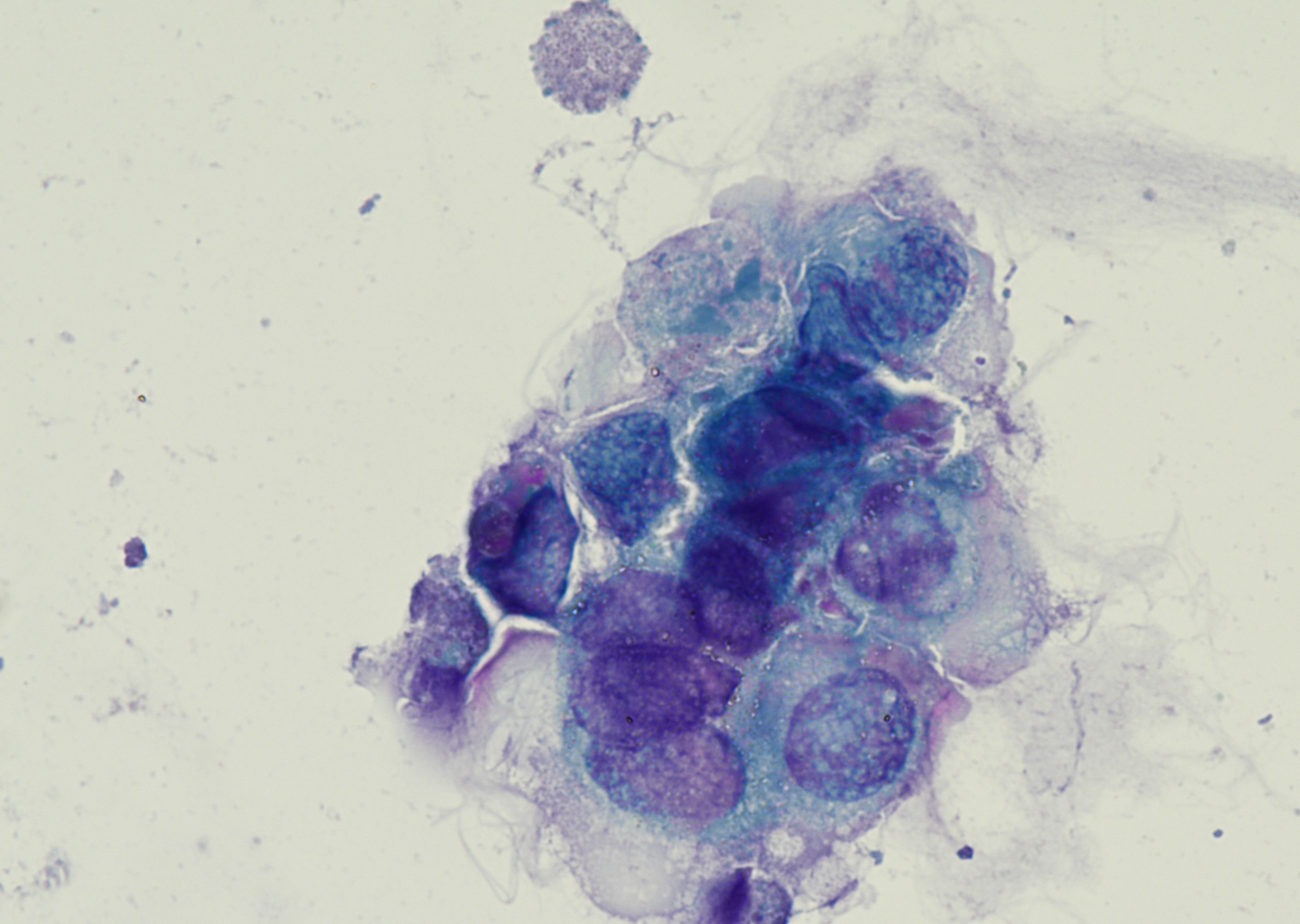 Voided Urine, cluster of multinucleated…
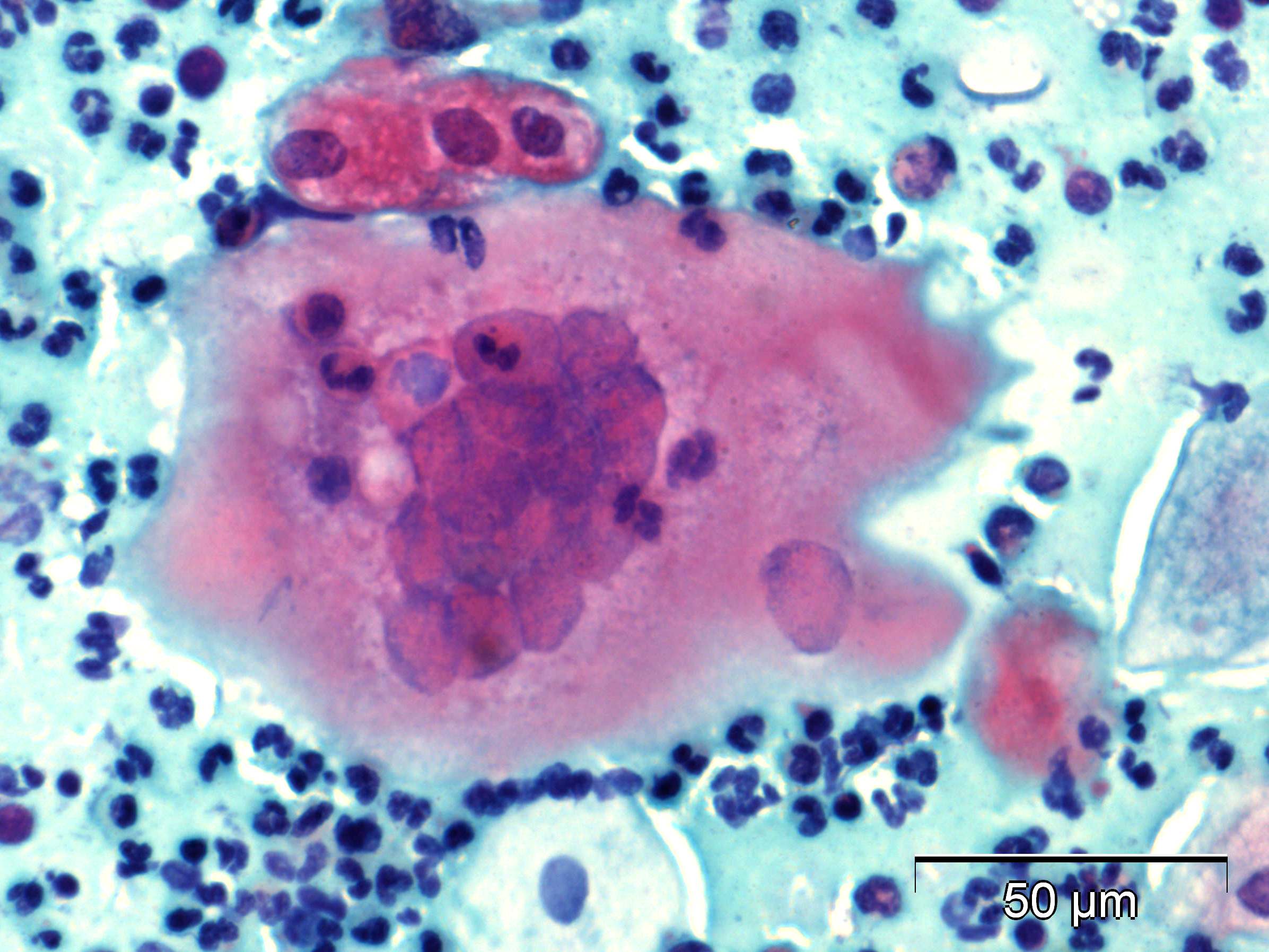 Cervical smear, inflammatory background…
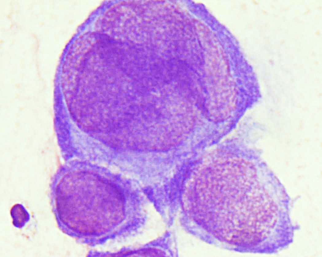 Bronchial secretion, cells with polymorphic…
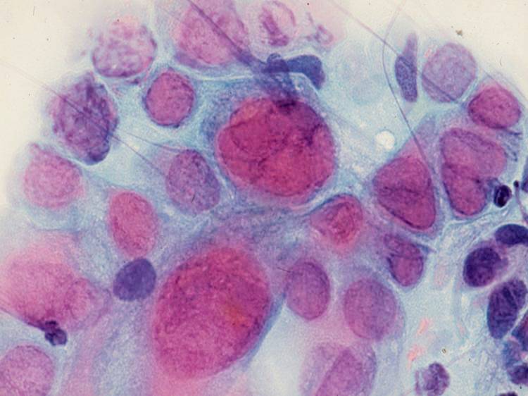 Lung, brush cytology, cell cluster, some…
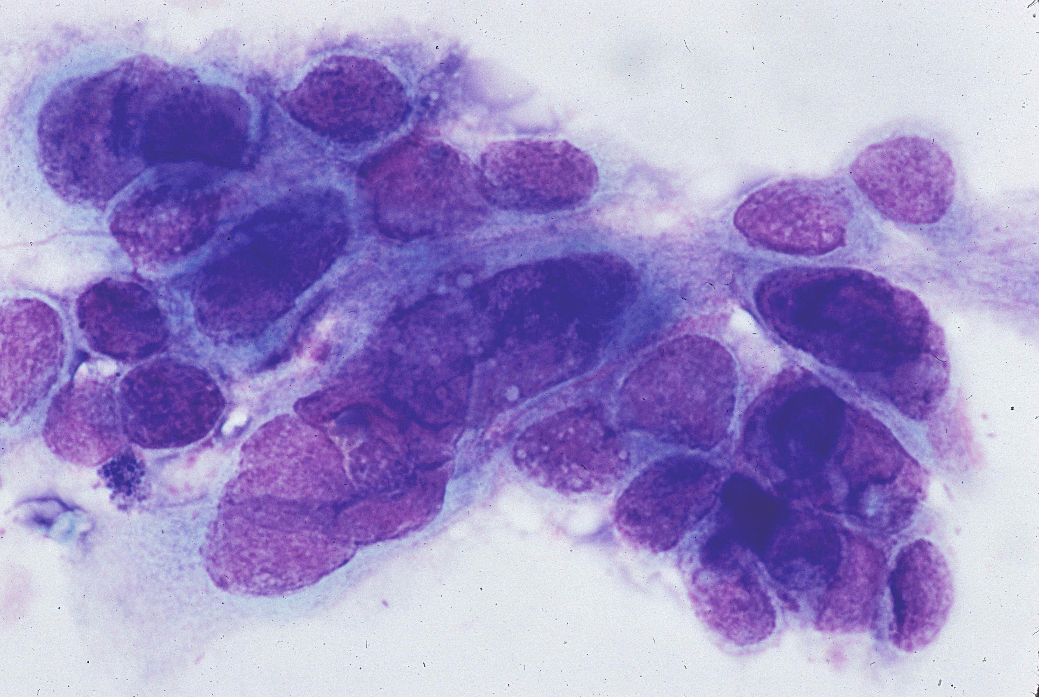 Oropharynx, brush cytology, group of cells…
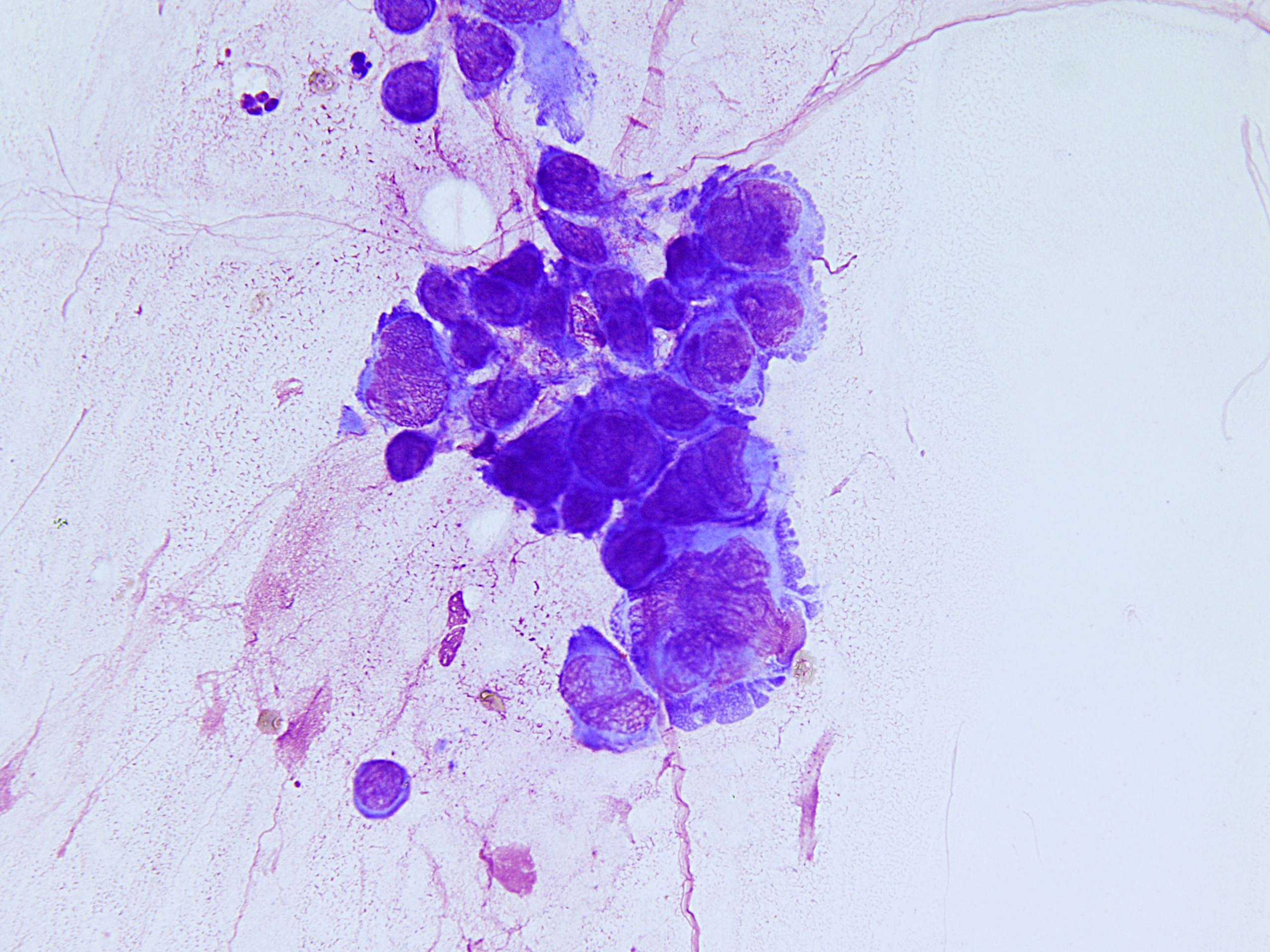 Oropharynx, brush cytology, group of cells…
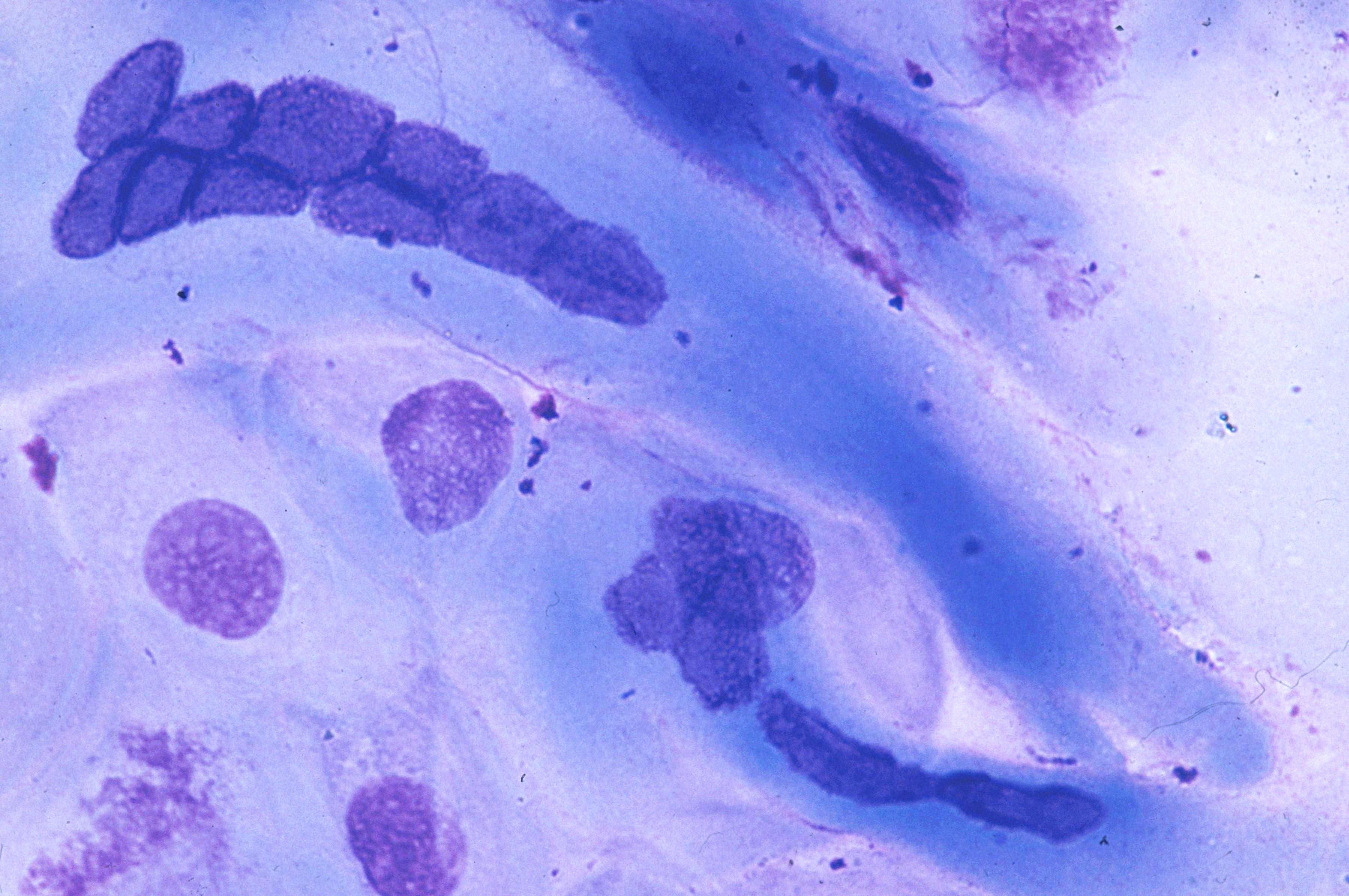 Esophagus, brush smear, multinucleated cell…
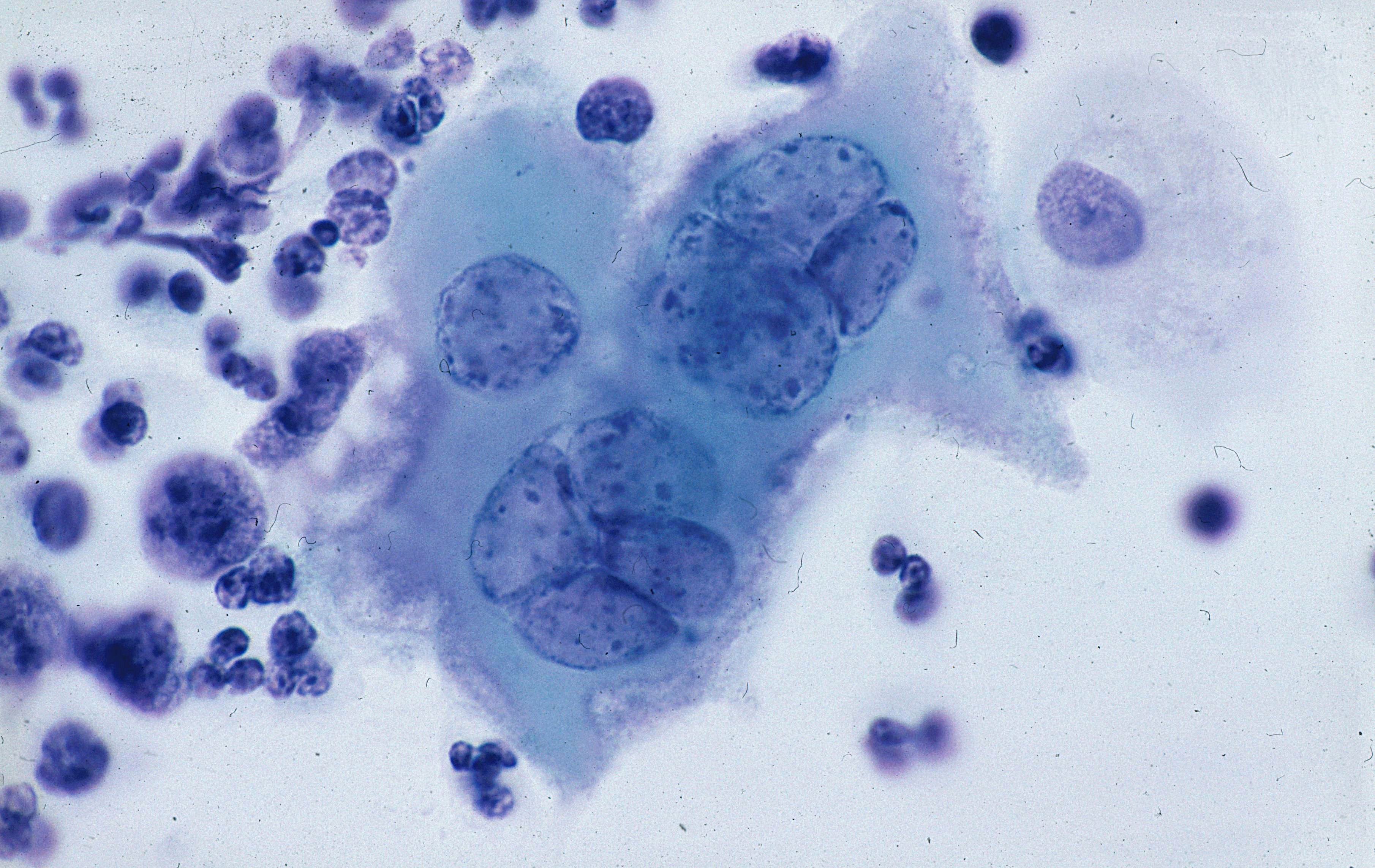 Esophagus, brush smear, multinucleated cells…
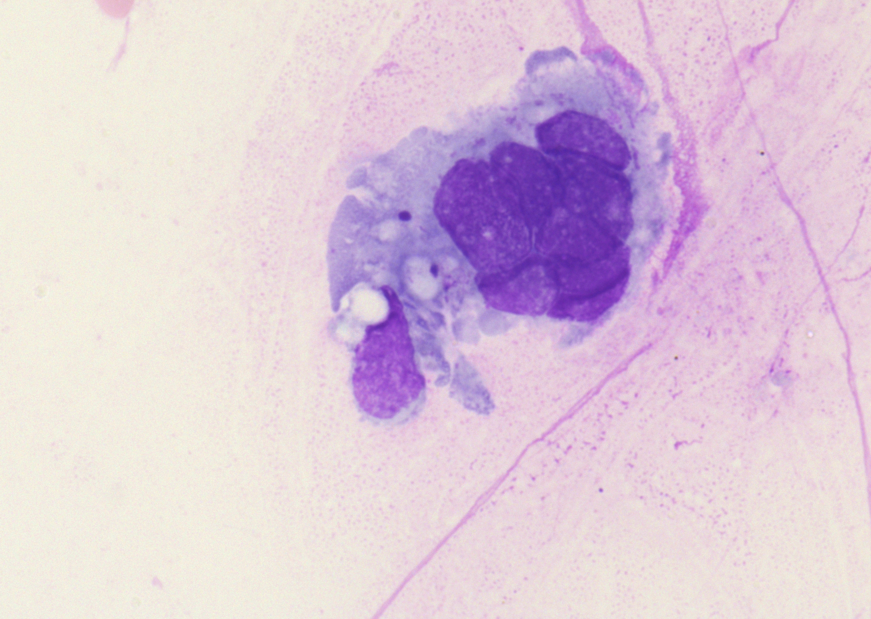 Lung, bronchial brushing, multinucleated…
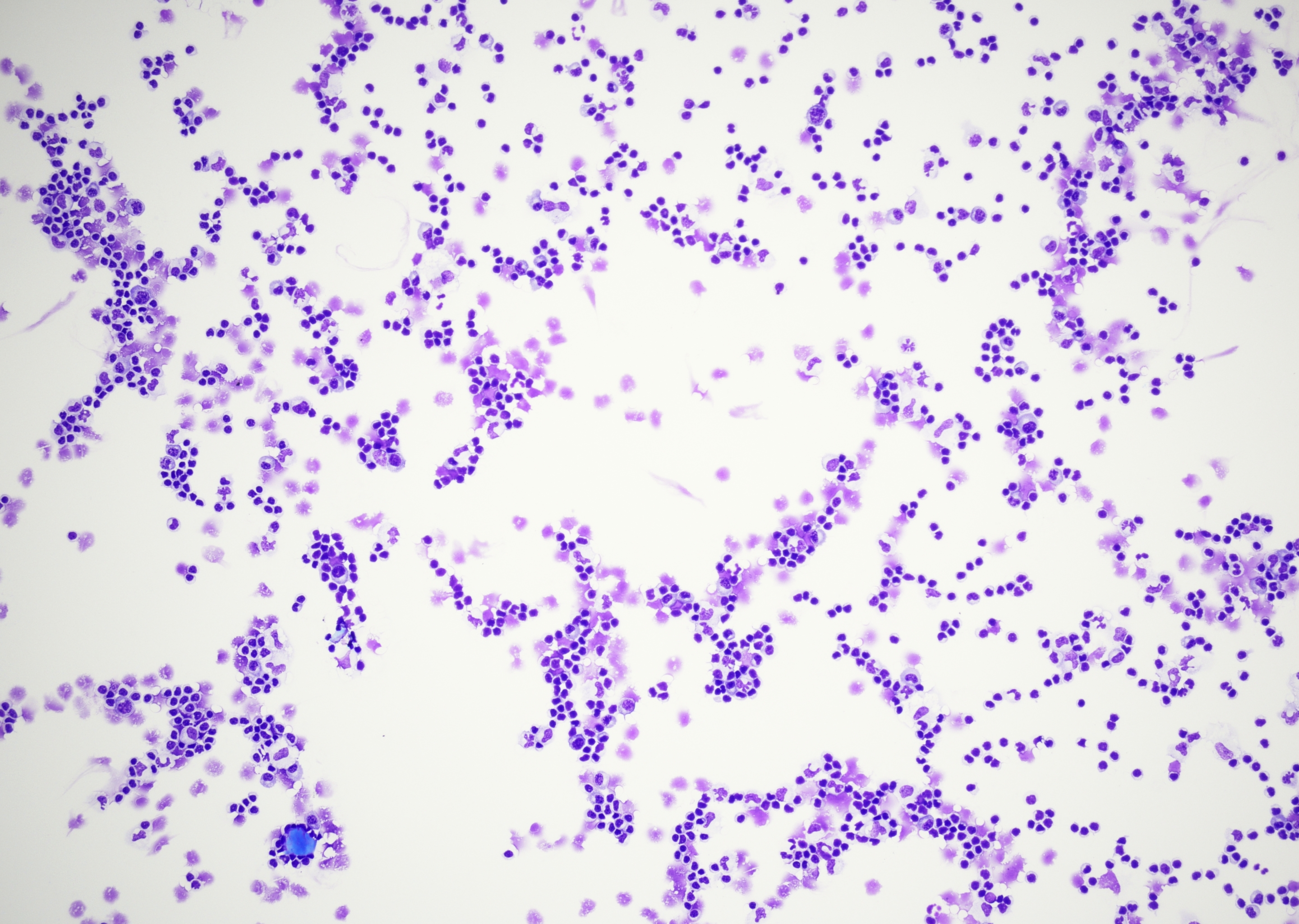 CSF, pleocytosis, nonspecific lymphocytic…
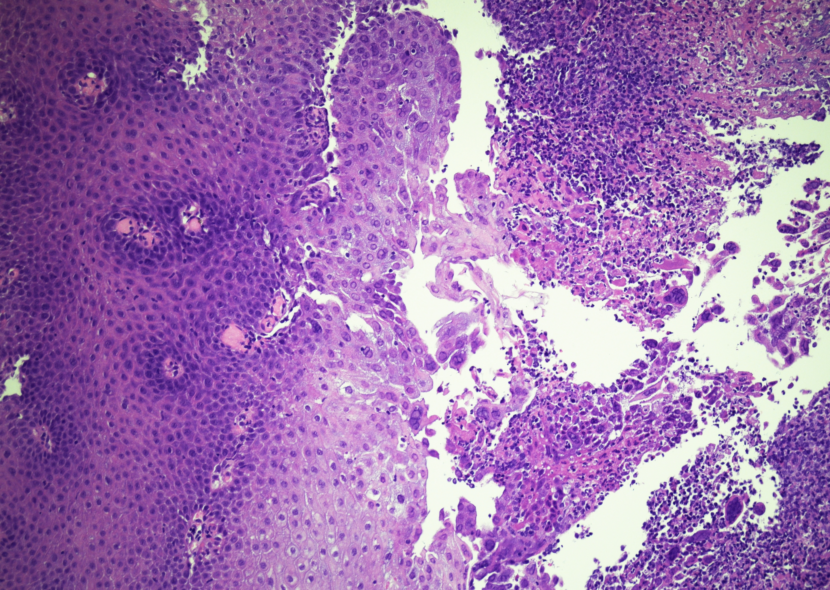 Esophageal biopsy, squamous epithelium with…
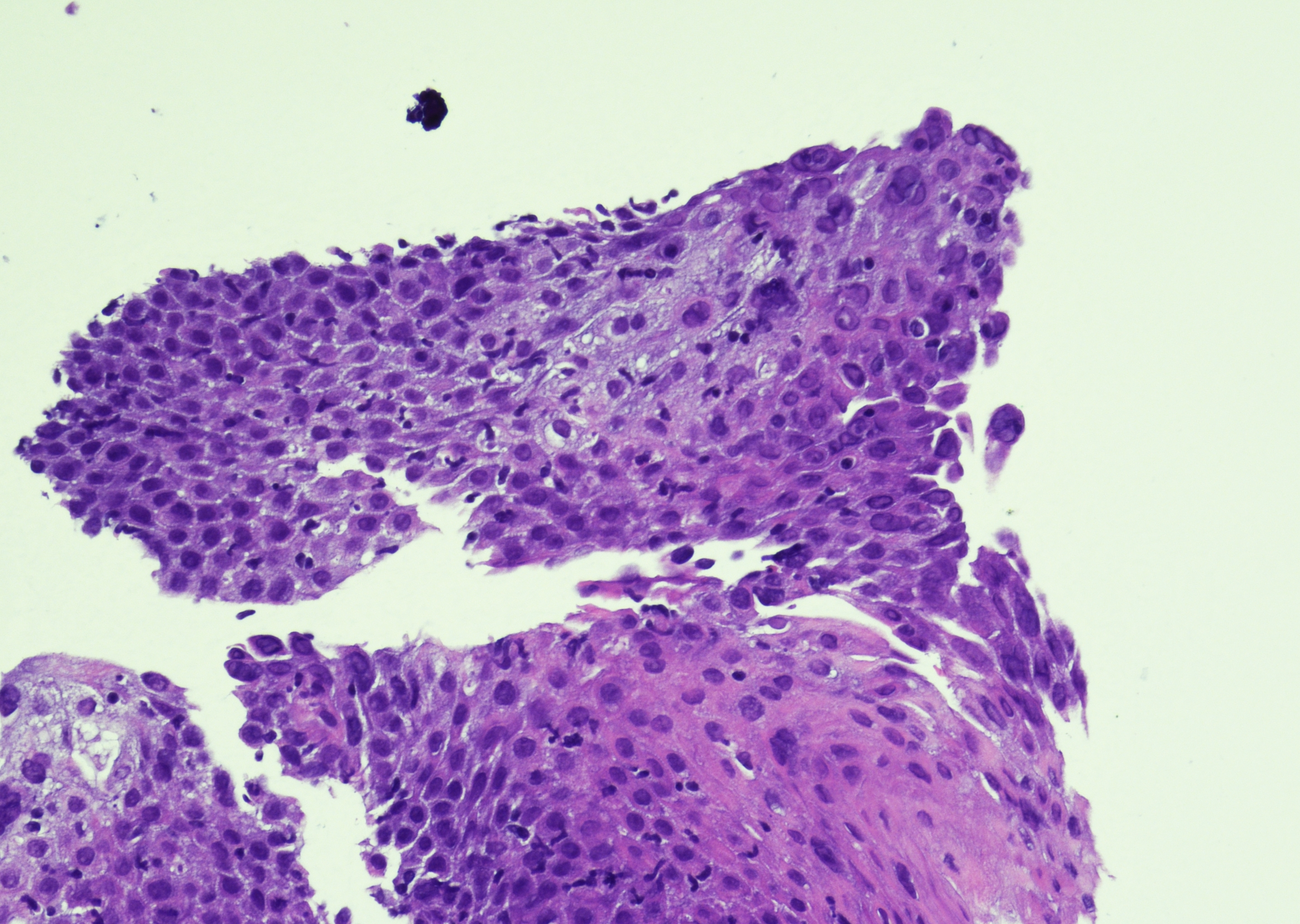 Esophageal biopsy, squamous epithelium with…
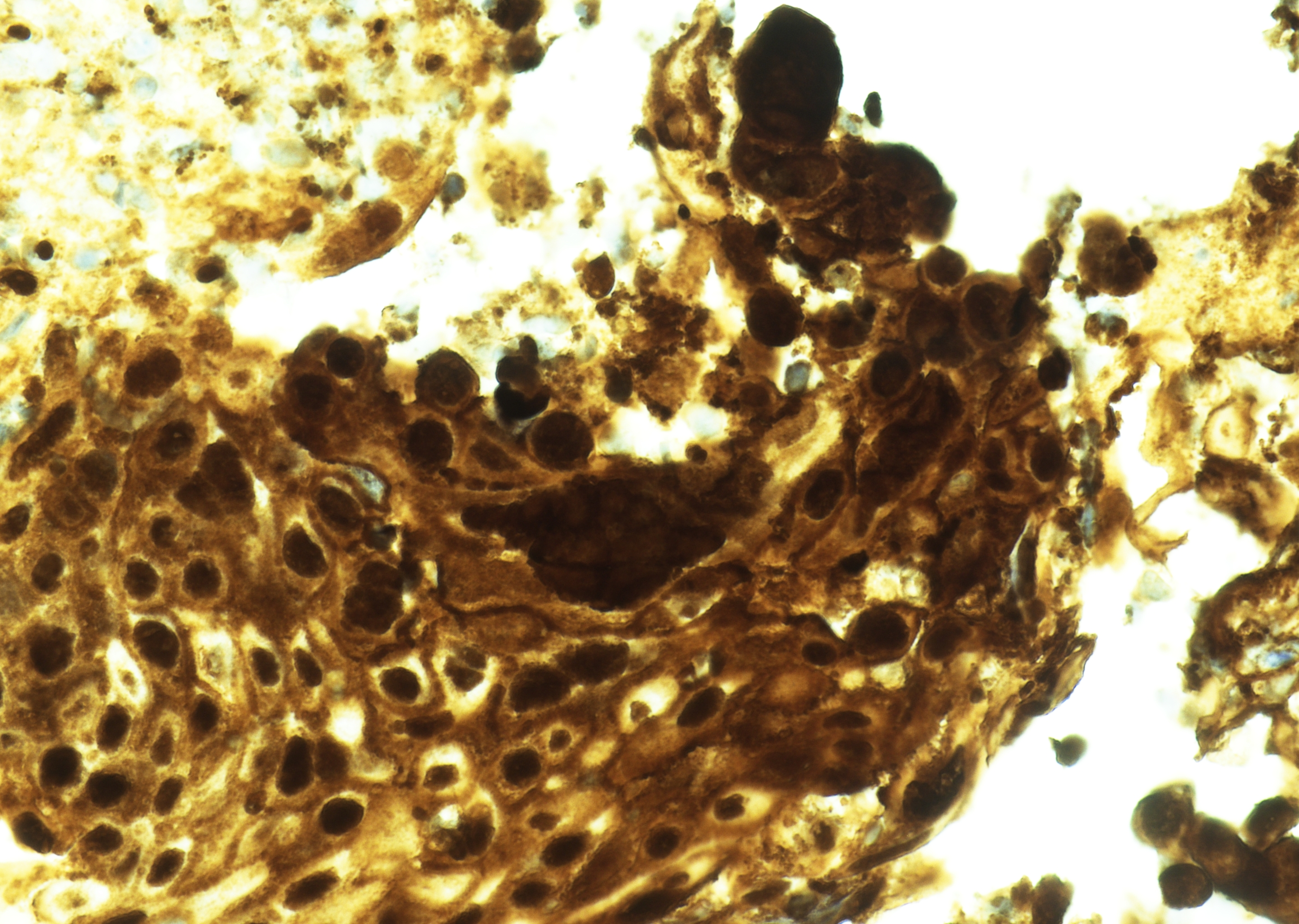 Esophageal biopsy, squamous epithelium…
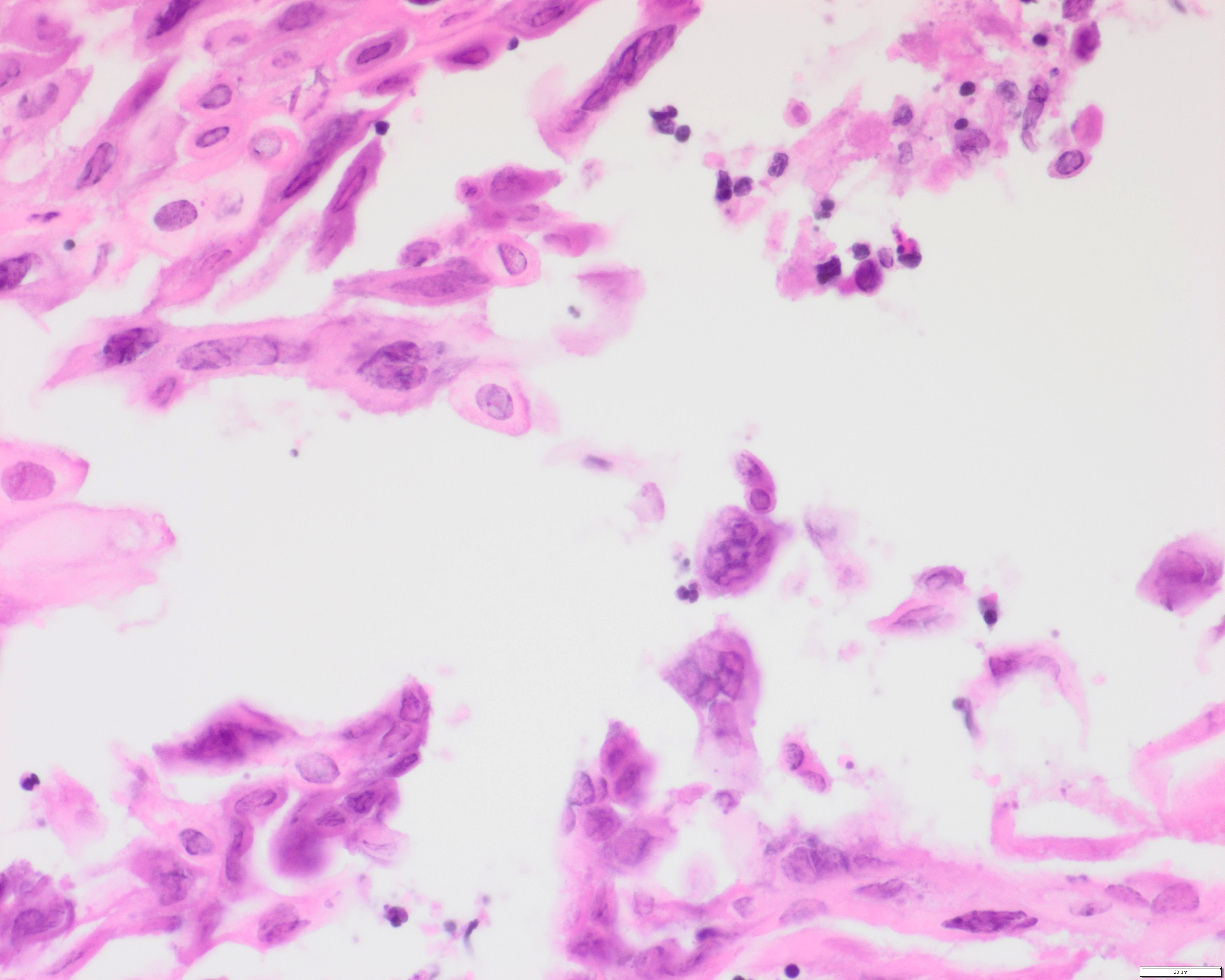 Esophageal biopsy, superficial squamous…
Morphology
Cytology:
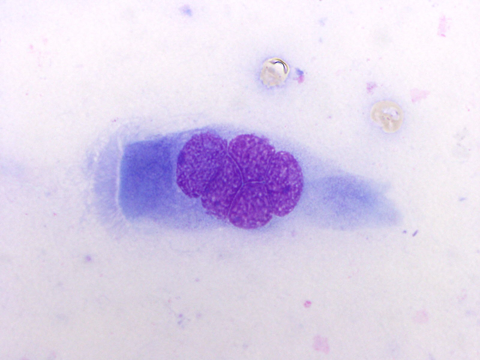
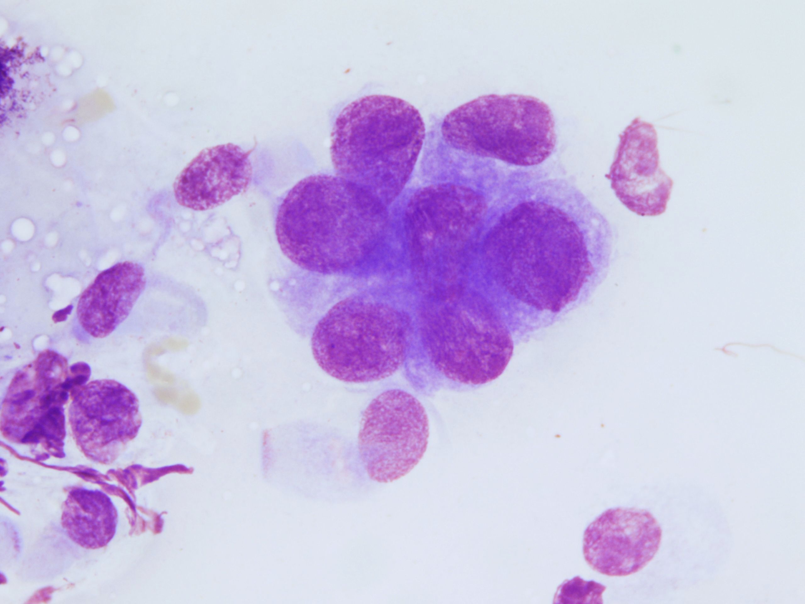
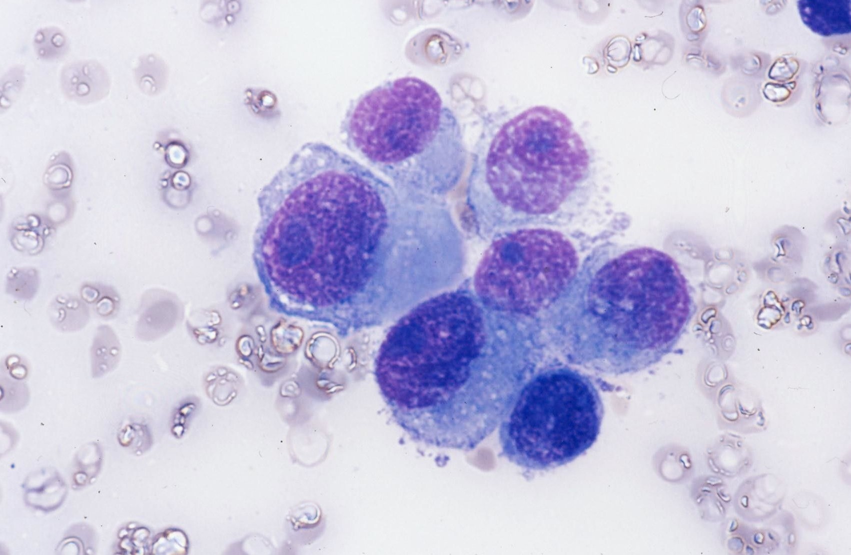
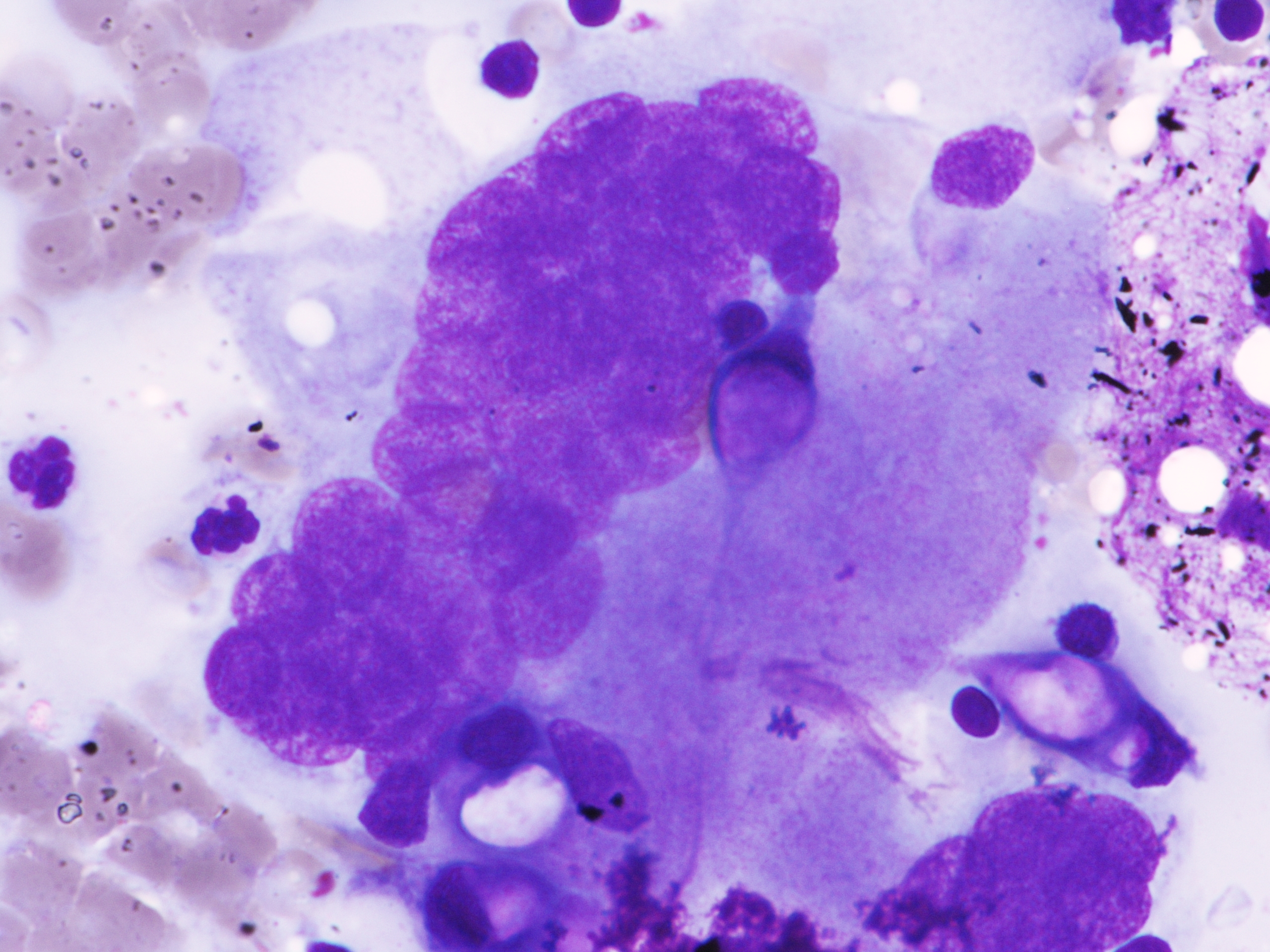
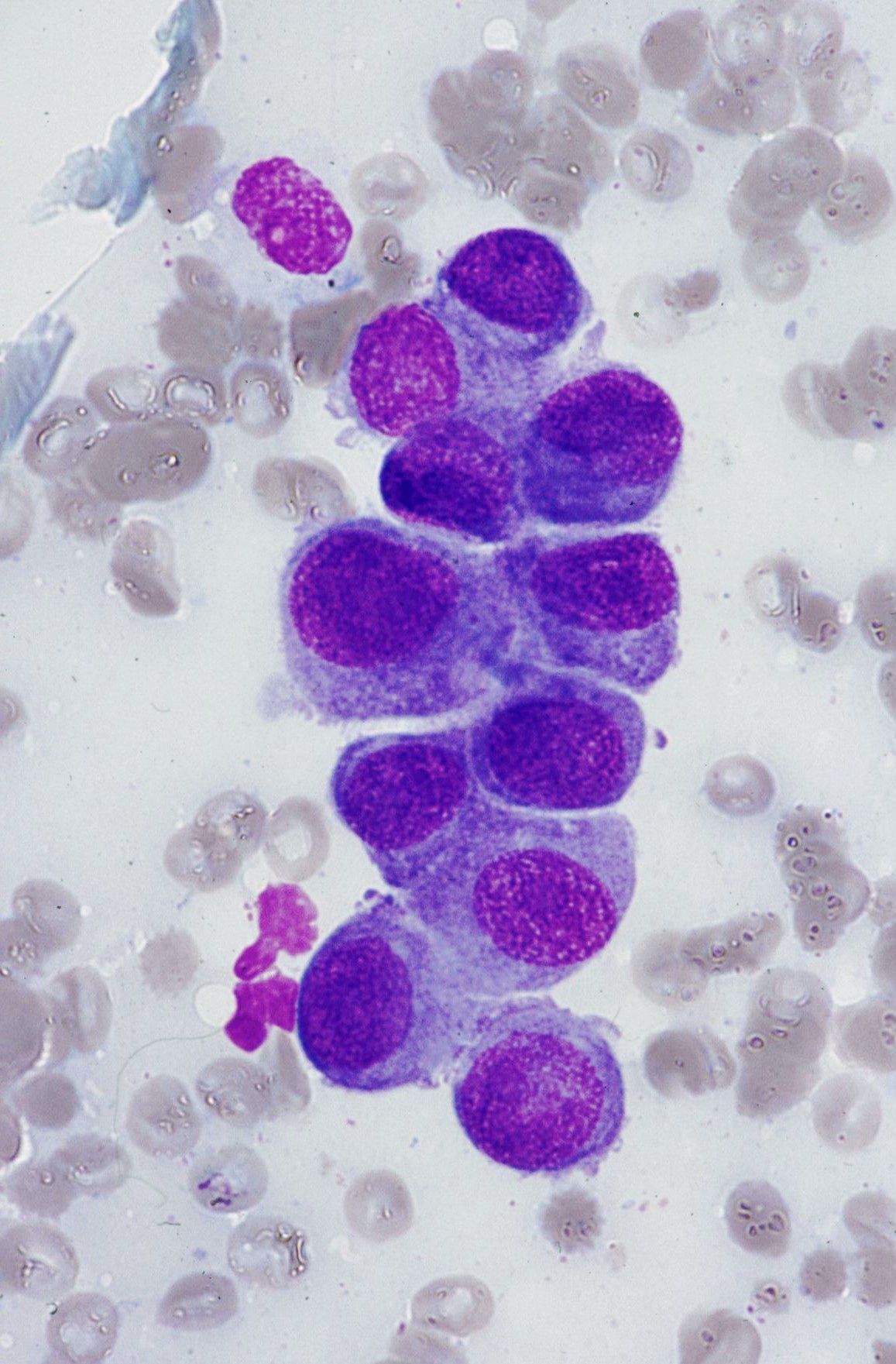
- Infected epithelial cells show enlarged nuclei with irregular contours, homogeneous, pale chromatin (ground-glass nuclei), and peripheral chromatin marginationIn (early lesions)
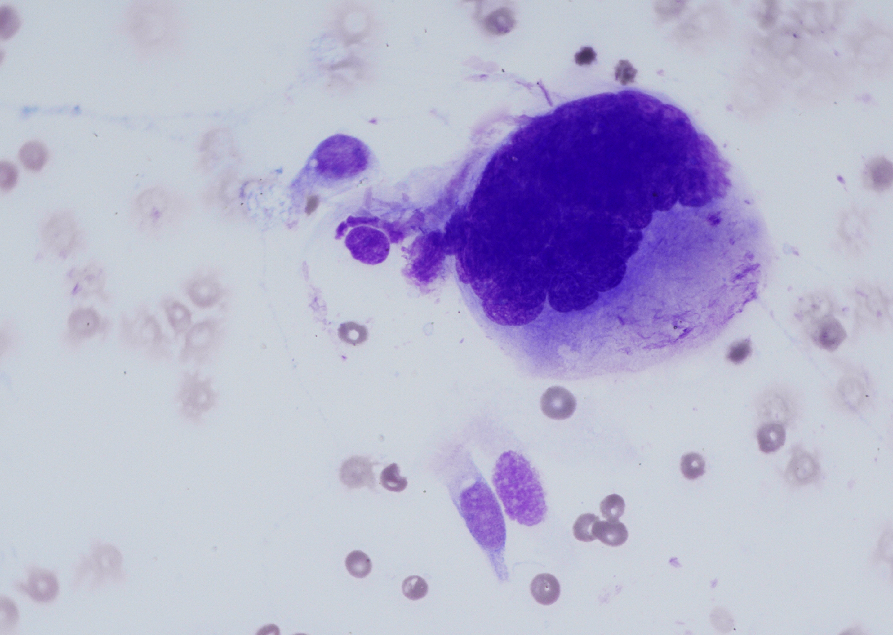
- Hyperchromatic epithelial cells but retain homogeneous chromatinIn, making them easy to differentiate from dysplastic cells (long-standing infections)
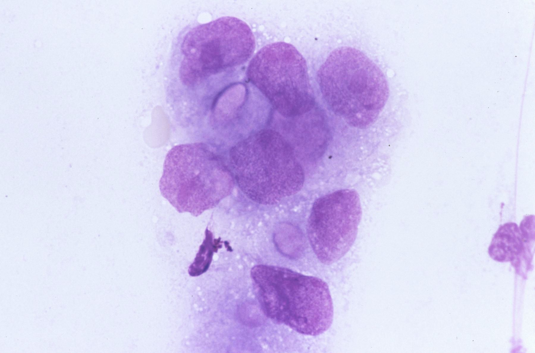
- Infected cells fuse to form multinucleated giant cells with typical nuklear morphology, nuclear crowding and molding. Some may show eosinophilic intranuclear inclusions with a clear halo
Surrounding Background:
- Clear or most commonly neutrophil-rich mixed inflammatory cell infiltrate
- Erosion, ulceration, necrosis
Histology:
- Skin/mucocutaneous lesions: intraepidermal basal ballooning and reticular degeneration with marked intracellular edema, leading to acantholysis and necrosis (macroscopically vesicle formation). Neutrophil-rich mixed inflammatory cell infiltrate. Characteristic viral cellular and nuclear cytopathic changes (as described above) are typically found at the periphery of the vesicle or ulcer
- Esophagus: ulceration with necrosis, sloughed epithelial cells, and acute cell infiltrate. Viral cytopathic changes found at the periphery of ulcer and in sloughed epithelial cells
- Lymph node: necrosis of interfollicular and/or paracortical regions with infected lymphocytes, histiocytes, and endothelial cells with typical viral cytopathic changes. It can occur simultaneously with lymphoma
- Organ involvement (liver, lung, brain): differences in histologic findings by location and immune status, but the most common features include necrosis, a mild or prominent mixed inflammatory infiltrate, and viral cytopathic changes at the periphery of necrosis
Differential Diagnosis:
- Varicella-Zoster virus - identical changes as in HSV-1/2, IHC or PCR for confirmation required
- Cytomegalovirus
- Other viral infections
- Reactive multinucleated epithelial cells
- Bacterial infections
Clinical notes
Typically, itchy, painful blisters on the lips/mouth or genitals; in severe cases, e.g., in immunocompromised individuals, it can also cause encephalitis, pneumonia, or disseminated disease.
HSV-1 is usually acquired during childhood. Only a small minority is showing clinical signs during primary infection. In contrast, primary infection with HSV-2 typically occurs in early adulthood and is asymptomatic in approximately two-thirds of cases. Herpes simplex virus is neurotropic. After primary infection, herpes simplex viruses persist in neurons and can be reactivated.
Ancillary Testing
HSV DNA detection by PCR or ISH (vesicle fluid, blood, CSF): direct immunofluorescence; Tzanck smear - Giemsa-stained cellular scrapings; IHC (HSV-1/2 antigens); serological testingDifferential Diagnosis
References
-
Nayar R., Wilbur DC. The Bethesda System for reporting cervical cytology; 3rd Ed. 2015, p84-5
Copyright ® 2025 Österreichische Gesellschaft für Zytologie