
- EN
- DE

Bacteria
Differential Diagnosis of Klebsiella granulomatis (Granuloma inguinale, Donovanosis):
Mucoid variants of Gram-negative rods

- Organ System
- Respiratory Tract
- Diagnostic Method
- Cytology, Microbiological Culture

- Geographical Region
- Dry Areas,
- Islands,
- Tropical and Subtropical Region,
- Polar and Subpolar Zone,
- Temperate Zone
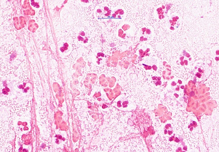
 Sputum, Pseudomonas aeruginosa:
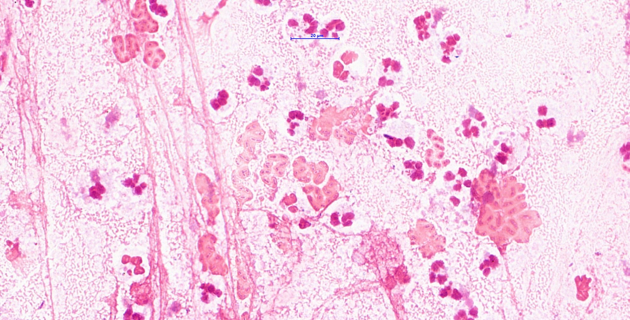
 Sputum, P. aeruginosa: The extracellular…
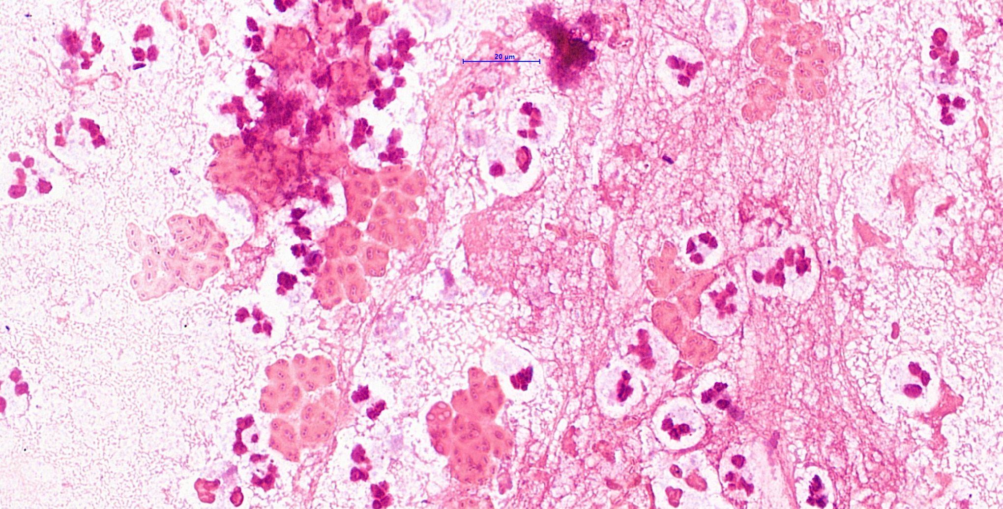
 Sputum, P. aeruginosa: The…
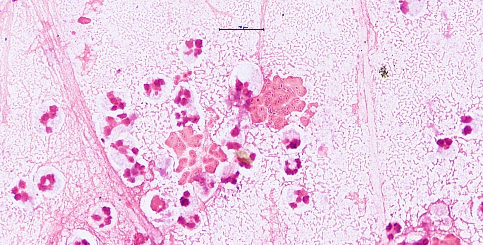
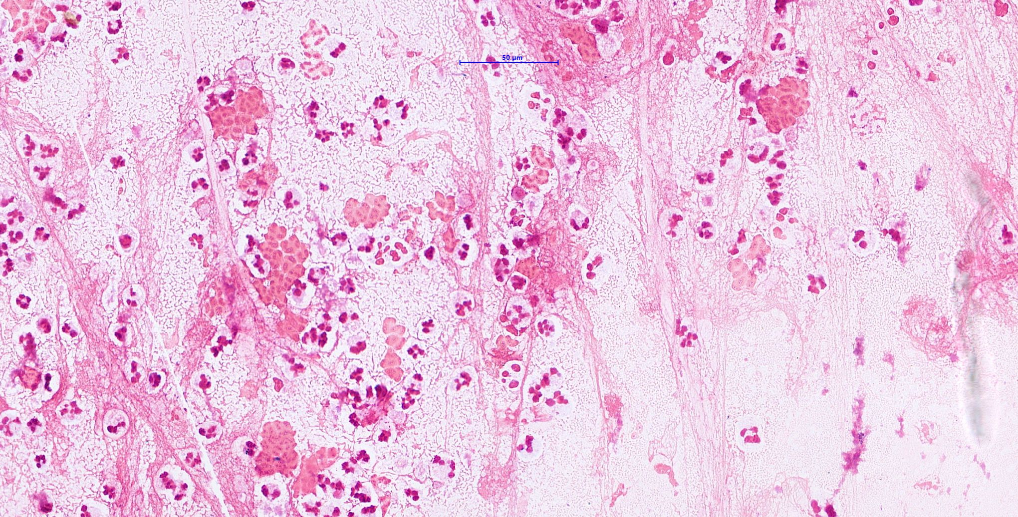 Sputum, P. aeruginosa: Inflammatory…
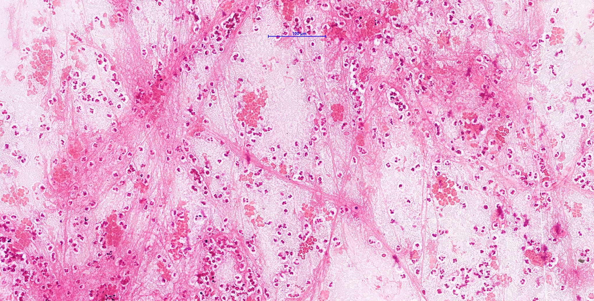 Sputum, P. aeruginosa (Gram, x200)
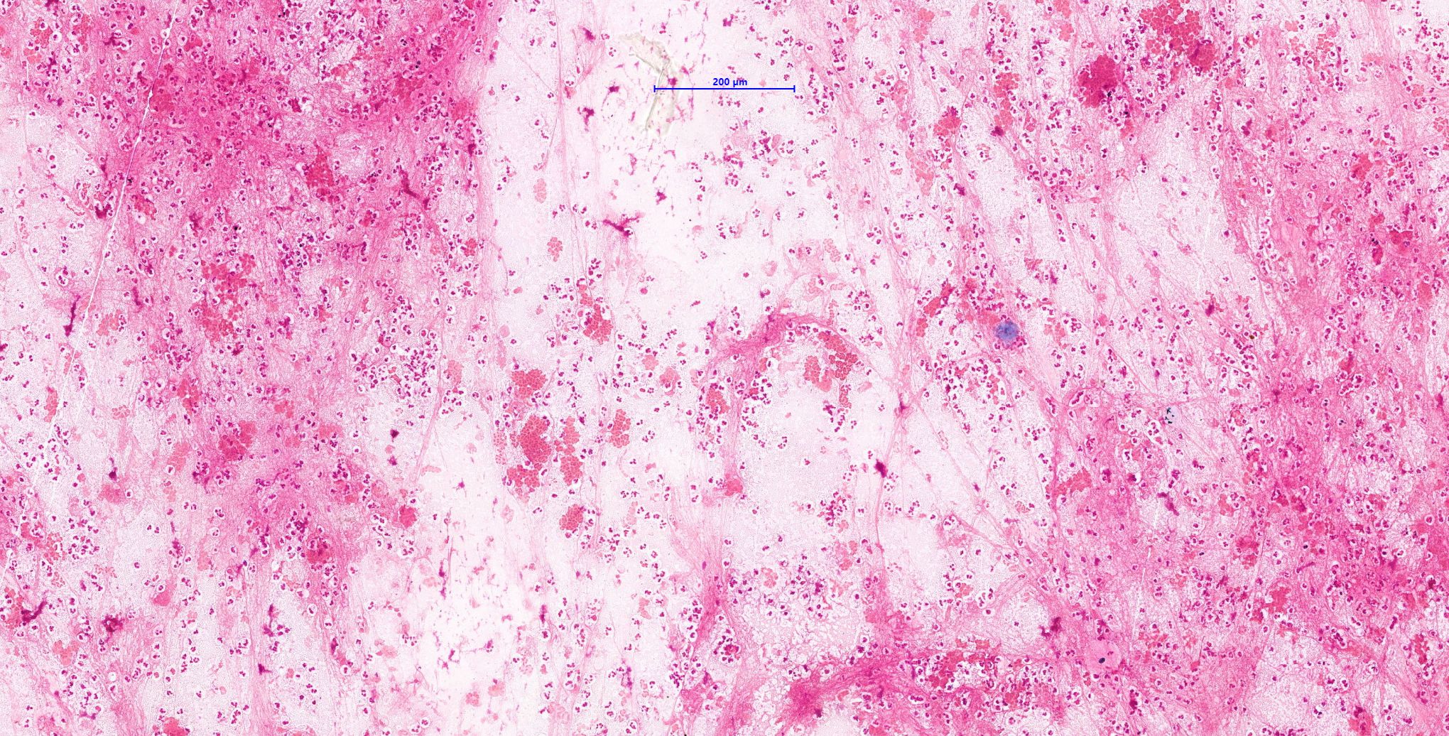 Sputum, P. aeruginosa (Gram, x100)
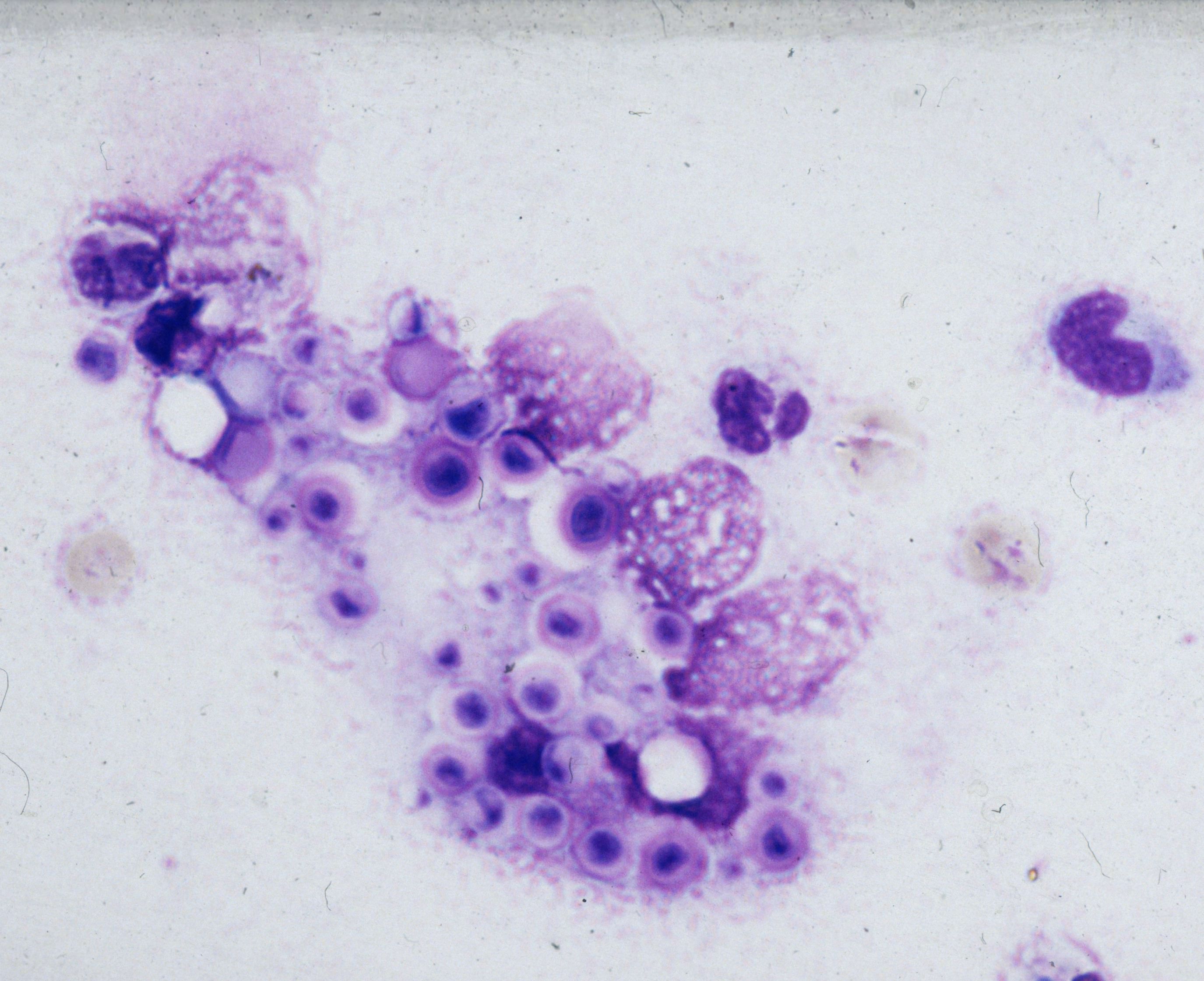
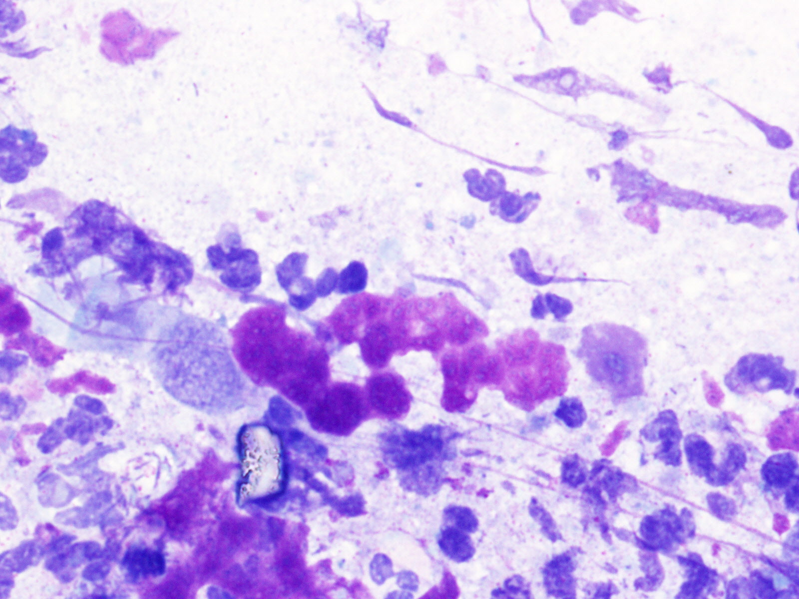 Brush cytology, P. aeruginosa (MGG)
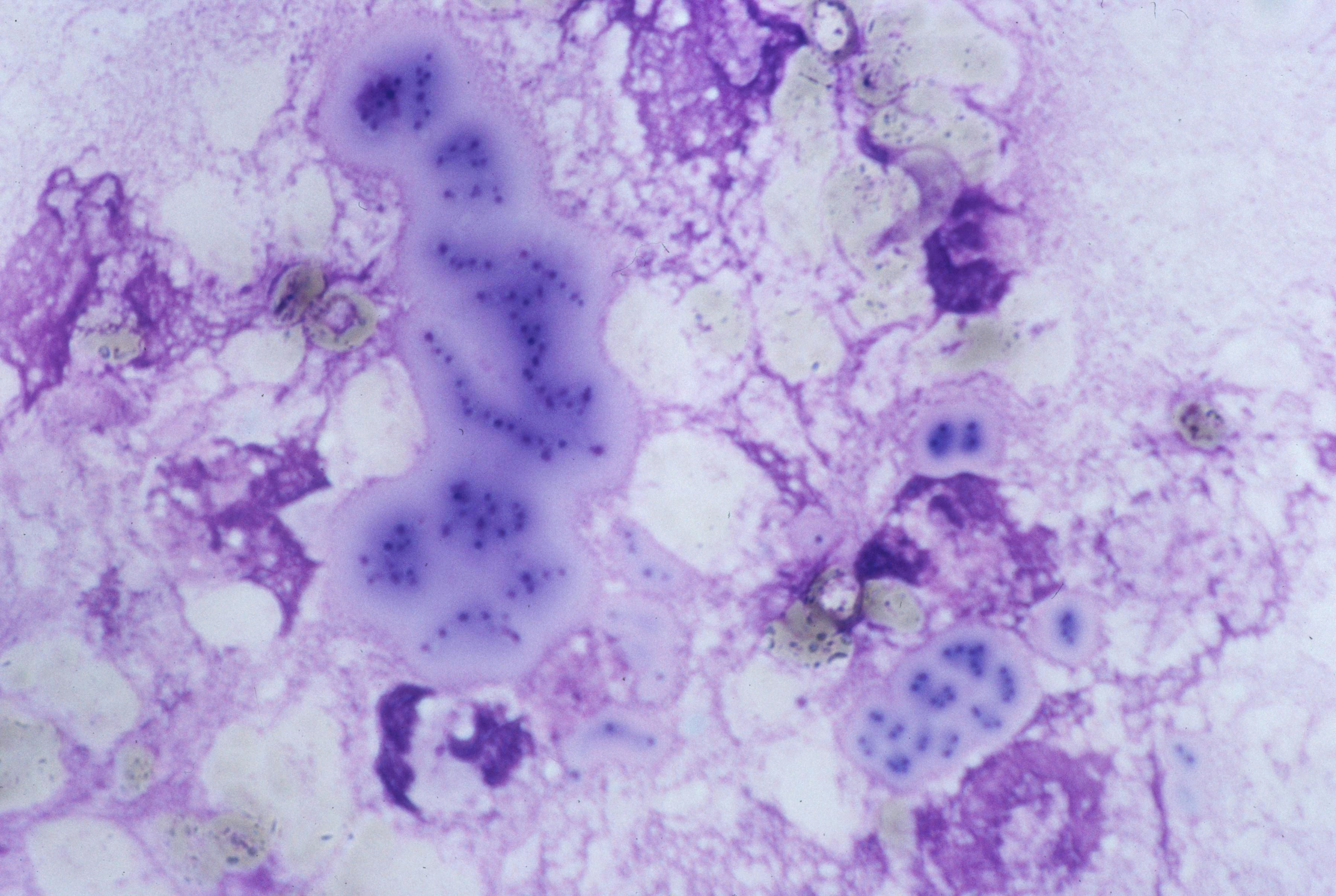 Klebsiella spp.: may oft appear as short…
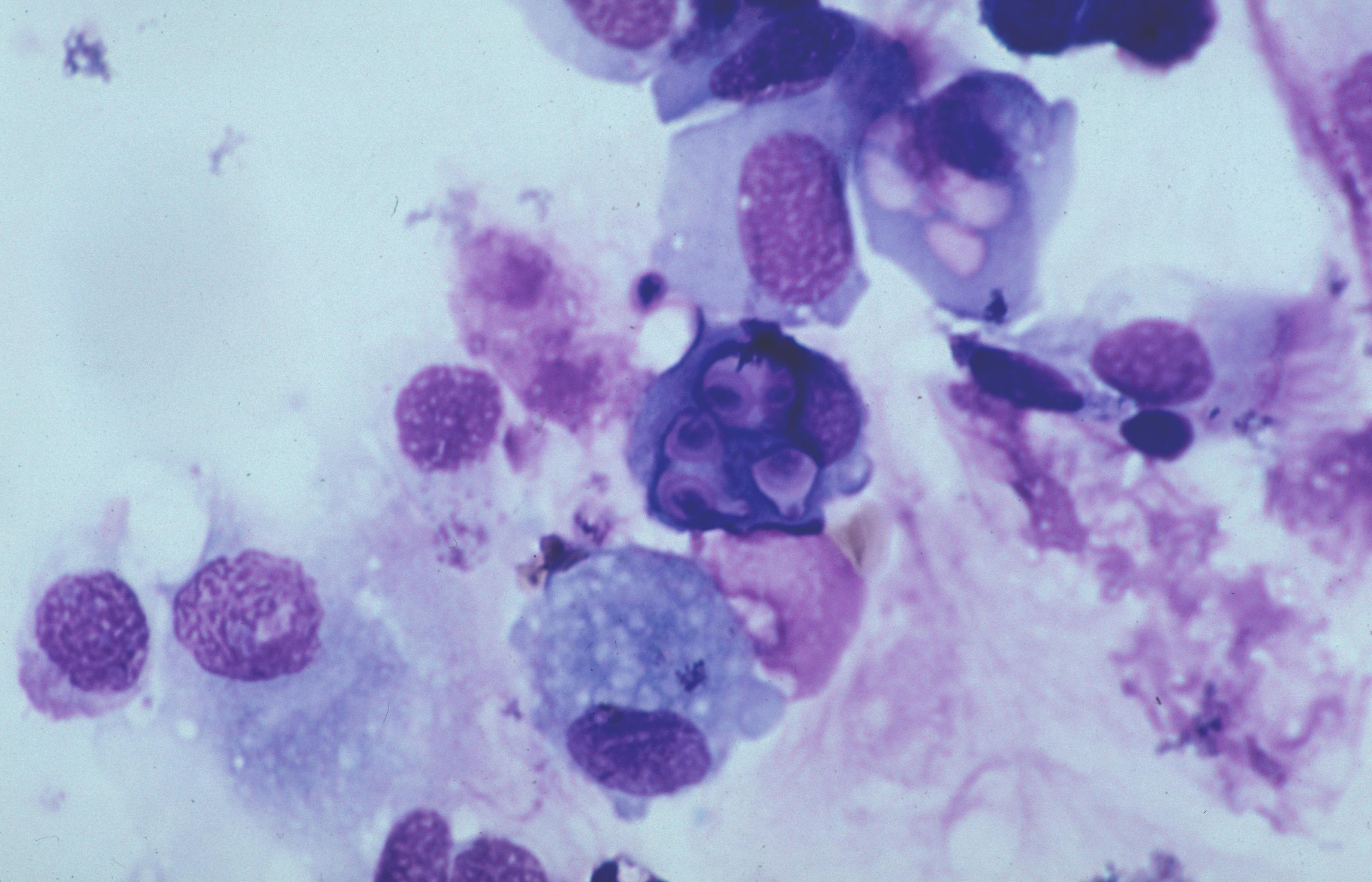
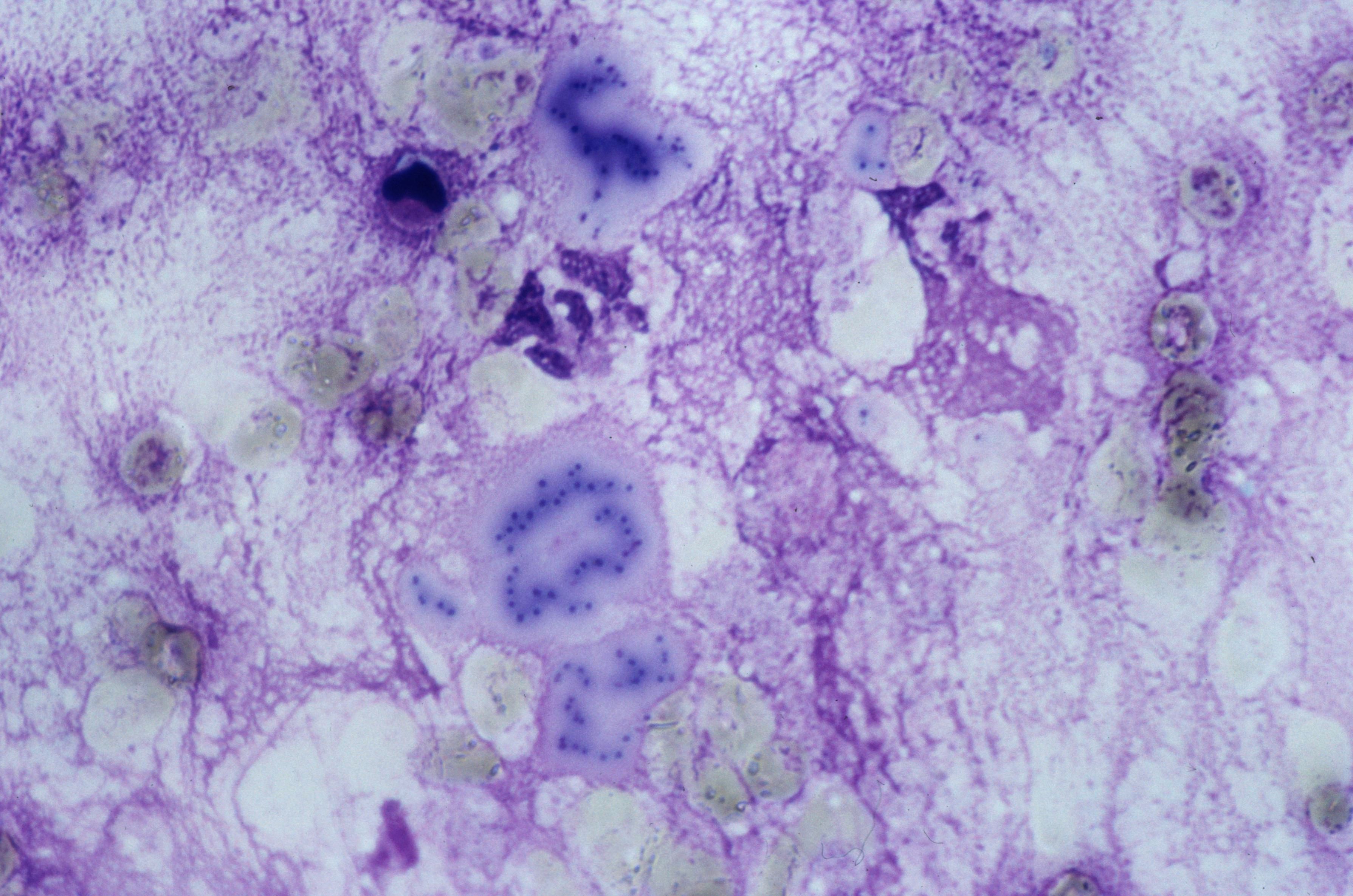 Klebsiella spp. (MGG)
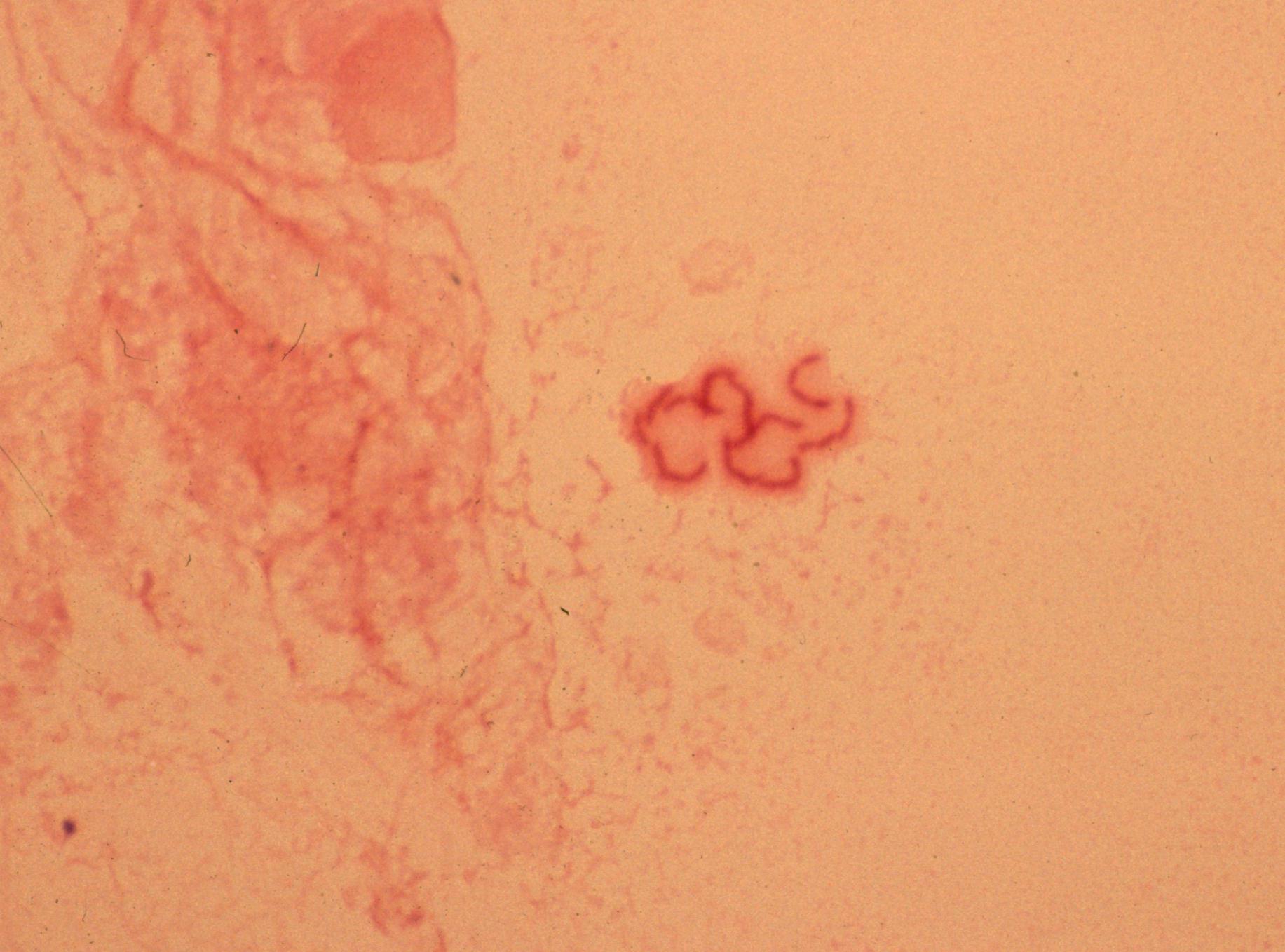 Bacteria with mucoid capsule; Klebsiella…
 The mucoid strain of Pseudomonas aeruginosa…
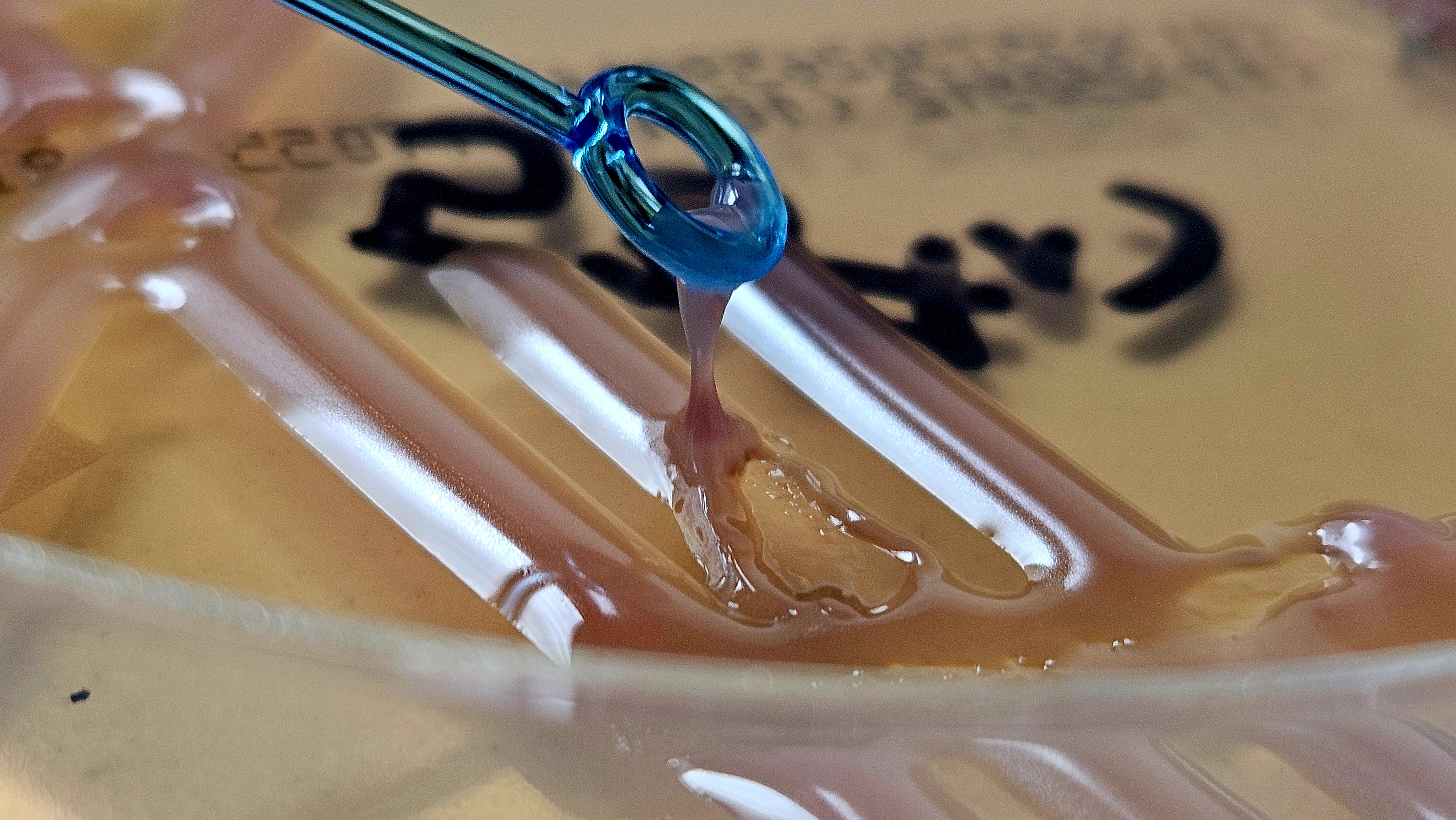 The term ‘mucoid’ is restricted to those…
Morphology
Cytology:
- Gram-negative rods (GNR) in a mucoid, viscous background; abundant extracellular slime
- often a capsule halo effect in Gram (this is not specific)
- intraneutrophilic bacteria in purulent material
- reliable separation from other GNR is not possible on morphology alone
Histology:
Suppurative and necrotizing inflammation; abscesses with copious, viscous exudate. In invasive liver abscess syndrome dense necrosis without obvious biliary source.
Clinical notes
Mucoid variants are associated with worse clinical outcomes and increased antibiotic resistance. They are difficult to eradicate and promote chronic infection and progression.
Hypervirulent Klebsiella can cause primary liver abscess with metastatic spread to eye or CNS, often in diabetes but also in otherwise healthy adults. Mucoid Pseudomonas is typical of chronic airway infection in cystic fibrosis and bronchiectasis. In the lungs of patients with cystic fibrosis, diverse subpopulations of P. aeruginosa often coexist - including mucoid, non-mucoid, and antibiotic-resistant variants.
This microbial heterogeneity poses significant challenges for both treatment and interpretation of culture results.
Contamination: Possible from oropharyngeal or GI flora in non-sterile samples. In sterile sites, clinically significant.
Ancillary Testing
Isolation, bacterial culture, oxidase test, lactose fermentation profile, string test for hypermucoviscosityautomated identification and susceptibility testing systems, MALDI-TOF, AST with attention to ESBL and carbapenemases in Klebsiella and MDR profiles in PseudomonasAdditional Information
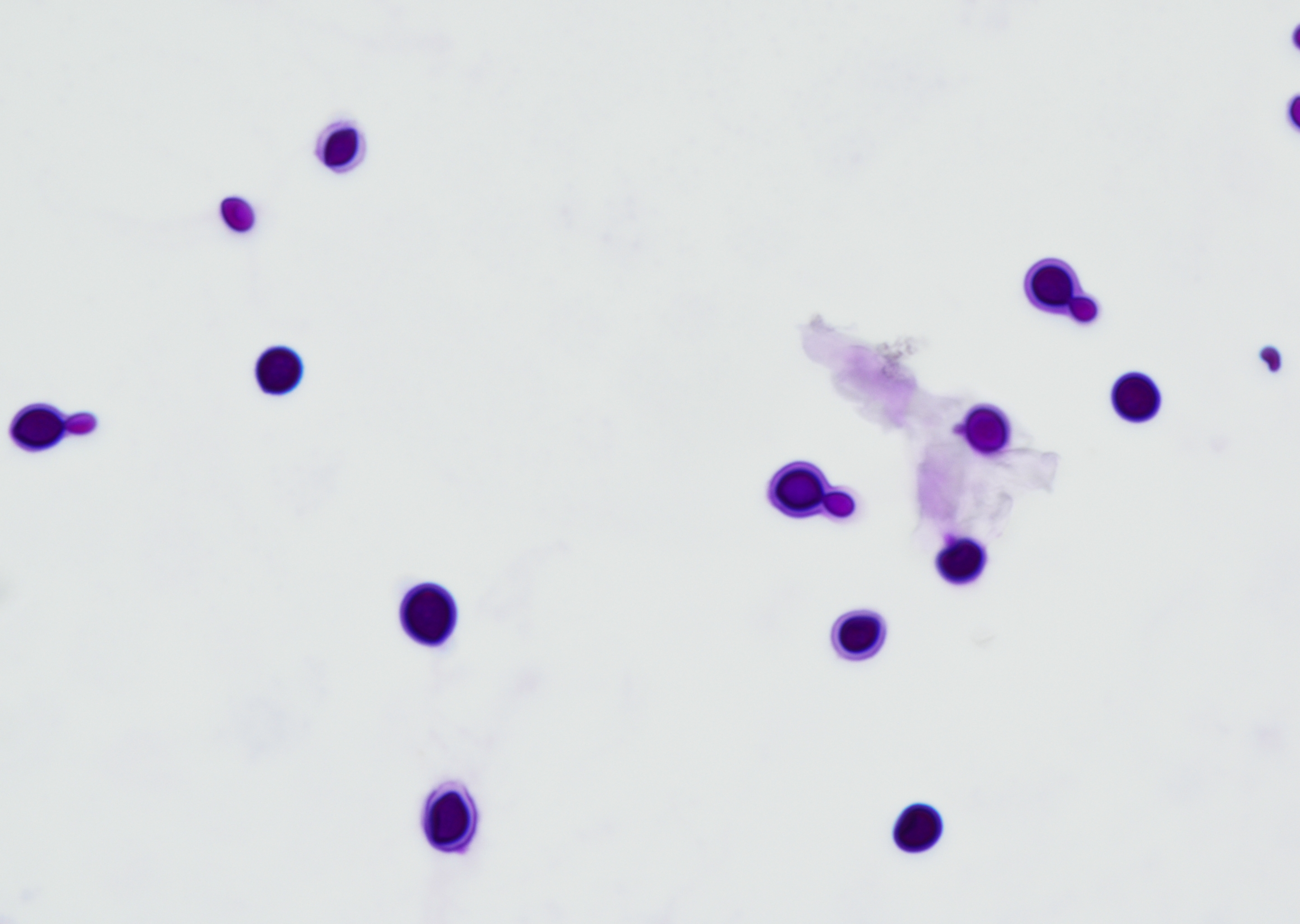
Mucoid appearance reflects abundant extracellular polysaccharide capsule in Klebsiella, alginate in Pseudomonas. Cytomorphology and Gram cannot reliably distinguish Klebsiella from Pseudomonas and other GNR; use ancillary testing. Differential Diagnosis: Non-mucoid Enterobacterales; Serratia/Enterobacter/Escherichia; non-fermenters such as Stenotrophomonas and Achromobacter; yeast with capsule such as Cryptococcus in mucinous background.
The mucoid phenotype of Pseudomonas aeruginosa may be unstable and can revert to a non-mucoid phenotype in vitro or in vivo. Therefore, non-mucoid colony morphology in culture does not reliably exclude mucoid behavior in vivo, particularly in chronic airway or biofilm-associated infection (1).
Differential Diagnosis
References
- Malhotra S, Limoli DH, English AE, Parsek MR, Wozniak DJ2018.Mixed Communities of Mucoid and Nonmucoid Pseudomonas aeruginosa Exhibit Enhanced Resistance to Host Antimicrobials. mBio9:10.1128/mbio.00275-18.https://doi.org/10.1128/mbio.00275-18