
- EN
- DE

Fungi
Differential Diagnosis of Pneumocystis jirovecii:
Histoplasma Species

- Organ System
- Respiratory Tract
- Diagnostic Method
- Cytology

- Geographical Region
- Dry Areas,
- Islands,
- Tropical and Subtropical Region,
- Polar and Subpolar Zone,
- Temperate Zone
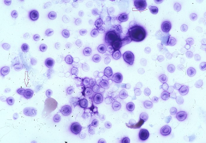
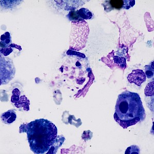
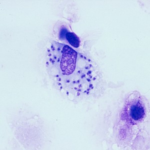
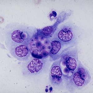
 Lung, BAL, predominantly small oval…
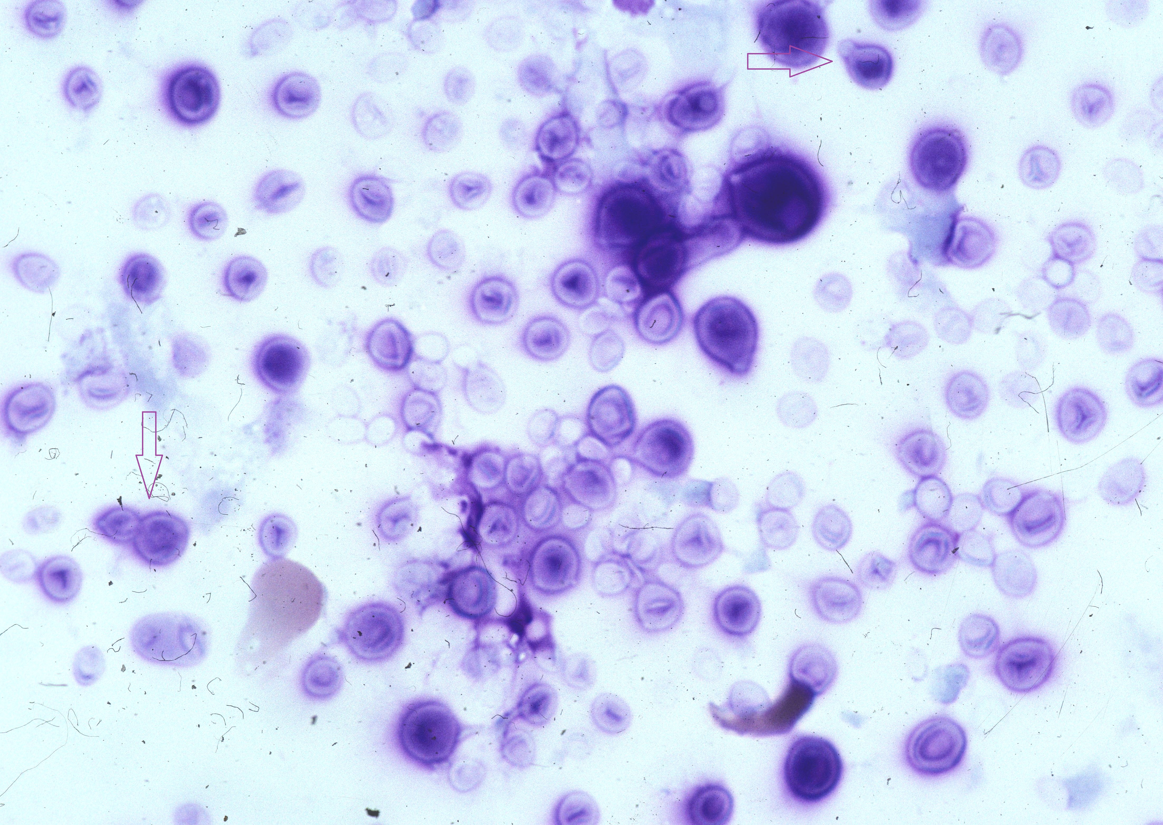
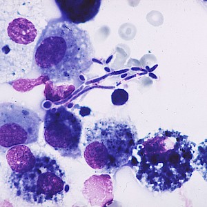
 Lung, bronchial secretion, round…
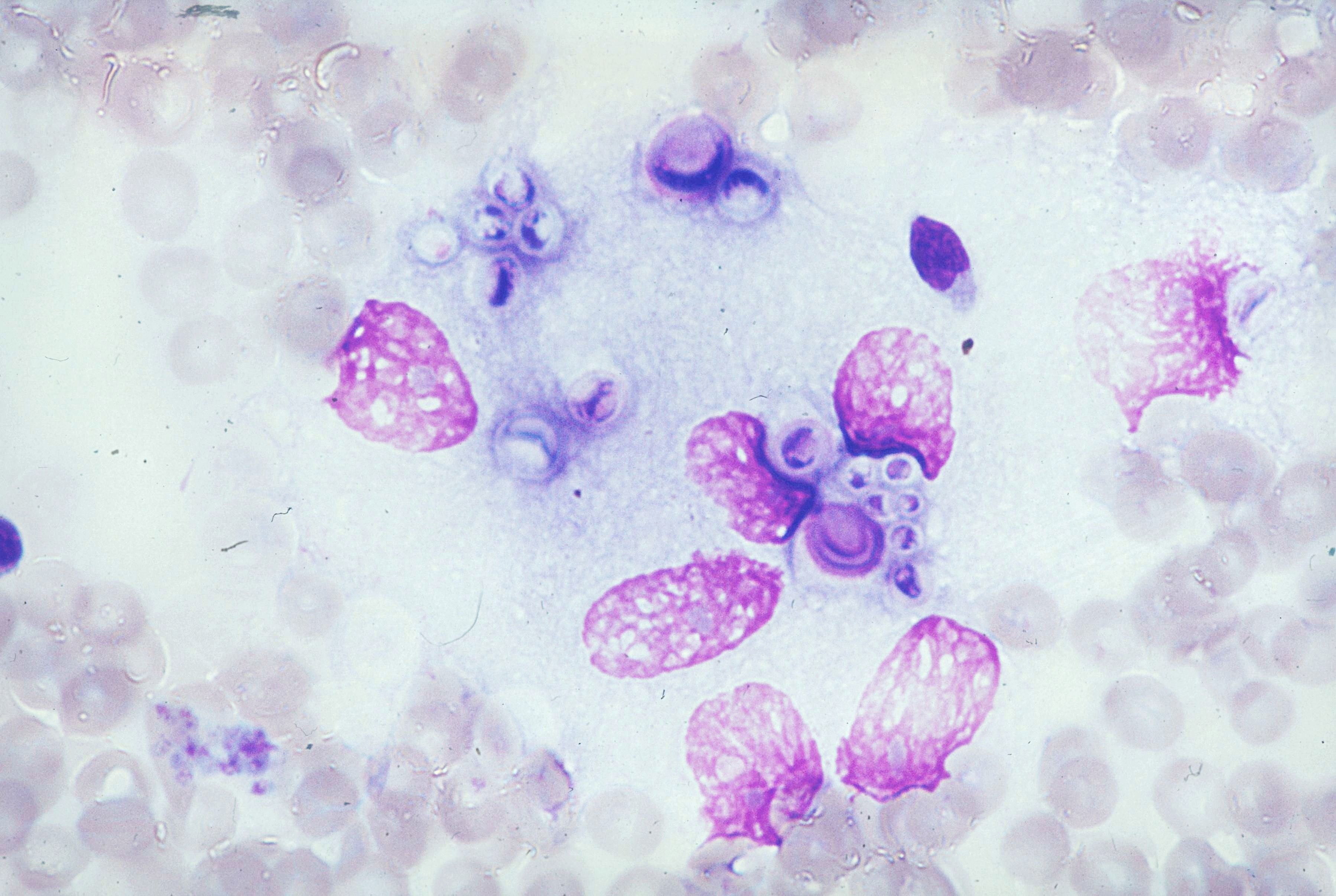
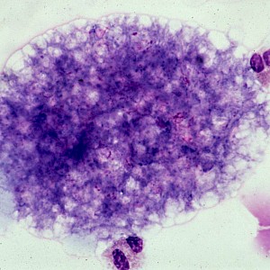
 Lung, bronchial secretion, macrophages…
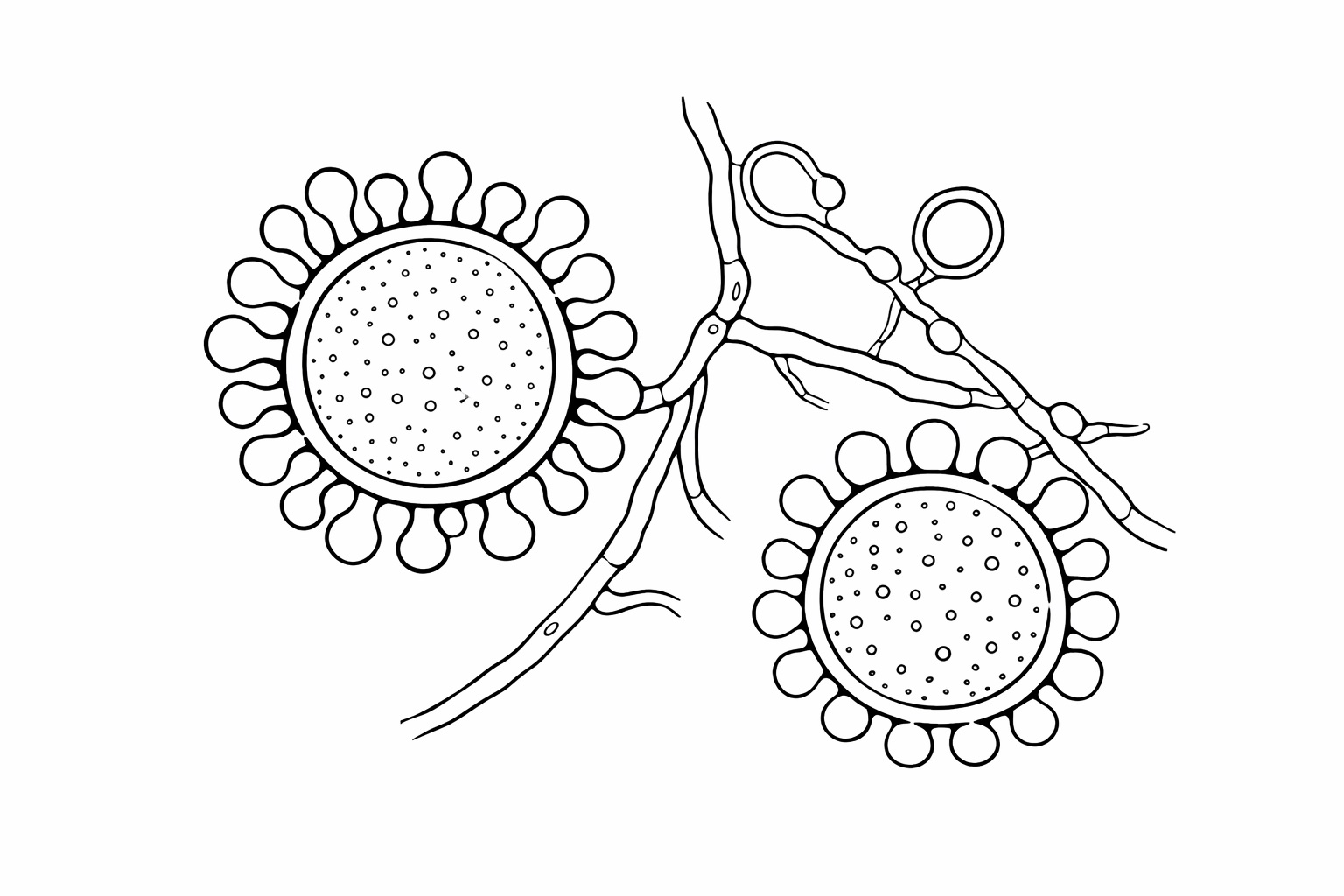 Schematic line drawing of Histoplasma…
Morphology
H. capsulatum: Small oval, non-encapsulated yeast cells with narrow-based budding, measuring 2-6 µm in diameter.
H. duboisii: Small to large, round to oval yeast cells with narrow-based budding, and with a range of 8 to 15 µm in diameter.
Cytology:
- Numerous small yeasts are usually found intracellularly in macrophages.
- Extracellular yeast cells are more commonly found in disseminated disease.
- The cell wall appears as a clear rim surrounding the yeast cells, and can mimic a pseudocapsular appearance.
- A granulomatous reaction may be seen in the background.
Histology: Yeast cells morphologically similar to those seen in cytology. The histological reaction pattern depends on the immune status and duration of infection. Tuberculoid, anergic, mixed/intermediate and fibrotic sequelae forms have been described: (1)
- Tuberculoid form: granulomatous, partly necrotizing inflammation with usually few intracytoplasmic yeast cells within histiocytes.
- Anergic form: typically associated with severe immunosuppression or disseminated disease; numerous intra- and extracellular yeast cells with minimal or variable tissue reaction.
- Mixed/intermediate form: transitional pattern between tuberculoid and anergic reaction.
- Sequelae form: predominant fibrosis with mild inflammation and, occasionally, rare yeast cells.
Special stains: Yeast cells not readily seen in HE; PAS and fungal silver stains (Grocott/GMS) highlight the cell wall.
Differential Diagnosis:
Tuberculosis - granulomatous inflammation; no yeast forms. Ziehl-Neelsen, auramine or mycobacterial PCR/culture support the diagnosis.
Sarcoidosis - compact, usually non-necrotizing granulomas; no organisms on fungal or mycobacterial stains. Diagnosis only after exclusion of infection.
Other fungi such as C. glabrata - can be very similar in size, but usually more variable in size and more often extracellular; may stain better on H&E/Gram, often without the typical intrahistiocytic clustering of Histoplasma. Culture or molecular confirmation may be needed.
Cryptococcus spp. - usually larger yeasts with a true mucopolysaccharide capsule highlighted by mucicarmine or Alcian blue. Narrow-based budding may be present. Capsule-deficient forms can mimic Histoplasma.
Blastomyces spp. - larger yeasts, typically about 8-15 µm, with thick refractile wall and broad-based budding; highlighted by GMS/PAS.
Pneumocystis jirovecii - no true budding yeast cells; classically foamy or honeycomb-like alveolar casts in BAL. GMS highlights small cup-shaped cysts, often with central dot-like staining.
Protozoans such as Toxoplasma spp. - intracellular tachyzoites or tissue cysts with bradyzoites; no fungal wall, therefore not GMS-positive like yeasts.
Leishmania spp. - small intracellular amastigotes in macrophages, with nucleus and rod-shaped kinetoplast; Giemsa is helpful. GMS negative, unlike fungal yeasts.
Trypanosoma cruzi - amastigotes with nucleus and kinetoplast, often in pseudocysts within muscle, especially myocardium; morphologically similar to Leishmania, so topography, clinical context and PCR/serology are important.
Clinical notes
Histoplasma spp. are thermally dimorphic fungi that exist as mould in the environment and convert to the yeast form in vitro and in tissue.
Infection via inhalation of microconidia.
Histoplasmosis shows a broad clinical spectrum with pulmonary, mediastinal and disseminated manifestations. In immunocompetent patients, infection is often asymptomatic or self-limiting. Symptomatic disease is mainly observed after high exposure or in patients with impaired cell-mediated immunity. Severely immunosuppressed patients are at increased risk of disseminated disease, with possible involvement of various organ systems. (2, 3)
Relevant humapathogenic species are H. capsulatum and H. duboisii.
Ancillary Testing
Fungal culture, MALDI-TOF, antigen detection (EIA; urine and serum; in extrapulmonary diesaese), serology (Complement fixation test - CAVE: cross reactions; Immunodiffusion assay - detection of M and H antigens), RT-PCR, broad-spectrum assays.Additional Information
Temperature-dependent dimorphism: H. capsulatum grows in mycelial form at room temperature (25°C) and in yeast form at body temperature (37°C).
H. capsulatum shows a global distribution, but is most commonly found in North and Central America, and H. duboisii occurs only in Central and West Africa; found in nitrogen-rich soil and frequently in caves, associated with bird and bat guano.
Differential Diagnosis
References
- Drak Alsibai K, Couppié P, Blanchet D, Adenis A, Epelboin L, Blaizot R, Louvel D, Djossou F, Demar M and Nacher M (2020) Cytological and Histopathological Spectrum of Histoplasmosis: 15 Years of Experience in French Guiana. Front. Cell. Infect. Microbiol. 10:591974. doi: 10.3389/fcimb.2020.591974
- Clinical Overview of Histoplasmosis | Histoplasmosis | CDC
- Arnold, S. R., Spec, A., Baddley, J. W., Pappas, P., Lentz, R. J., Wolf, J., Kauffman, C. A., Ardura, M. I., Bahr, N. C., Bloch, K., Miller, R. A., Mocherla, S., Saccente, M., Schwartz, I., Stover, K. R., Wiederhold, N. P., & Loveless, J. (2025). 2025 clinical practice guideline update by the Infectious Diseases Society of America on histoplasmosis. Clinical Infectious Diseases.