
- EN
- DE

Parasites
Differential Diagnosis of Histoplasma Species:
Toxoplasma gondii

- Organ System
- Lymphatic System, Soft Tissue & Bone, Respiratory Tract, CNS and Sensory Organs
- Diagnostic Method
- Cytology

- Geographical Region
- Dry Areas,
- Islands,
- Tropical and Subtropical Region,
- Polar and Subpolar Zone,
- Temperate Zone
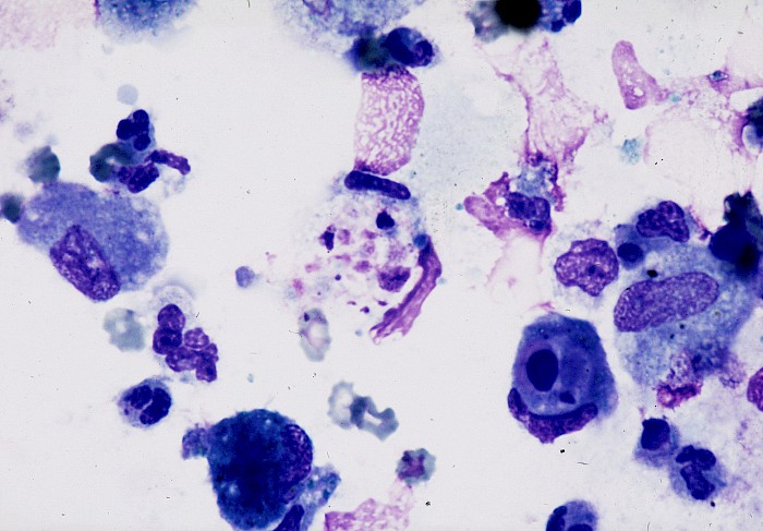
 Lung, BAL, tachyzoites, about 2 x 6 µm…
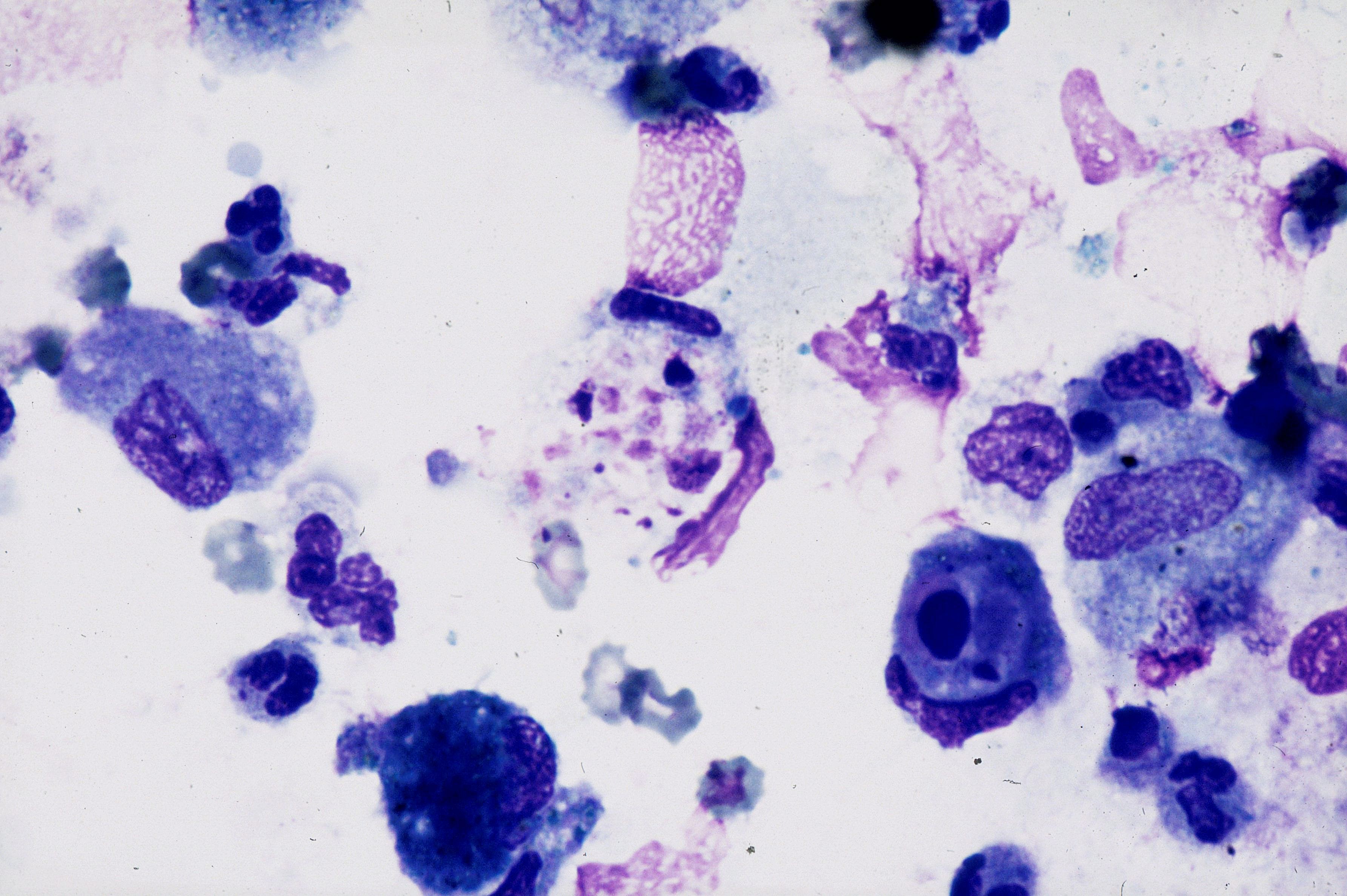
 Lung, BAL, tachyzoites crescent-shaped…
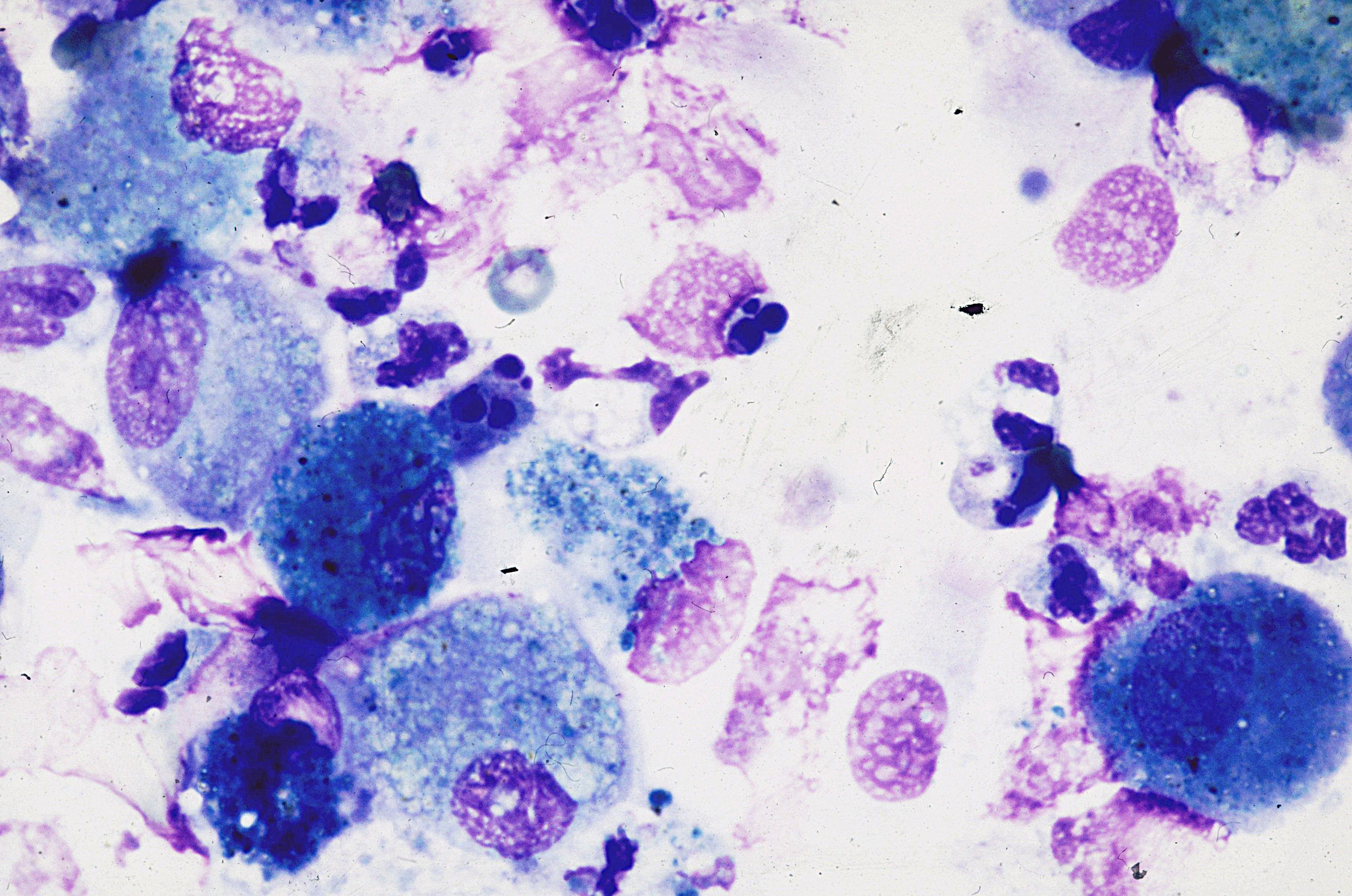
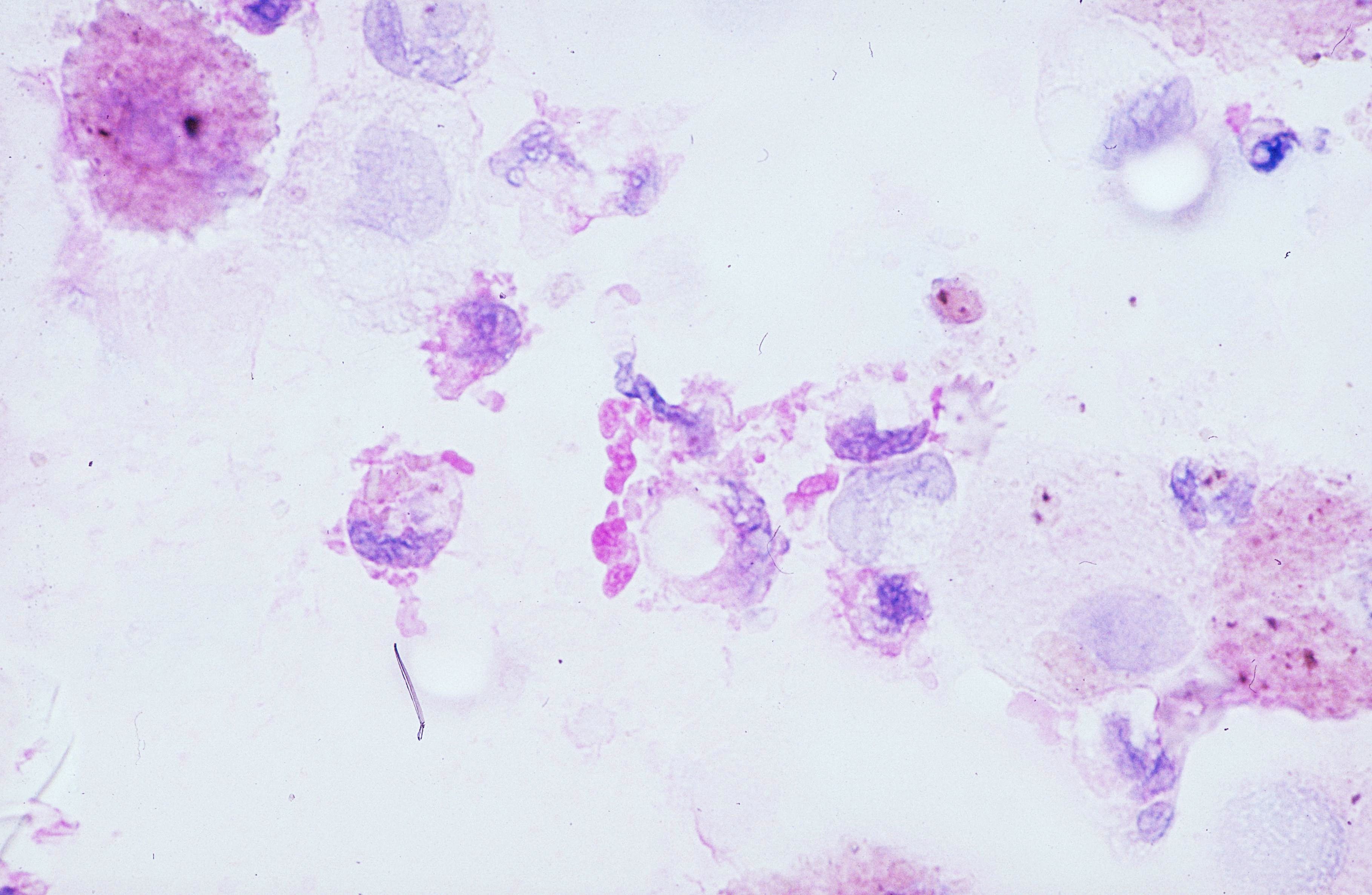 Lung, BAL, long slender bradyzoites…
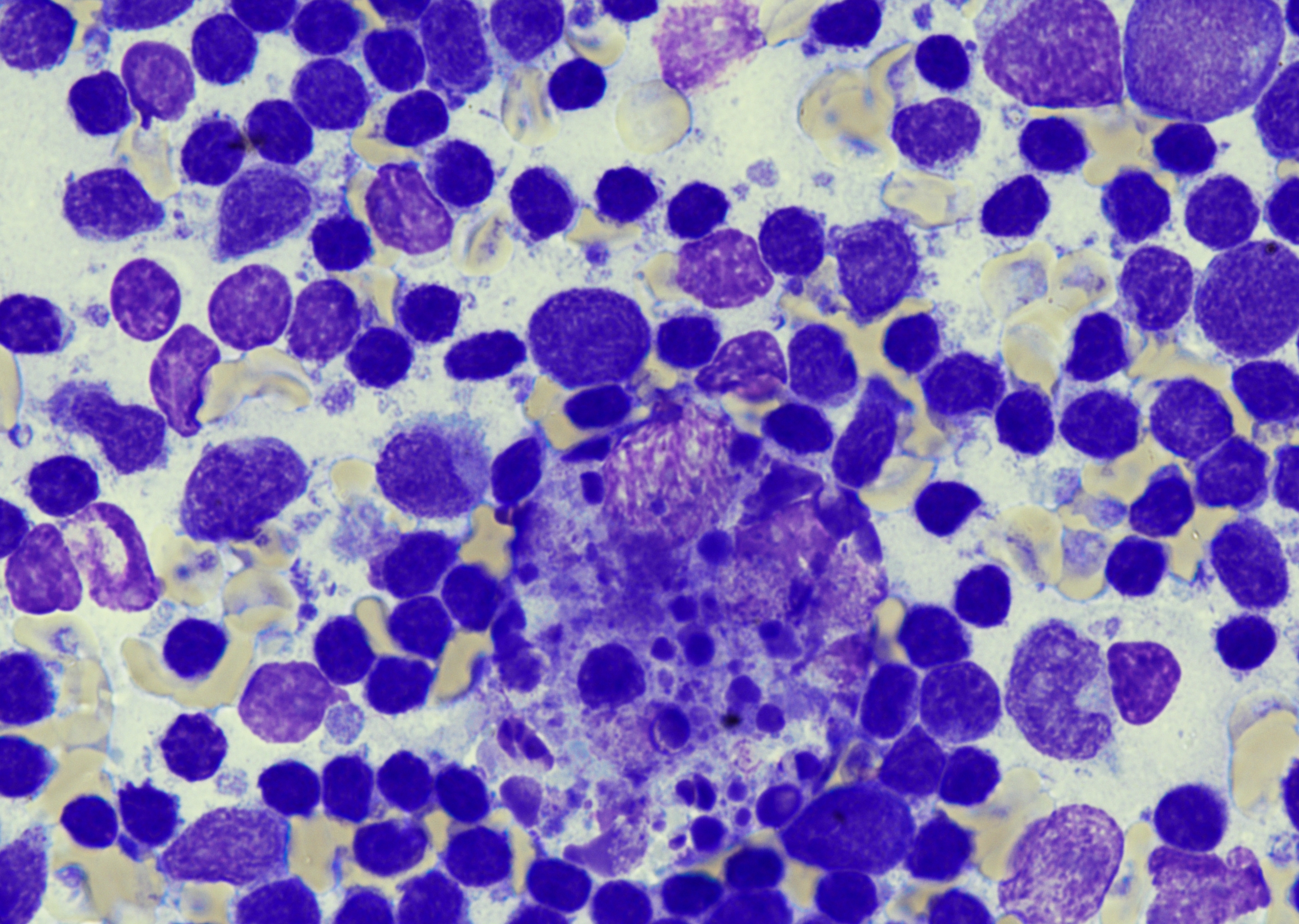 Lymph node, FNA, toxoplasma lymphadenitis…
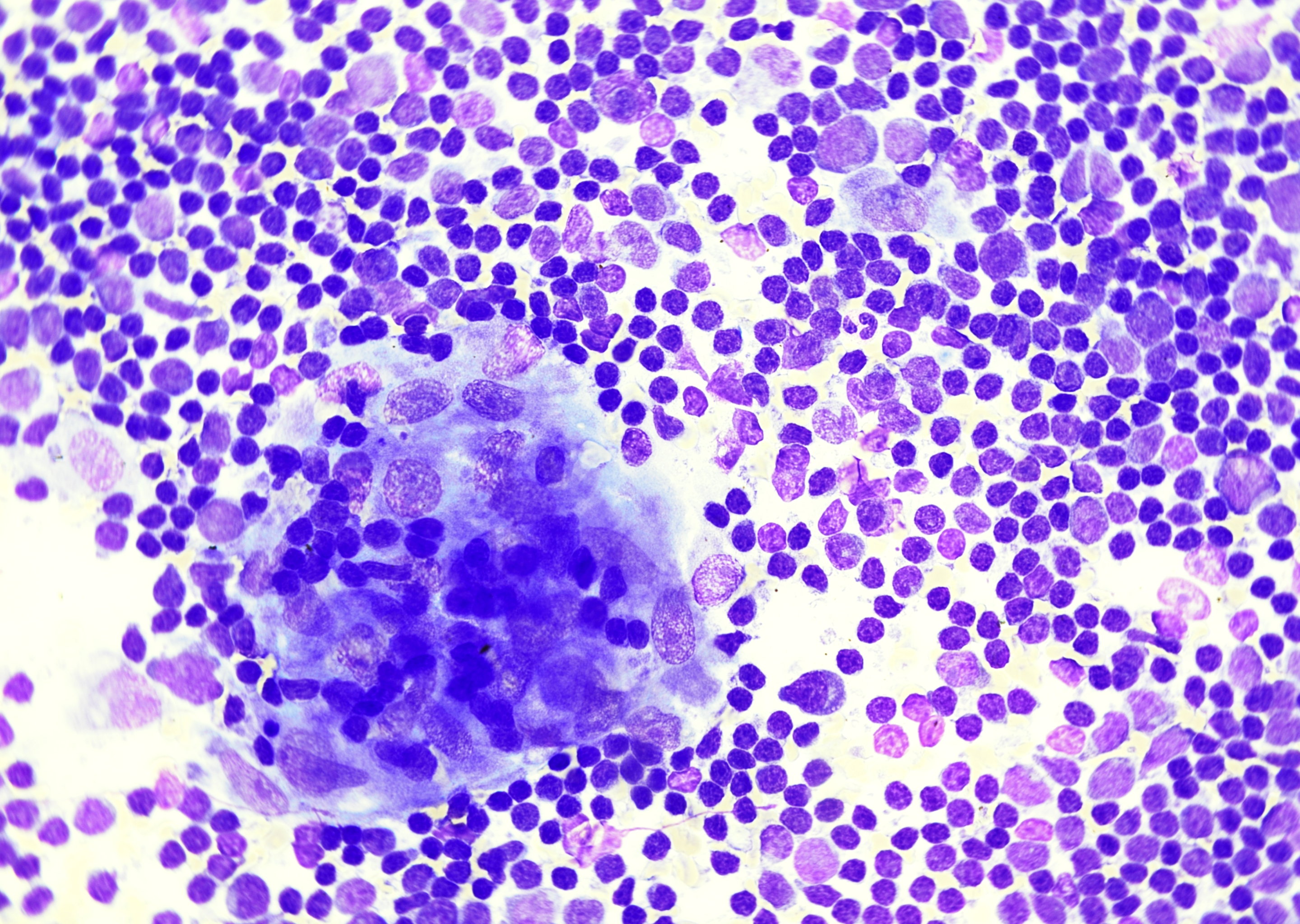 Lymph node, FNA, toxoplasma lymphadenitis…
Morphology
Cytology:
- Necrosis and microgranulomas can be seen in the background
- Tachyzoites crescent-shaped, about 6 x 2 µm, pointed at one end, often intracellular in macrophages but can be seen in the extracellular space
- Blue cytoplasm and red chromatin on Giemsa stain, often PAS negative; usually scarce and easy to miss
- Tissue cysts with bradyzoites are uncommon in cytology preparations
- The cyst wall and bradyzoites are typically PAS-positive
Histology:
- CNS tissue may show vasculitis and necrotizing encephalitis with tachyzoite clusters and cysts (no pictures available), often at the viable-necrotic interface
- In FFPE sections, tachyzoites may appear round to oval rather than distinctly crescentic; scattered spherical cysts, about 5 to 100 µm, are PAS positive and contain dozens of bradyzoites
- Ocular toxoplasmosis is characterized by necrotizing retinochoroiditis with vitritis
- Lymph nodes typically show reactive follicles, epithelioid microgranulomas, sinus histiocytosis, and monocytoid B-cell hyperplasia
- Organisms are rarely identified on routine HE stain
- T. gondii immunohistochemistry is useful
Differential Diagnosis:
- CNS: primary CNS lymphoma, tuberculosis, Nocardia, Aspergillus spp.: bacterial or fungal abscess
- Eye: CMV retinitis, herpetic retinitis
- Lymph node: EBV-associated reactive hyperplasia, infectious mononucleosis, Hodgkin lymphoma, HIV-related lymphadenopathy, Bartonella henselae lymphadenitis (cat-scratch disease), sarcoidosis, Rosai-Dorfman disease
- Cysts: Sarcocystis spp. (larger, thicker cyst wall, often compartmentalized, cytoplasm of bradyzoites PAS negative), Trypanosoma cruzi (heart tissue, amastigotes with kinetoplast)
Clinical notes
Immunocompetent: Usually asymptomatic or presenting with mild lymphadenitis; ocular toxoplasmosis (usually posterior uveitis) may cause floaters and decreased vision.
Immunosuppressed (patients with HIV and CD4 counts below 100 cells/µL or transplant recipients): Encephalitis with ring-enhancing brain lesions, fever, headache, and focal neurological deficits.
Congenital: Hydrocephalus, intracranial calcifications, chorioretinitis.
Ancillary Testing
Serology IgG and IgM; IgG avidity to time infection in pregnancy; PCR (CSF, blood, amniotic fluid, tissue), in ocular disease PCR on aqueous or vitreous fluid; radiology (brain); Loop-mediated isothermal amplification (LAMP)Additional Information
Lymph nodes may show a reactive pattern compatible with toxoplasmosis, with clinicopathologic correlation and, where appropriate, confirmation by serology or PCR.
In HIV, reactivation and prophylaxis are relevant considerations, particularly in Toxoplasma-IgG-positive patients with CD4 counts below 100 cells/µL.
During pregnancy, relevant exposures include undercooked meat, unwashed contaminated produce, soil, and cat litter. A recent infection is suggested by a positive IgM with low IgG avidity, with PCR used in selected settings, especially in amniotic fluid.
References
Madireddy S, Mangat R. Toxoplasmosis. [Updated 2024 Oct 14]. In: StatPearls [Internet]. Treasure Island (FL): StatPearls Publishing; 2026 Jan-. Available from: https://www.ncbi.nlm.nih.gov/books/NBK563286/