- EN
- DE

Fungi
Differential Diagnosis of Histoplasma Species:
Cryptococcus Species

- Organ System
- Respiratory Tract, CNS and Sensory Organs
- Diagnostic Method
- Cytology

- Geographical Region
- Dry Areas,
- Islands,
- Tropical and Subtropical Region,
- Polar and Subpolar Zone,
- Temperate Zone
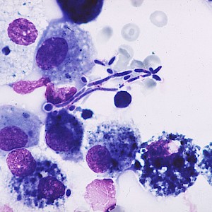
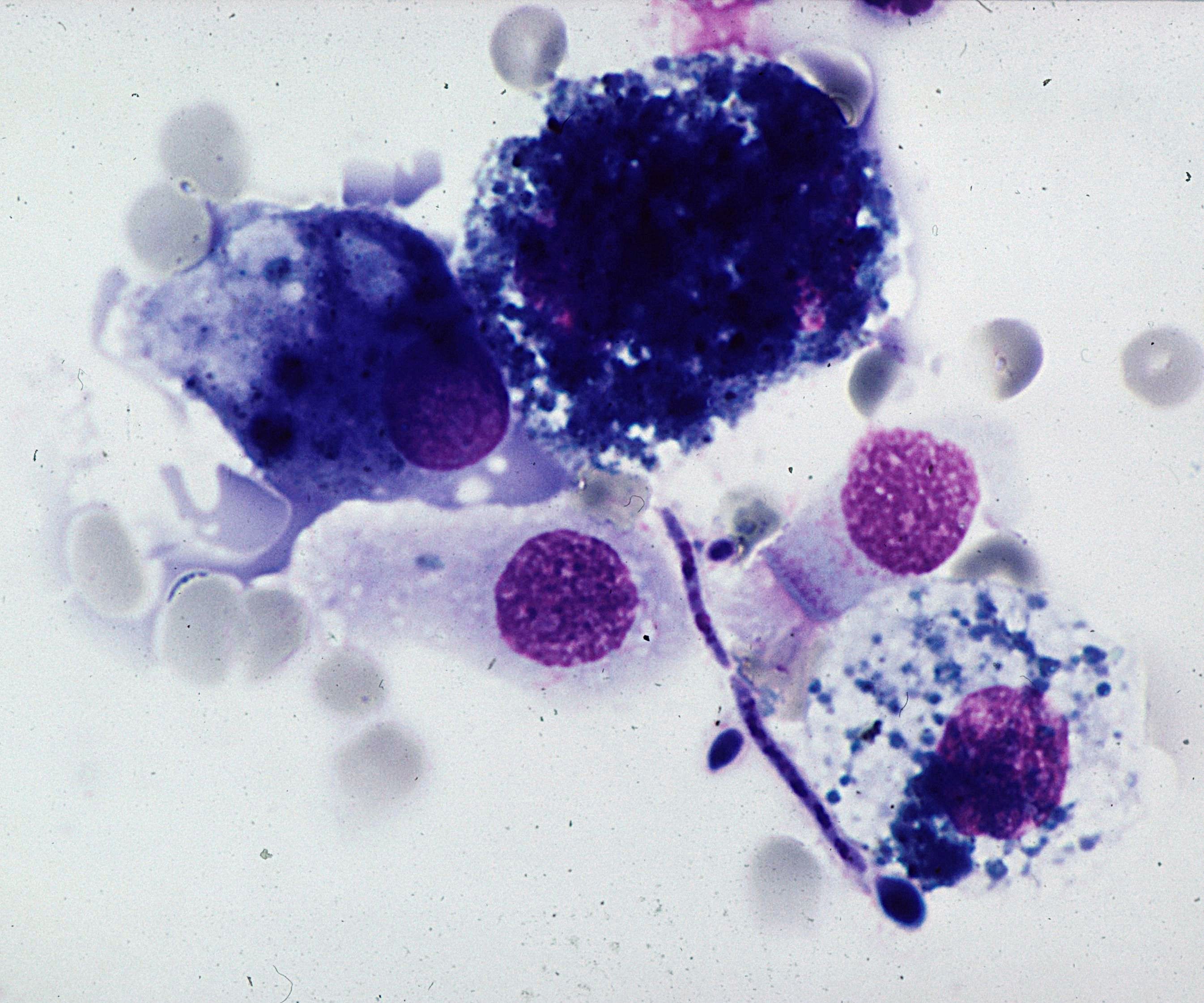
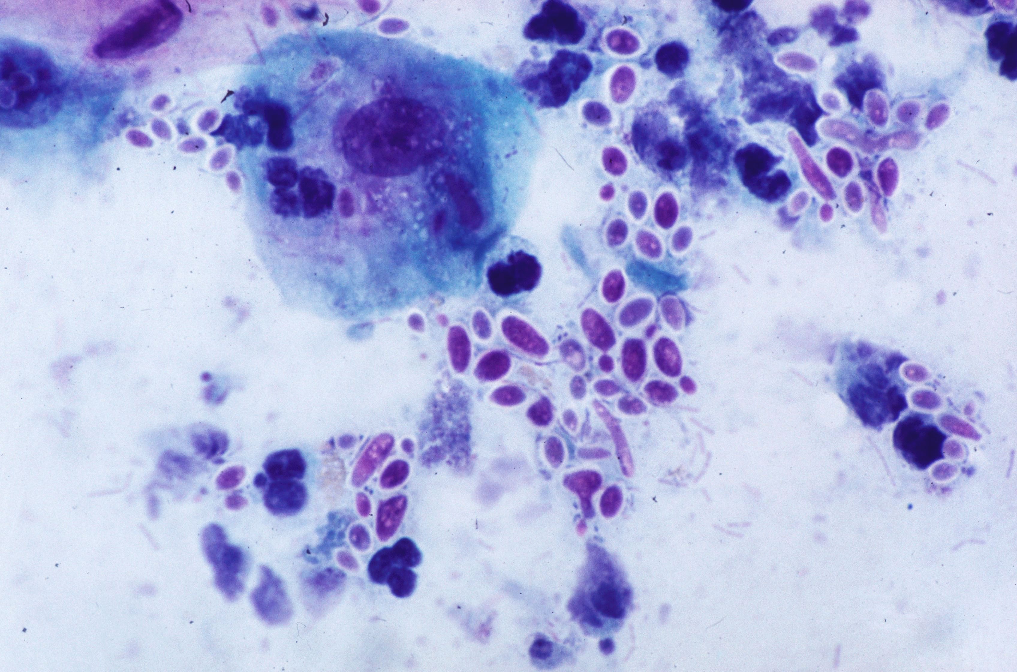
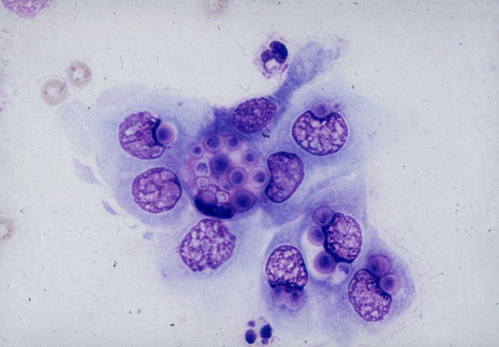
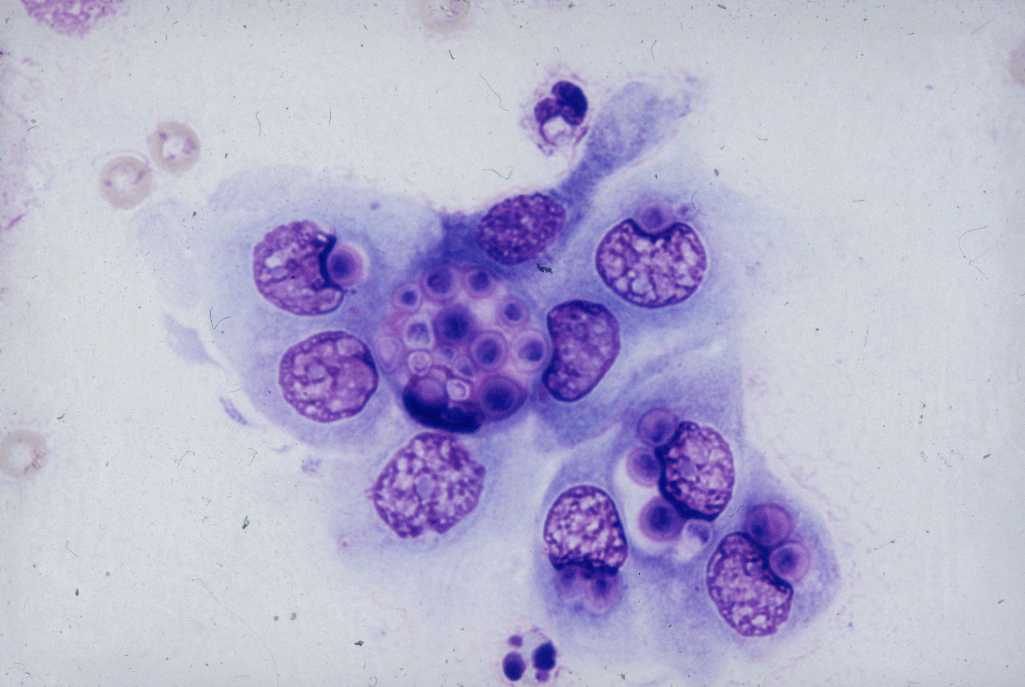 Intracytoplasmic round, spherical fungal…
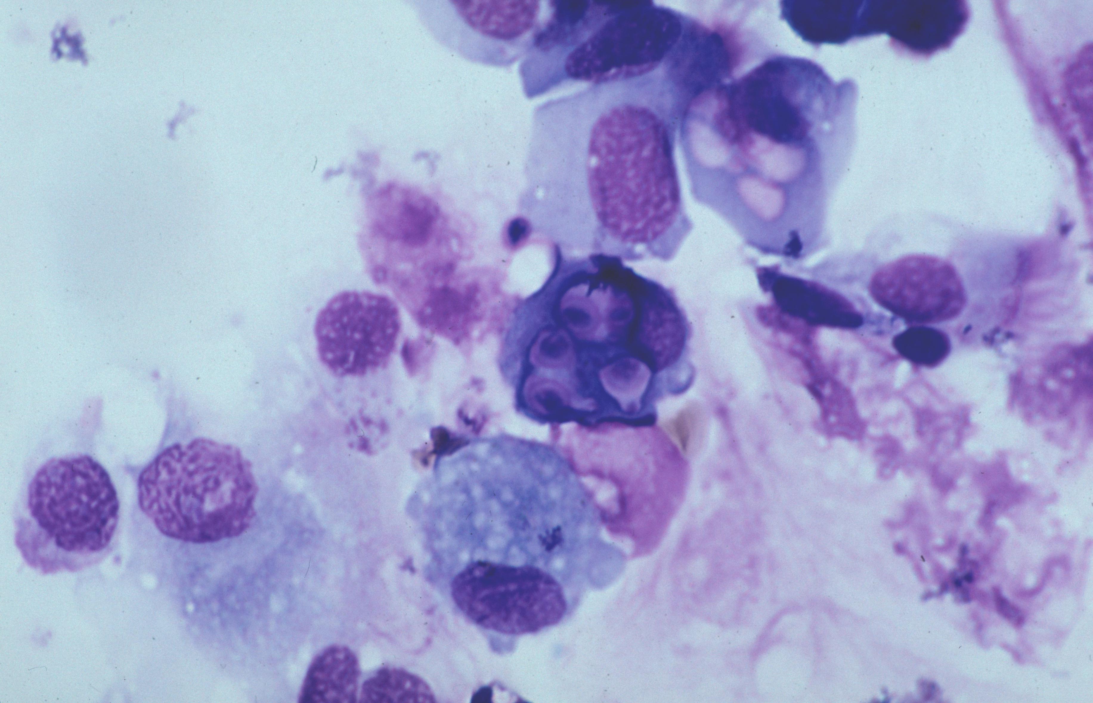 BAL, macrophages with intracytoplasmic…
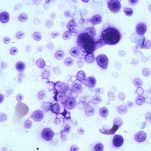
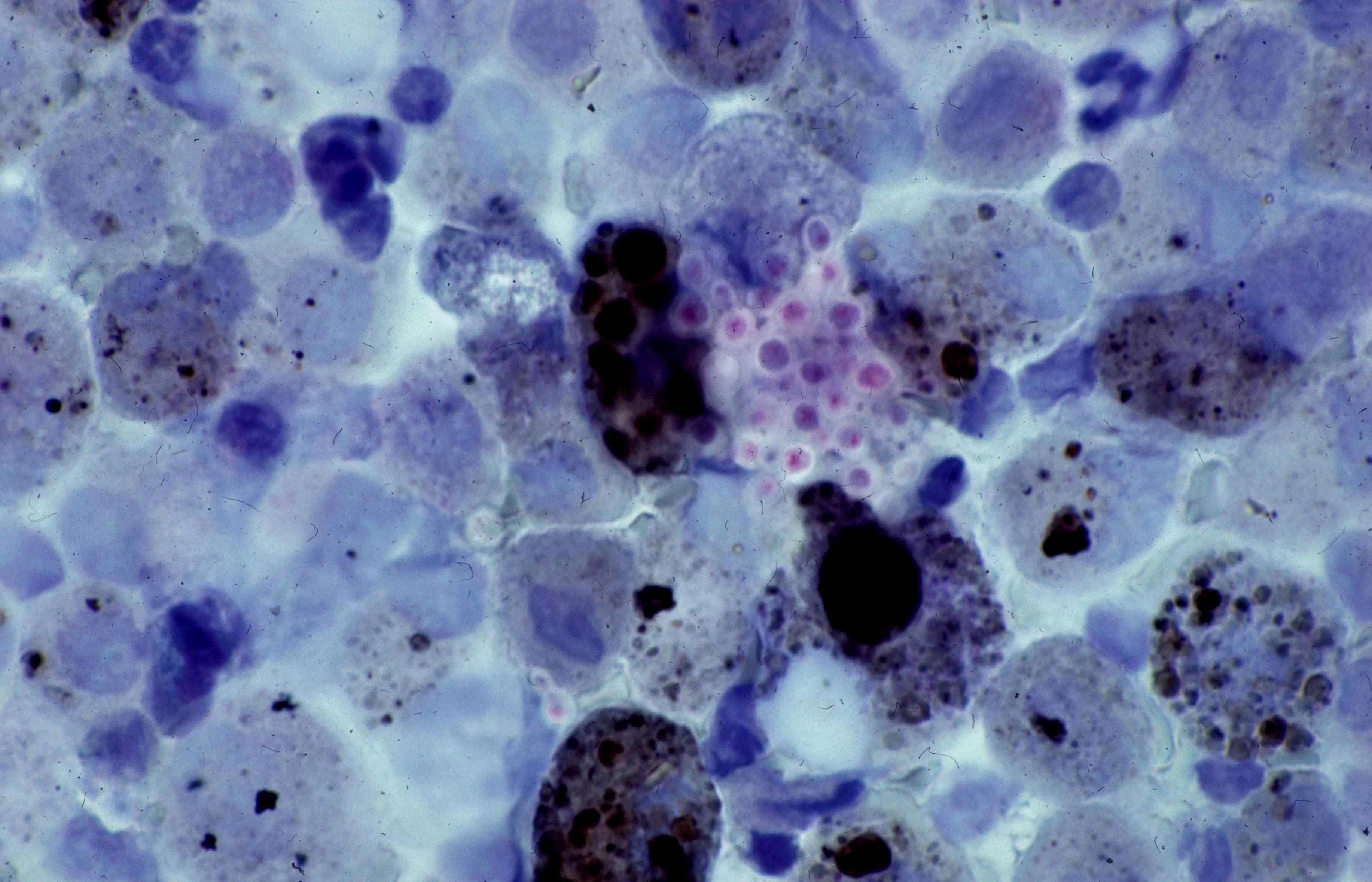 BAL with clusters of round, spherical fungal…
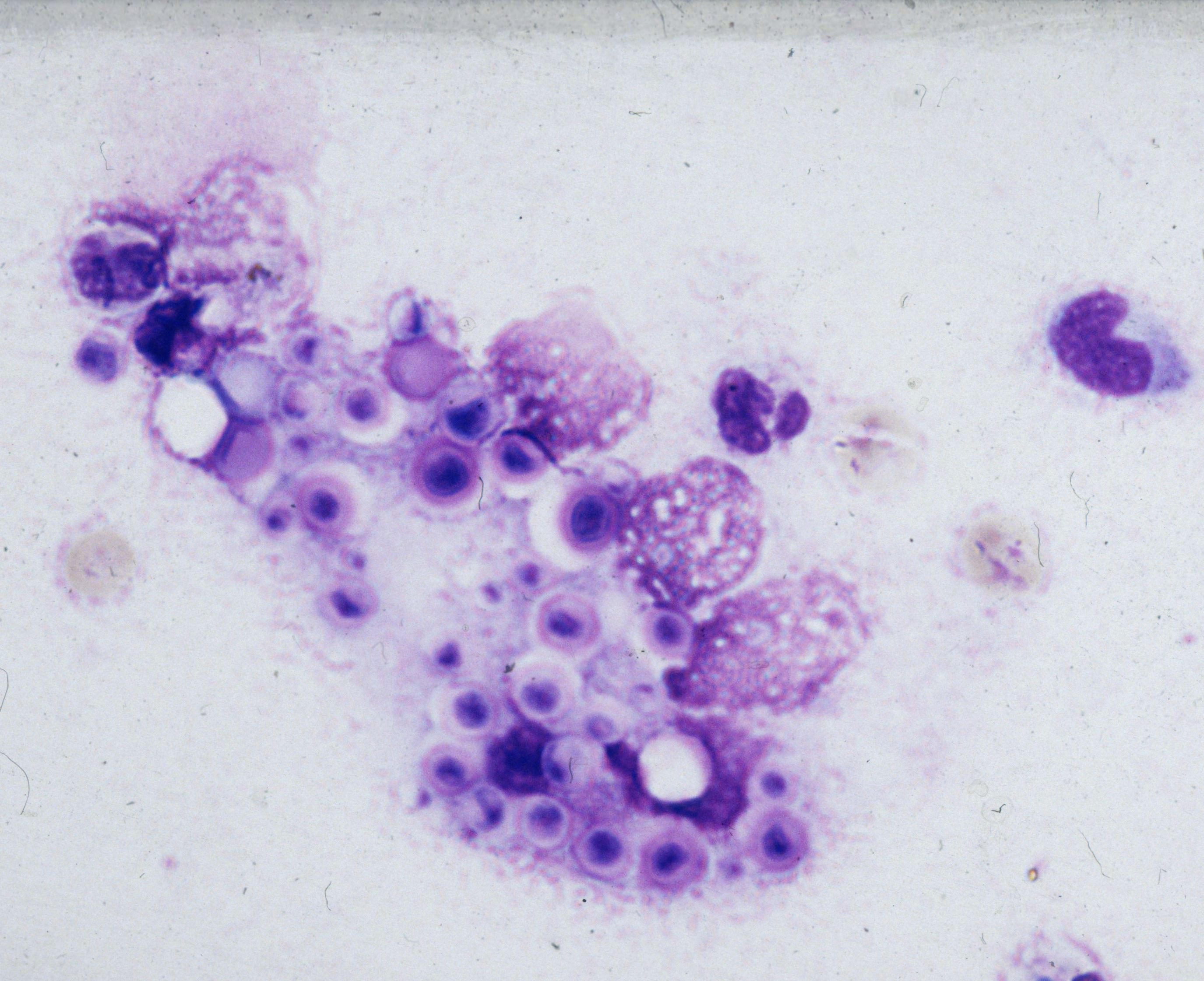 Clusters of round, spherical fungal cells…
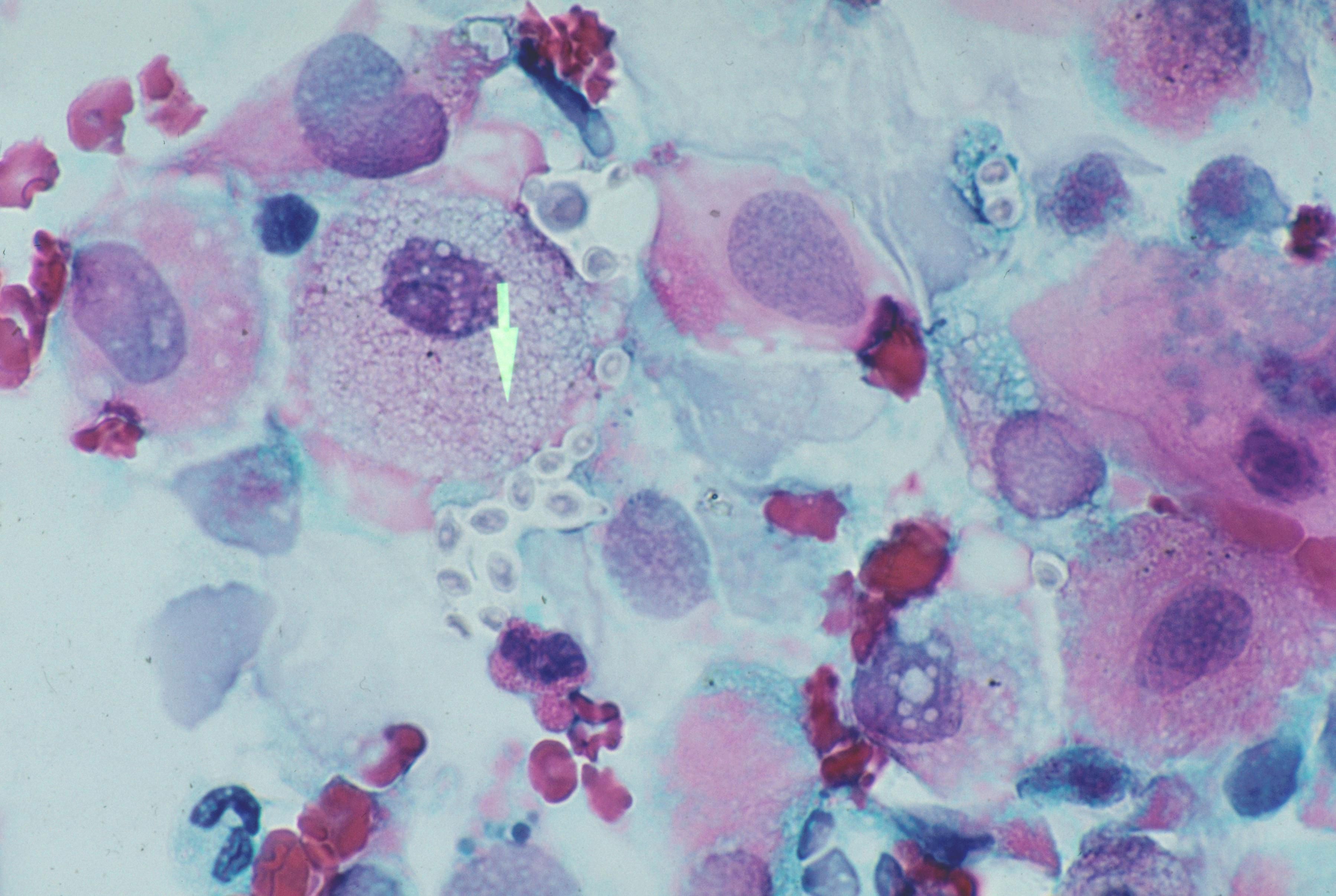 Clusters of round, spherical fungal cells…
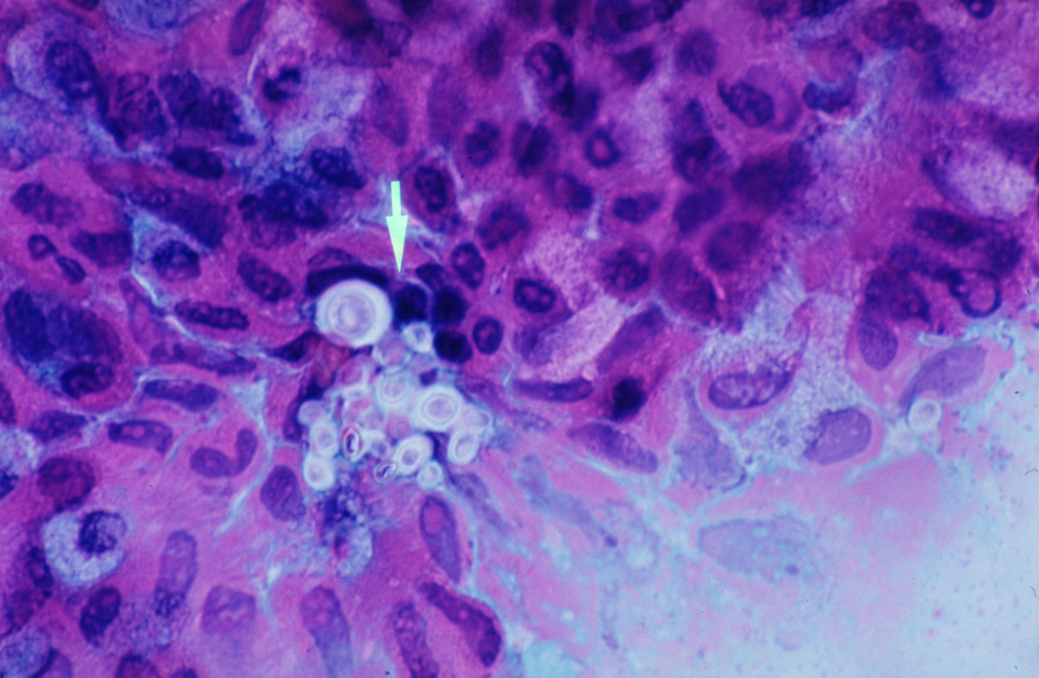 Clusters of round, spherical fungal cells…
 CSF, spherical fungal cells with a broad…
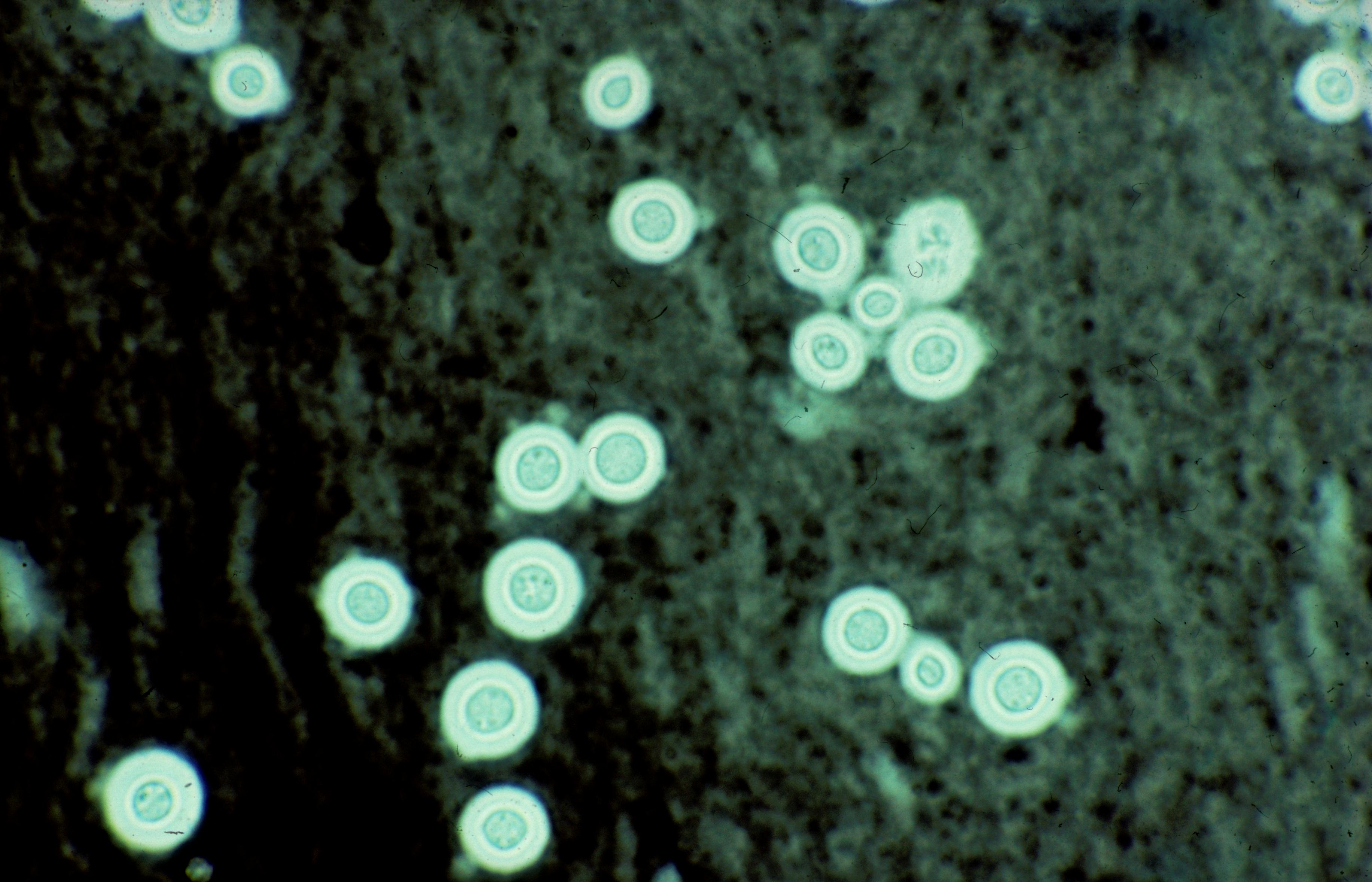 CSF, spherical fungal cells with a broad…
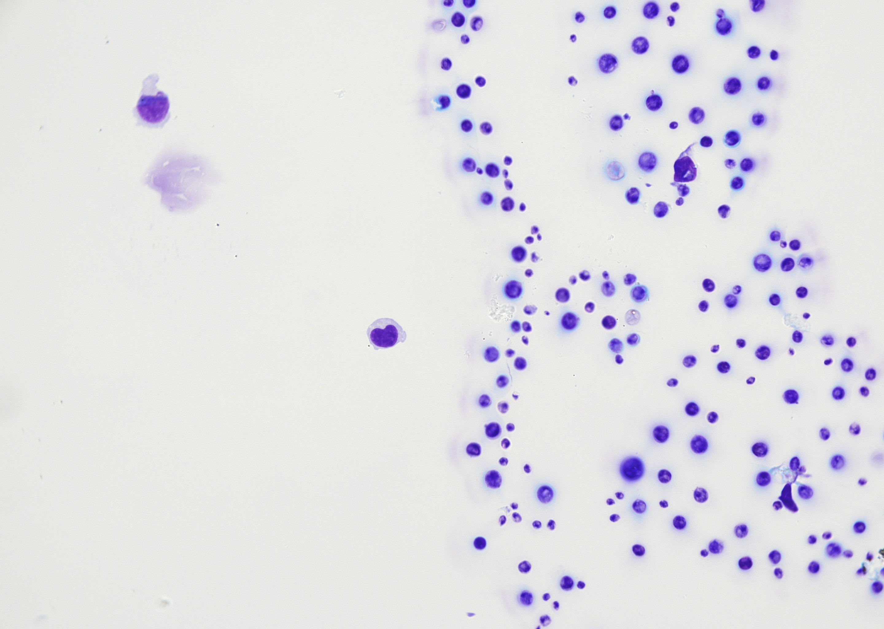 CSF, intracytoplasmic round, spherical…
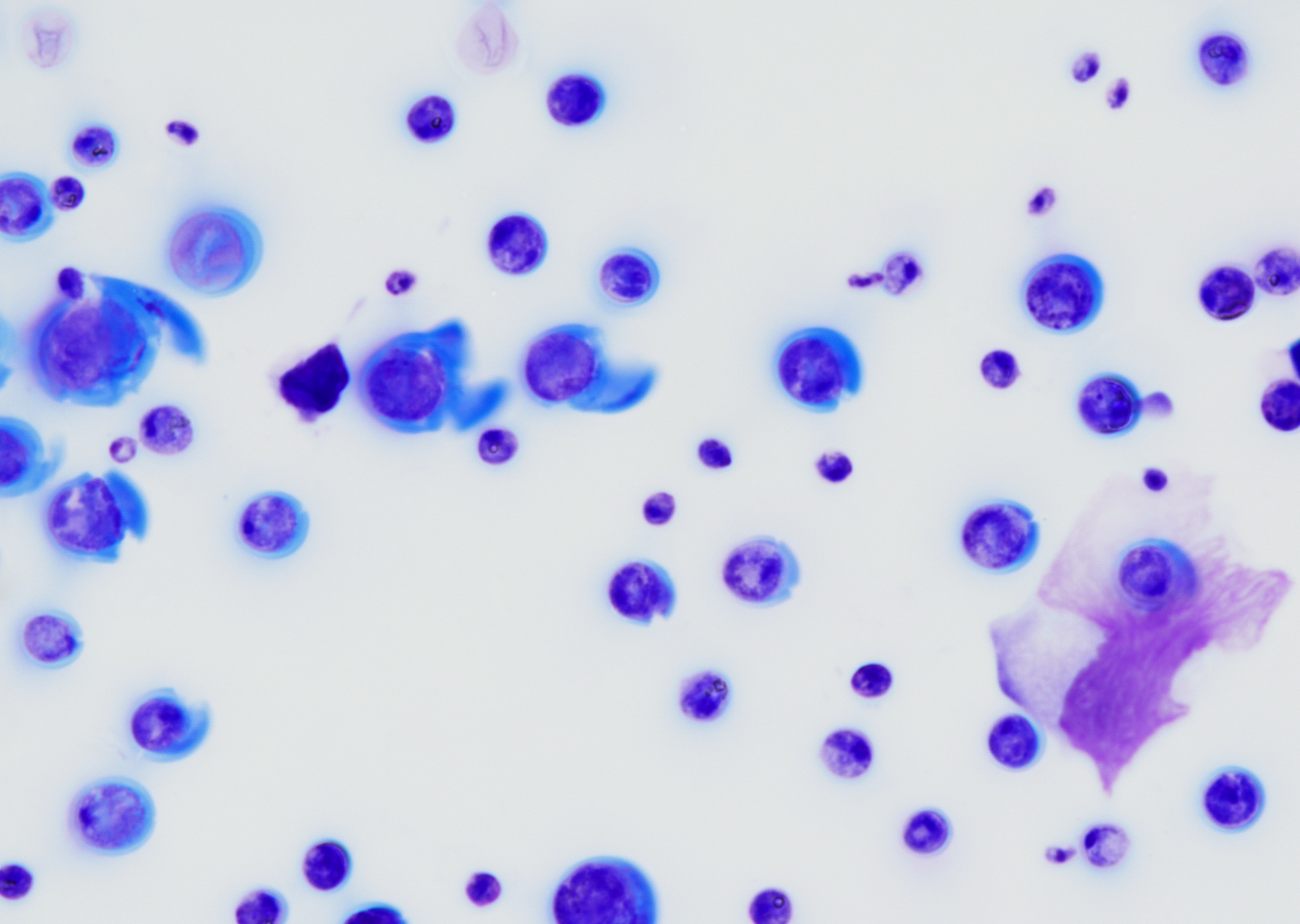 CSF, intracytoplasmic round, spherical…
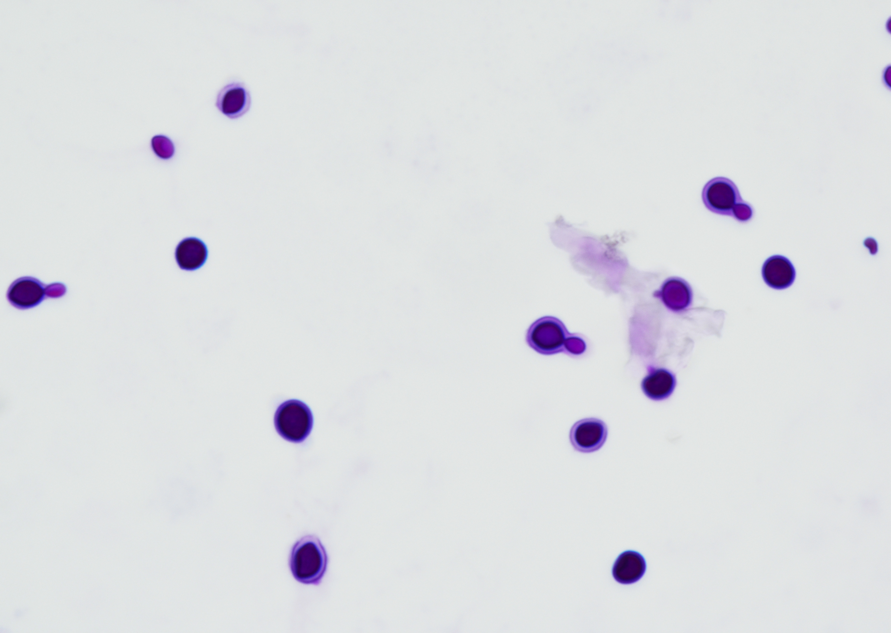 CSF, intracytoplasmic round, spherical…
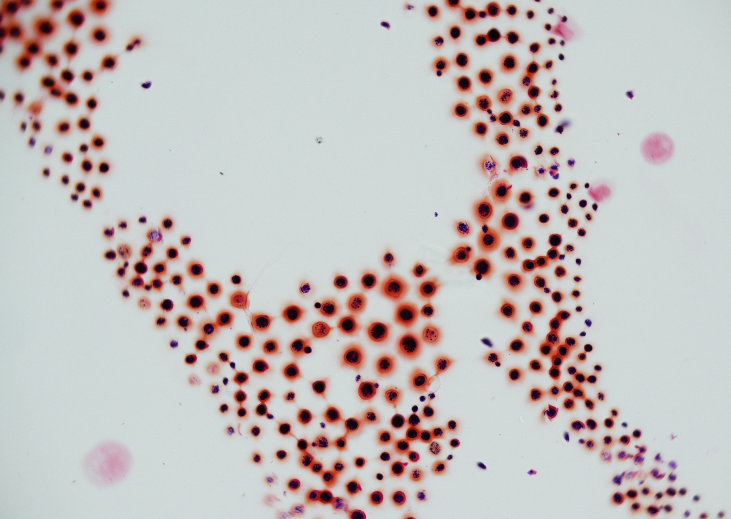 CSF, patient with acquired immunodeficiency…
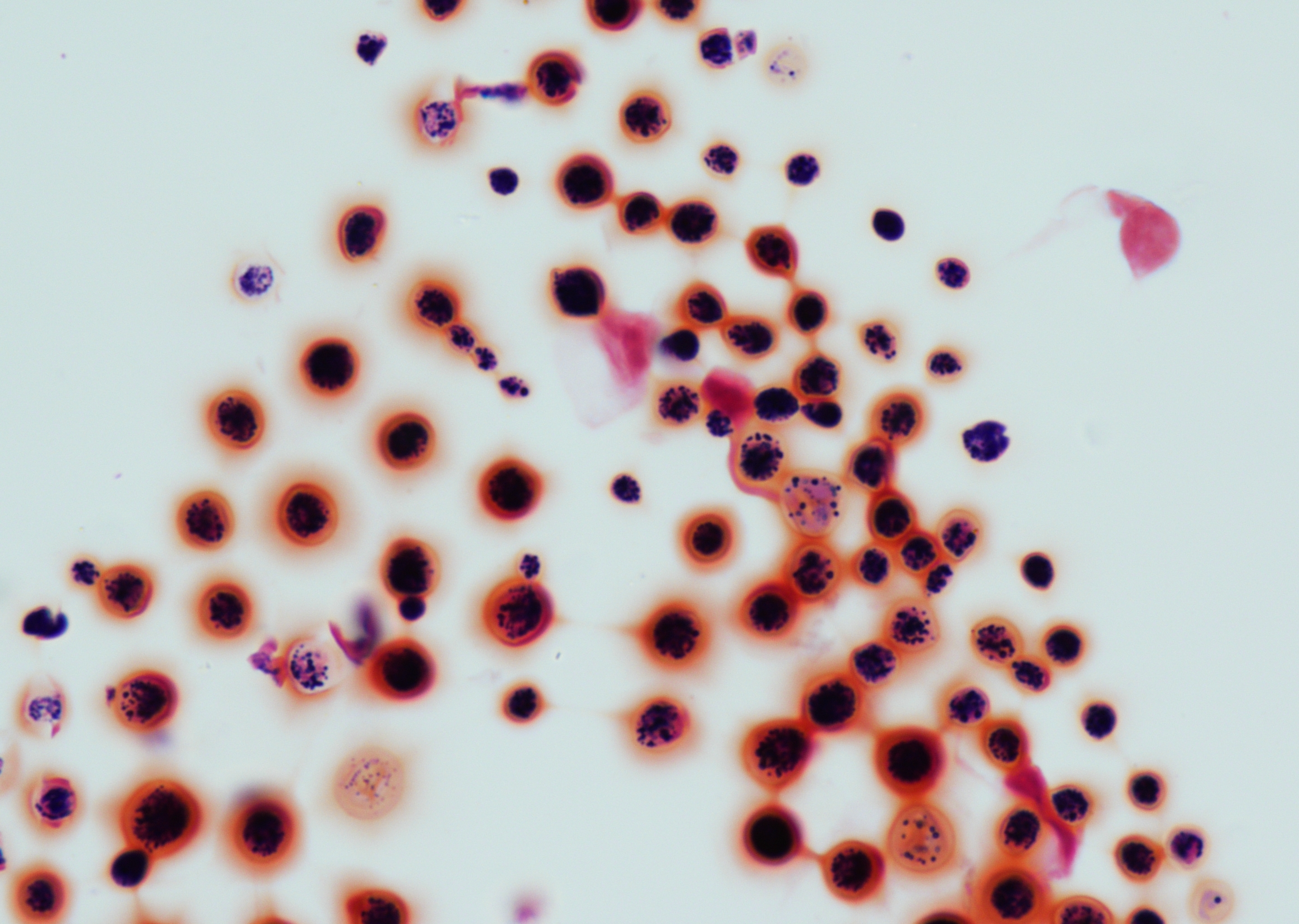 CSF, spherical fungal cells with a broad…
Morphology
Cytology:
- Encapsulated, spherical to ovoid yeasts with a broad double membrane (polysaccharide capsule)
- Narrow-based budding, and variable in size (4-20 µm). No pseudohyphae.
- Cryptococci are found extracellularly or intracellularly within macrophages.
- In liquid specimens, cryptococci can be detected using the India ink stain (punched-out round holes with a double contour). However, the detection of cryptococci in the India ink stain should be verified with conventional stains such as MGG, PAP, PAS, GMS/Grocott or Fontana-Masson stain.
- In immunosuppressed patients and patients who received antifungal therapy, the capsule can be absent (acapsular form).
Histology:
- In tissue sections, the polysaccharide capsule is best highlighted by mucicarmine and a combined PAS-Alcian blue stain; Grocott/GMS and PAS stain highlight the cell wall. When there are abundant yeast cells, they show a refractile appearance on HE and are characterized by copious extracellular mucoid material, which appears as a clear or empty halo.
Background:
- Numerous granulocytes and reactive changes in physiologically occurring cells
- Multinucleated giant cells
- Granulomatous reaction, often with central necrosis
- Limited inflammatory reaction in immunocompromised patients
Differential Diagnosis:
- Candida spp.
- Blastomyces spp.
- Histoplasma spp.
Clinical notes
Infection through inhalation of spores, which may stay latent in the airways.
Pulmonary cryptococcosis is either asymptomatic or associated with non-specific symptoms. Chest X-rays show various changes, such as solitary or multiple nodular structures, patchy infiltrates, less commonly interstitial infiltrates, and pleural effusions. Pulmonary infection can result from primary infection, reinfection with a new strain, or reactivation of latent infection, particularly in immunocompromised and most commonly in HIV-positive individuals due to decreased host T-cell immunity, with risk of disseminated disease due to hematogenous spread.
Cryptococci are able to penetrate the blood brain barrier, with risk of CNS involvement. The infection of the central nervous system (CNS) also initially proceeds without symptoms. In the CNS, multiplication and granuloma formation eventually lead to symptoms of meningitis or meningoencephalitis. The diagnosis of the mostly opportunistic infection requires immediate feedback with the treating physicians, due to high mortality rate.
Ancillary Testing
Culture, Cryptococcus antigen detection (CrAG; CSF, serum, plasma, urine), direct microscopy (liquid materials), PCR-based assays or DNA hybridization (species identification), immunohistochemistryAdditional Information
Cryptococcus neoformans is a fungus found in soil and bird droppings that can be inhaled into the lungs. The lungs may remain the sole site of manifestation, or dissemination may occur to other tissues, particularly in immunocompromised patients, potentially involving the central nervous system.
In severely immunocompromised patients, coinfection with other pathogens should be considered.
Two humanpathogenic species:
- C. neoformans (worldwide distribution, causing most of the infections)
- C. gattii (tropical/subtropical regions)
Cryptococcosis lacking a capsule can resemble Candida species morphologically. Conventional capsule‑specific stains are generally ineffective in such cases, whereas the Fontana‑Masson technique, by detecting melanin deposition, enhances accurate identification