
- EN
- DE

Parasites
Differential Diagnosis of Schistosoma Species:
Strongyloides stercoralis

- Organ System
- Digestive Tract, Respiratory Tract
- Diagnostic Method
- Cytology, Histology

- Geographical Region
- Tropical and Subtropical Region,
- Temperate Zone
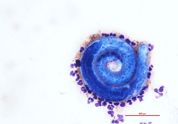
 Lung, bronchial secretion, Strongyloides…
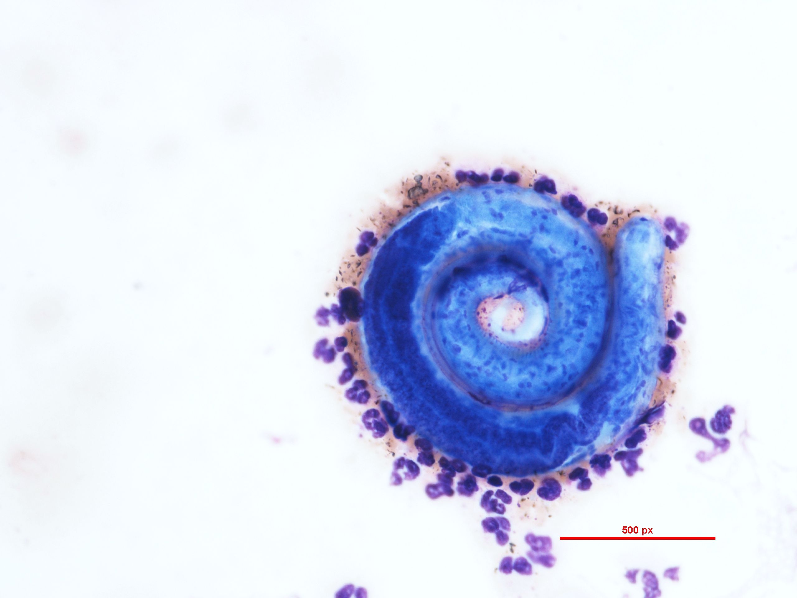
 Lung, bronchial secretion, Strongyloides…
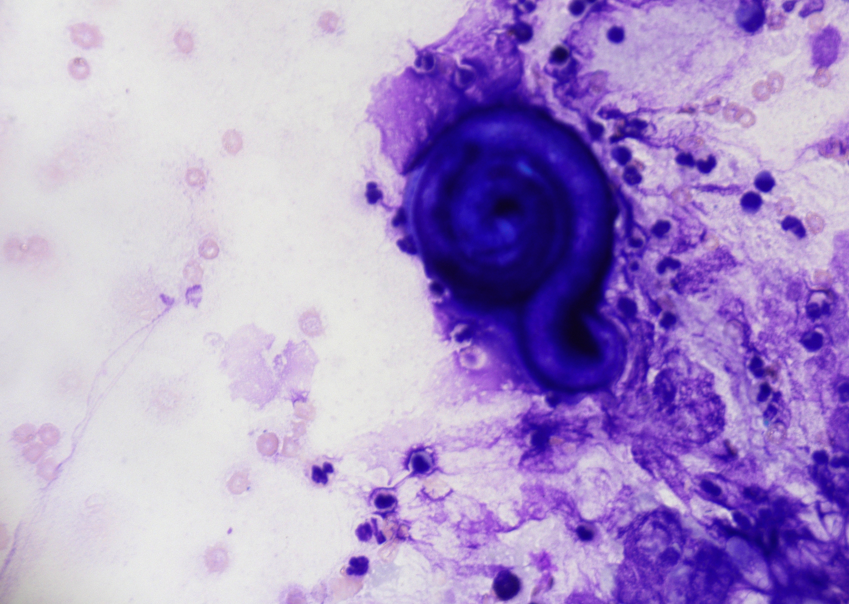
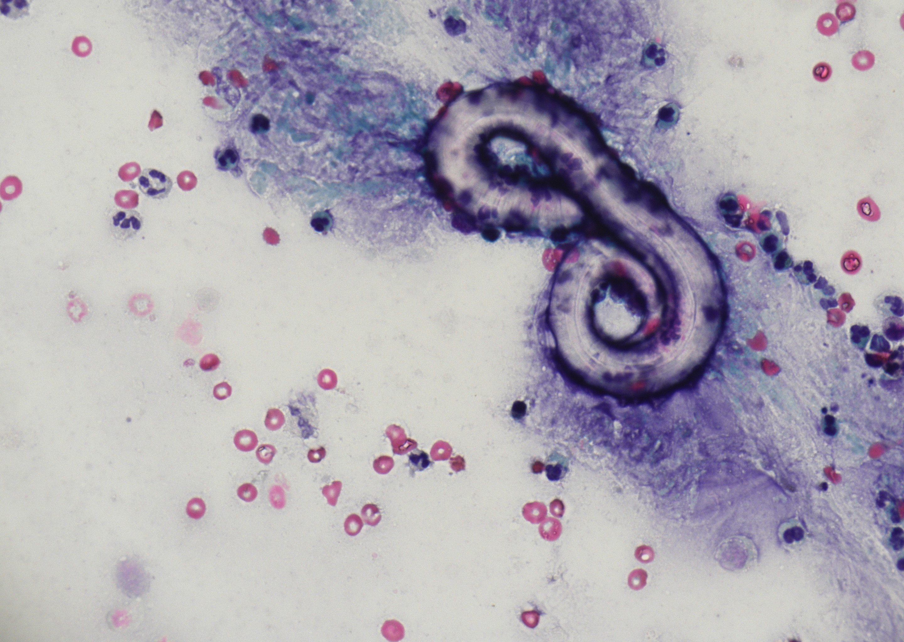 Lung, bronchial secretion, Strongyloides…
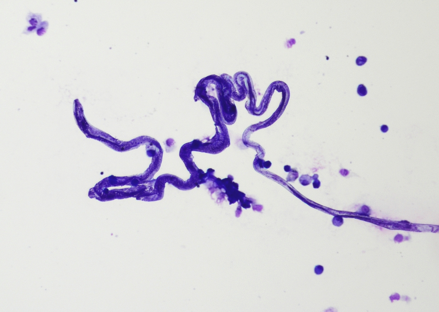 Lung, BAL, Strongyloides stercoralis (MGG)
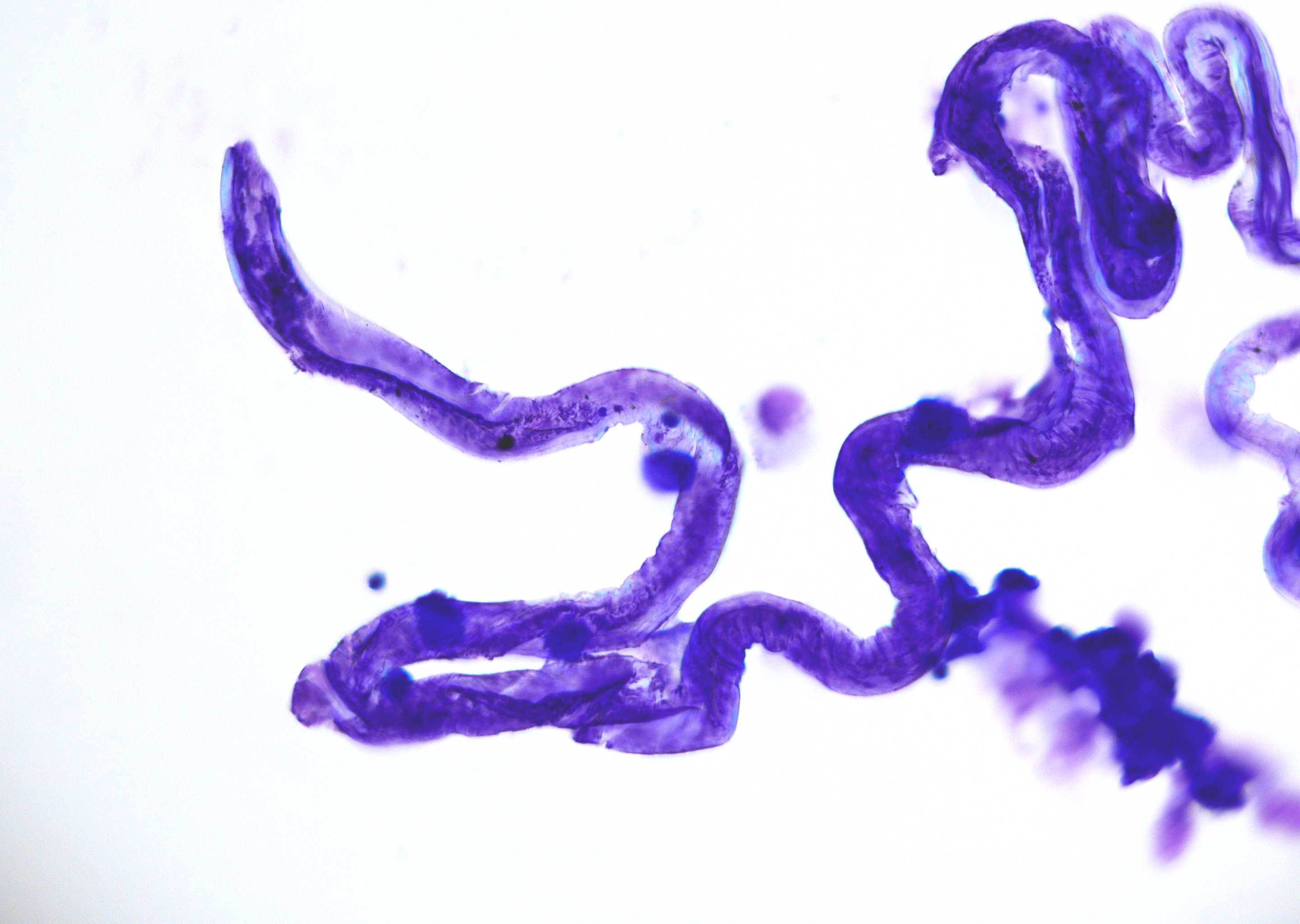 Lung, BAL, Strongyloides stercoralis with…
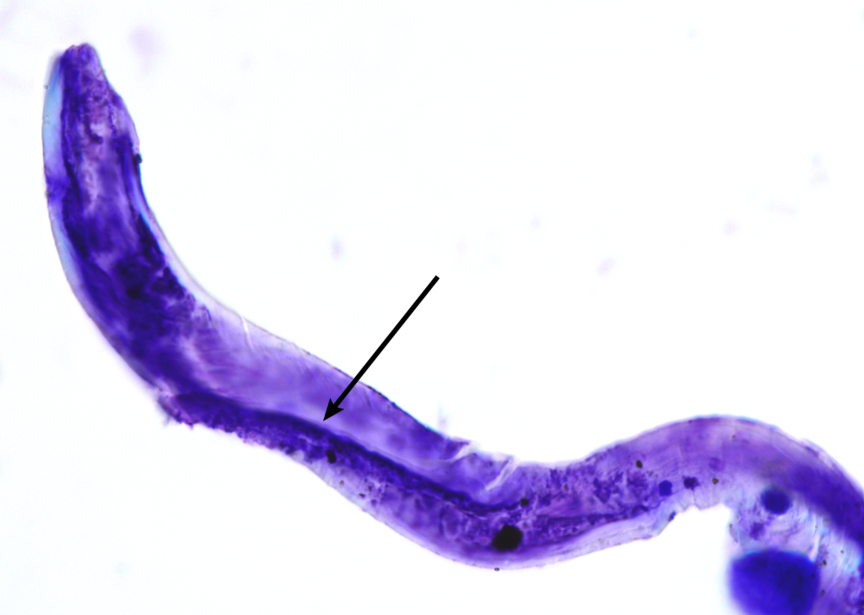 Lung, BAL, Strongyloides stercoralis…
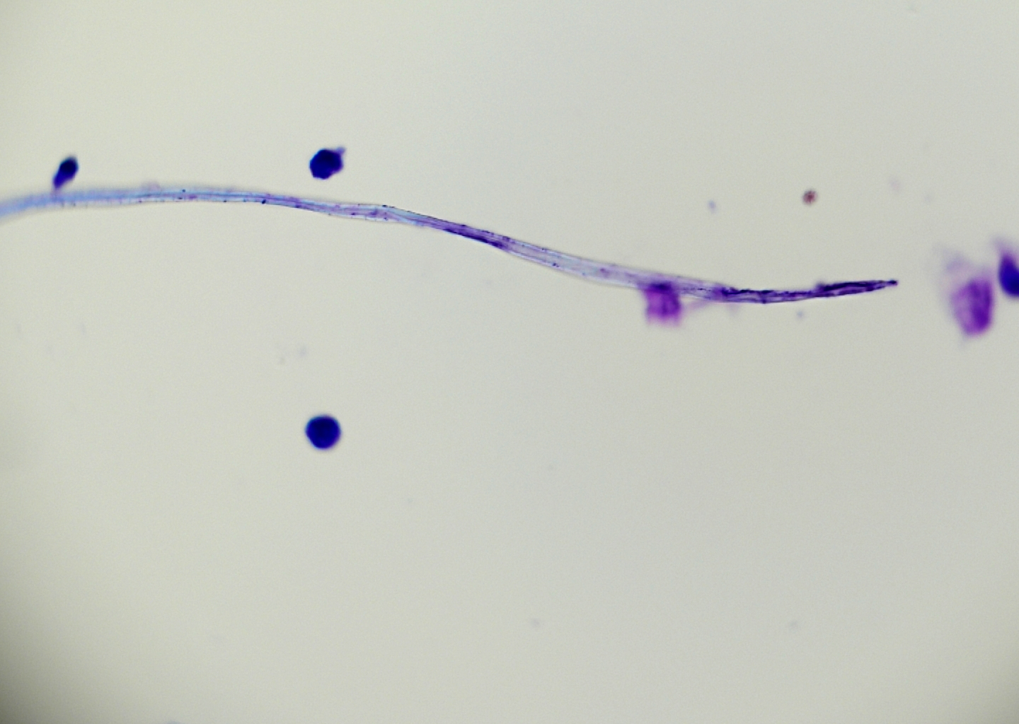 Lung, bronchial secretion, Strongyloides…
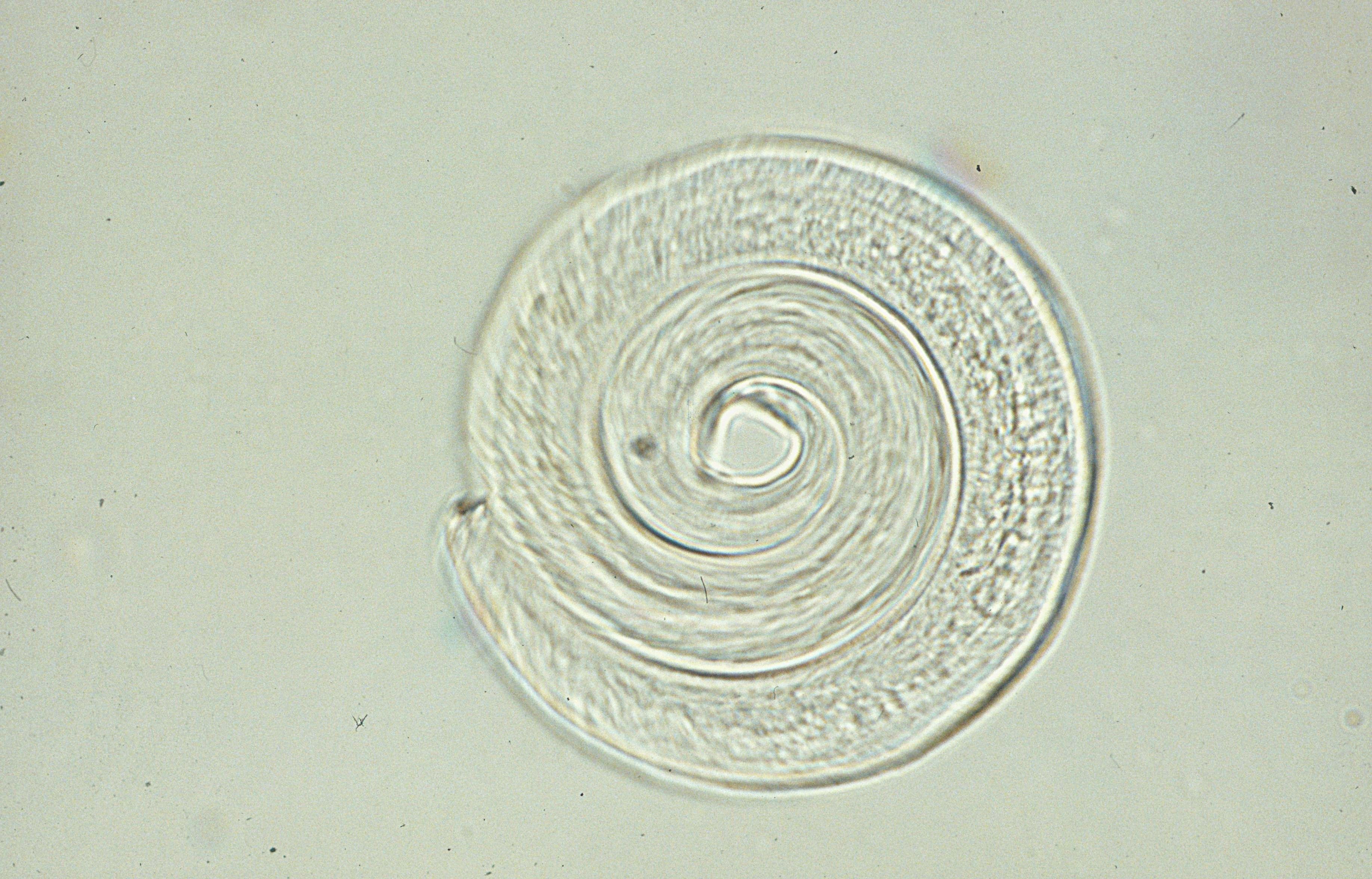 Lung, bronchial secretion, Strongyloides…
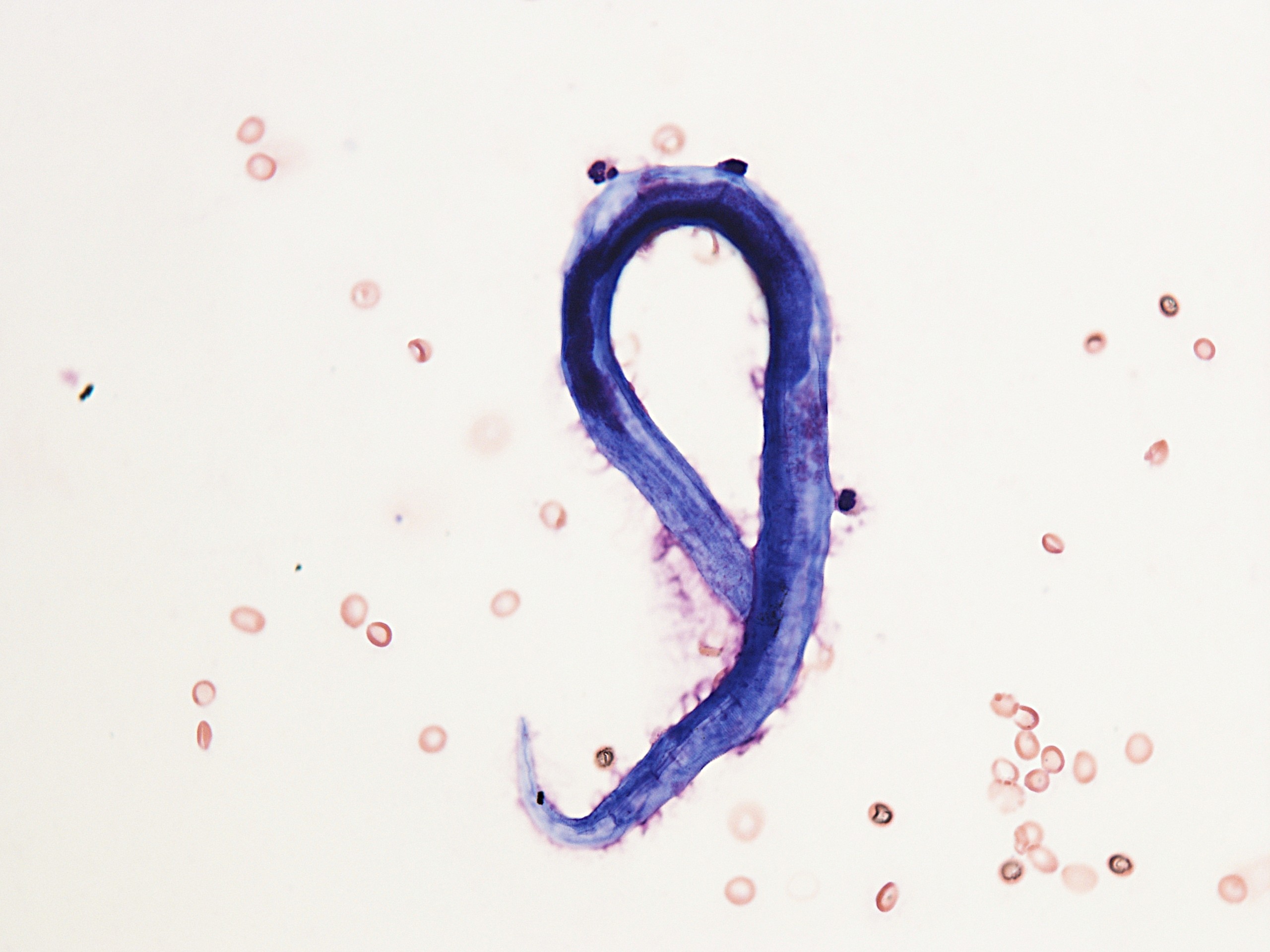 Lung, bronchial secretion, Strongyloides…
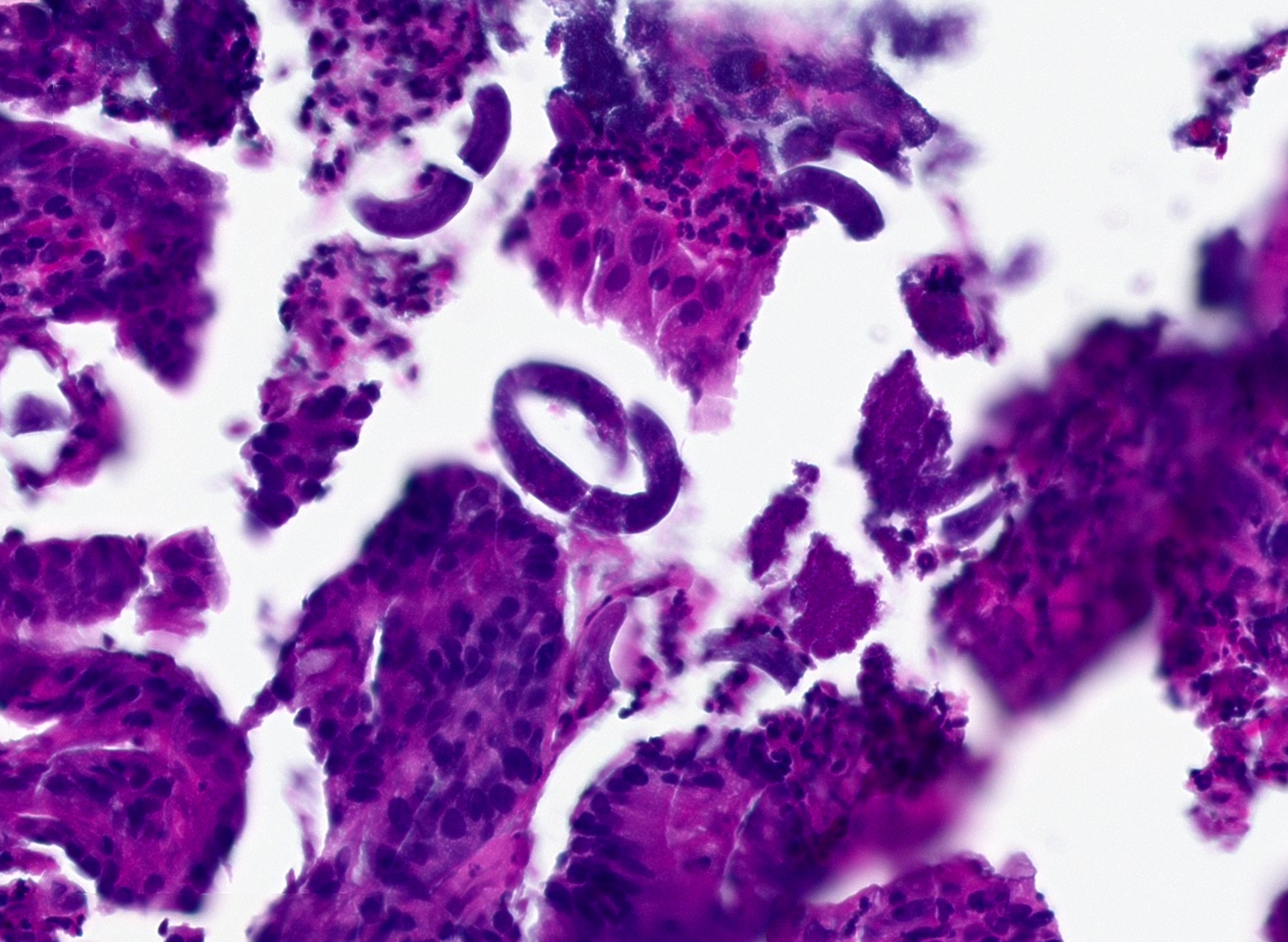
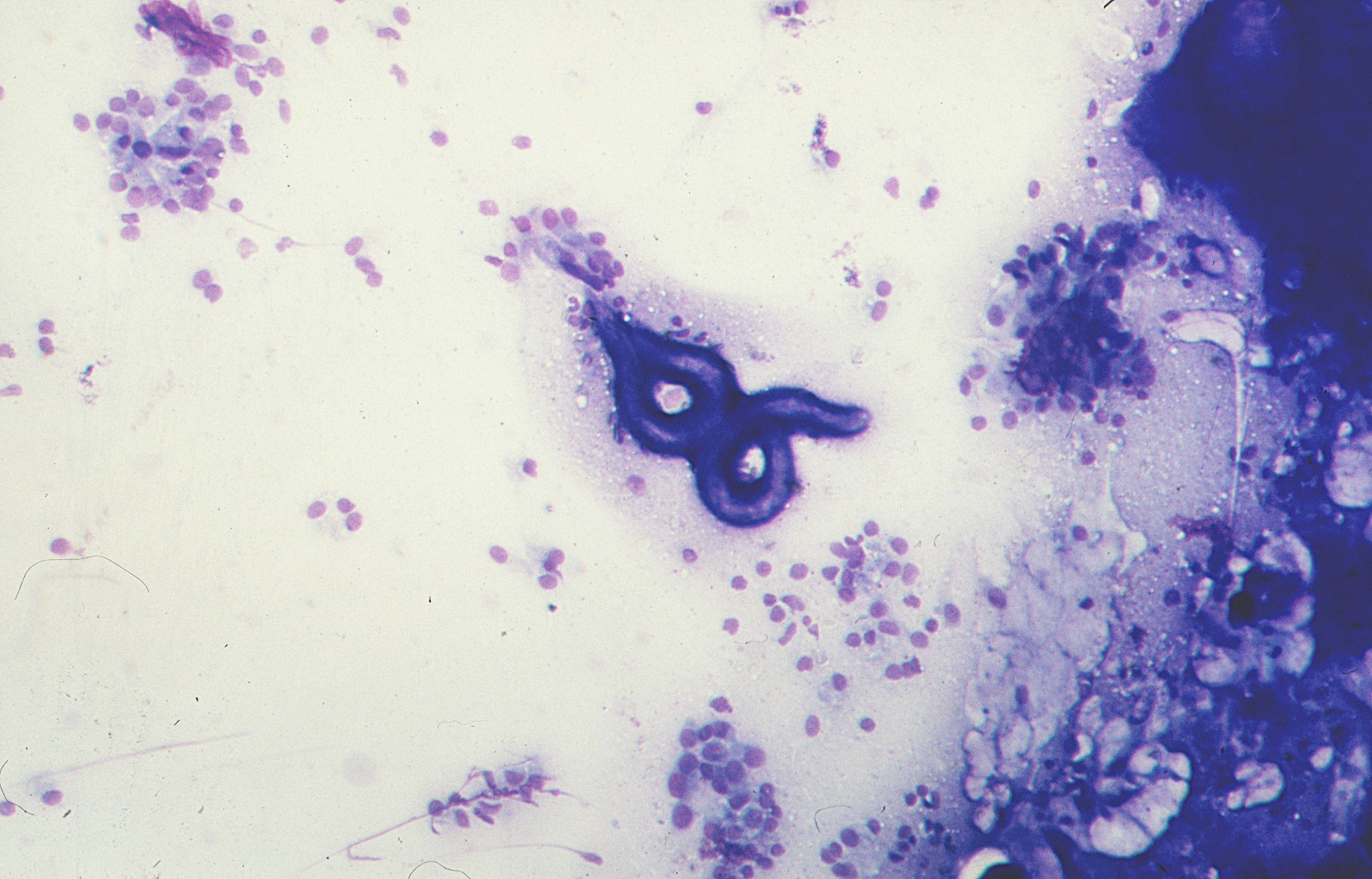 Stomach, brush cytology, Strongyloides…
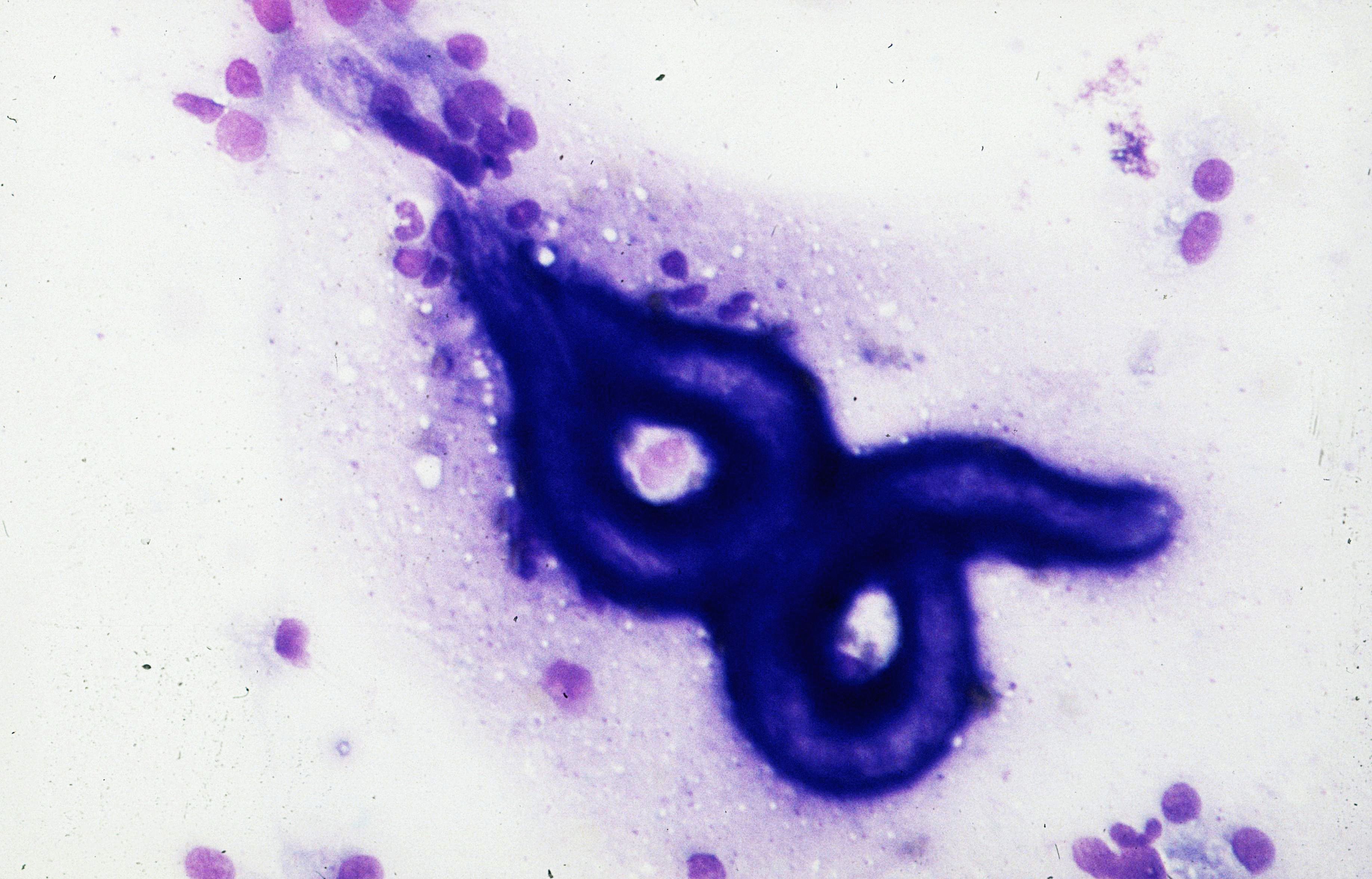 Stomach, brush cytology, Strongyloides…
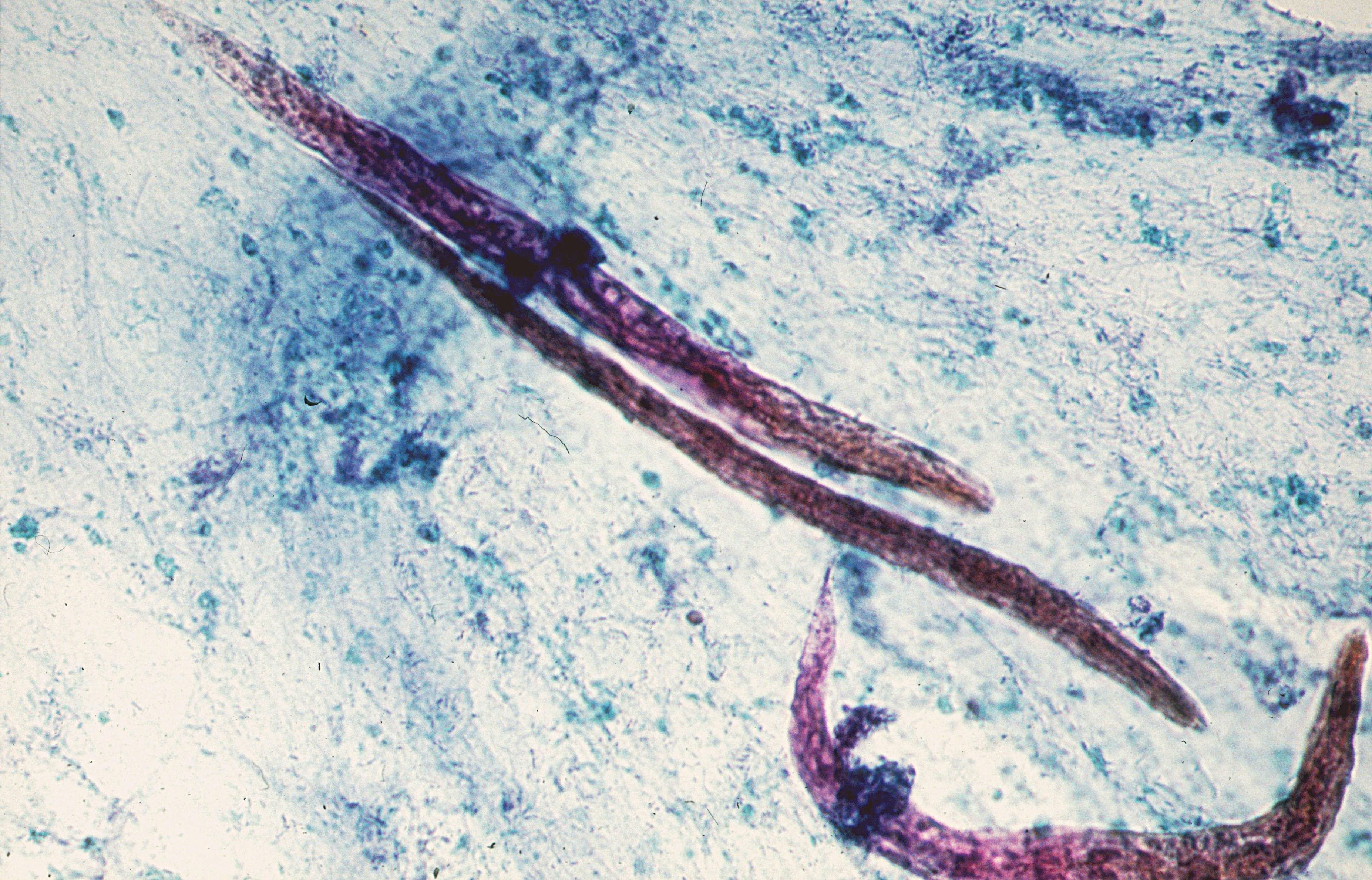 Feces, Strongyloides stercoralis (PAP, x400)
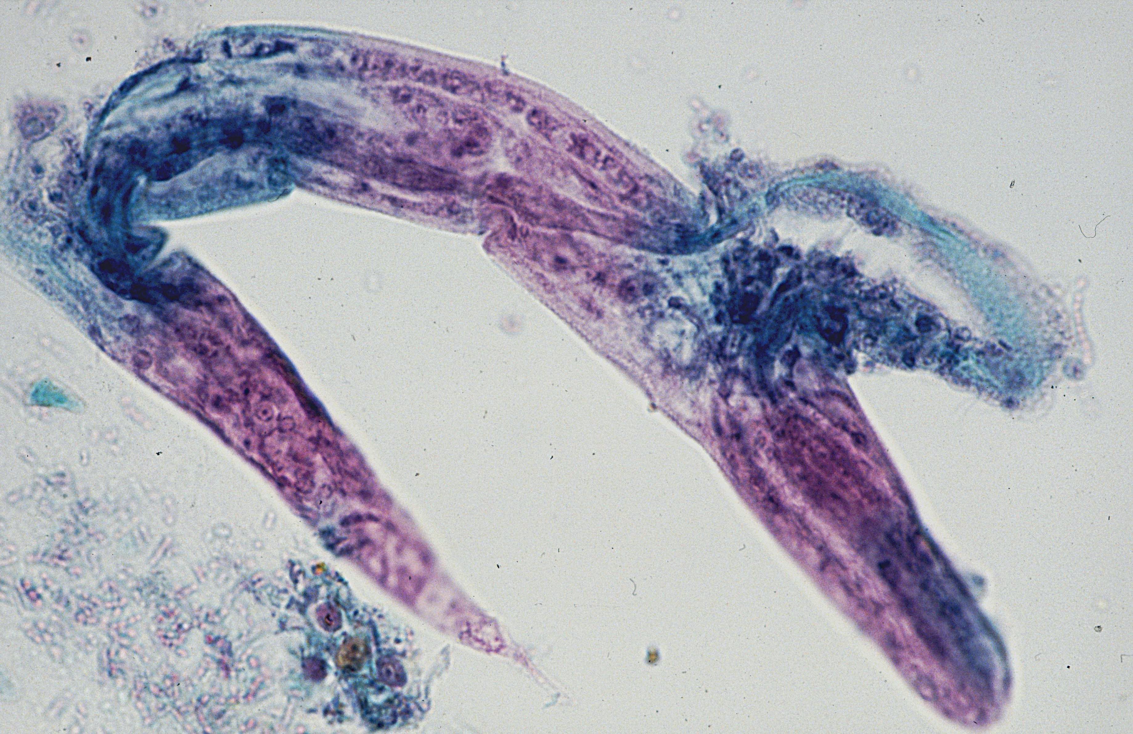 Feces, Strongyloides stercoralis (PAP, x600)
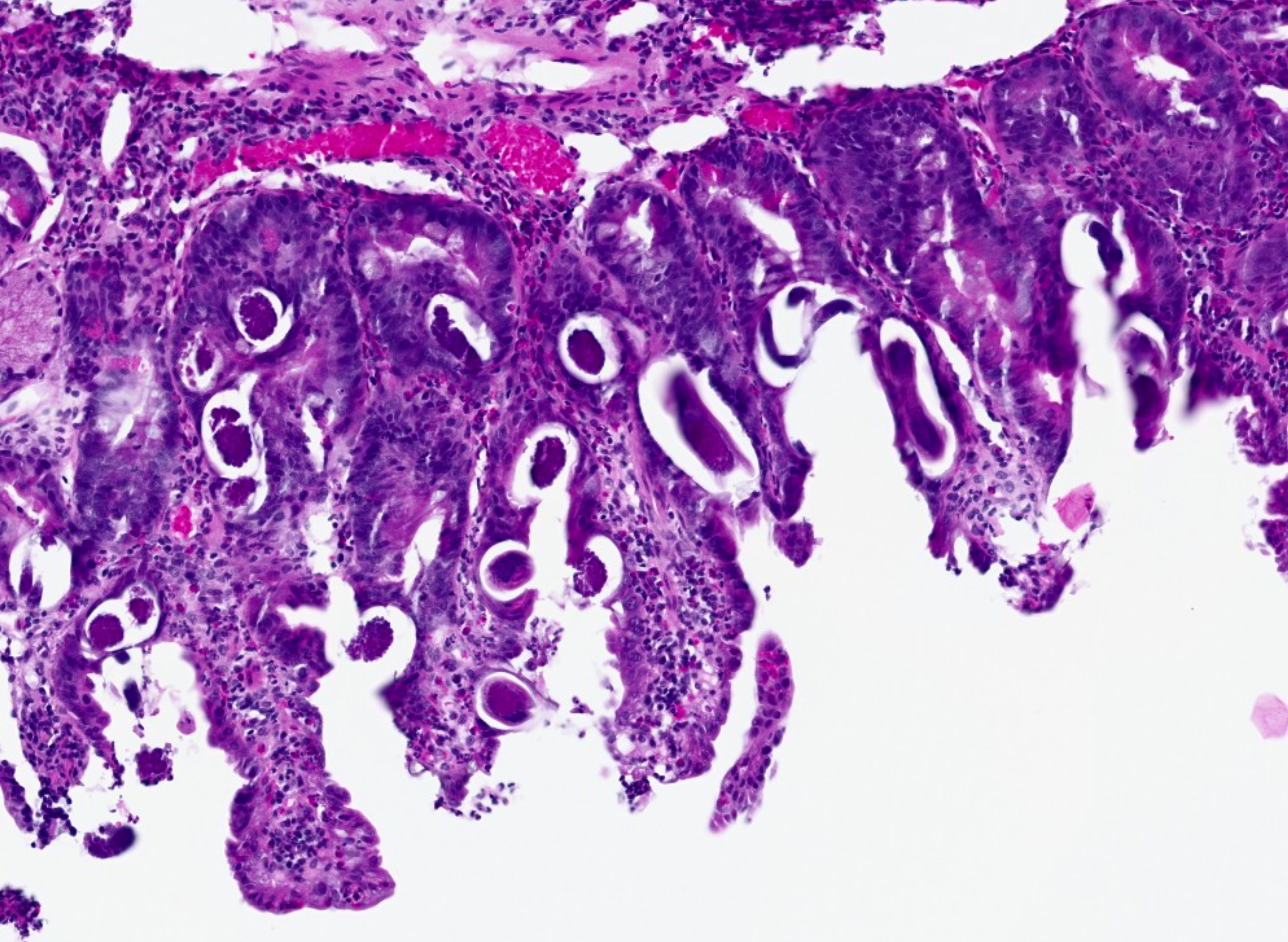 Reactive duodenitis with intraepithelial S.
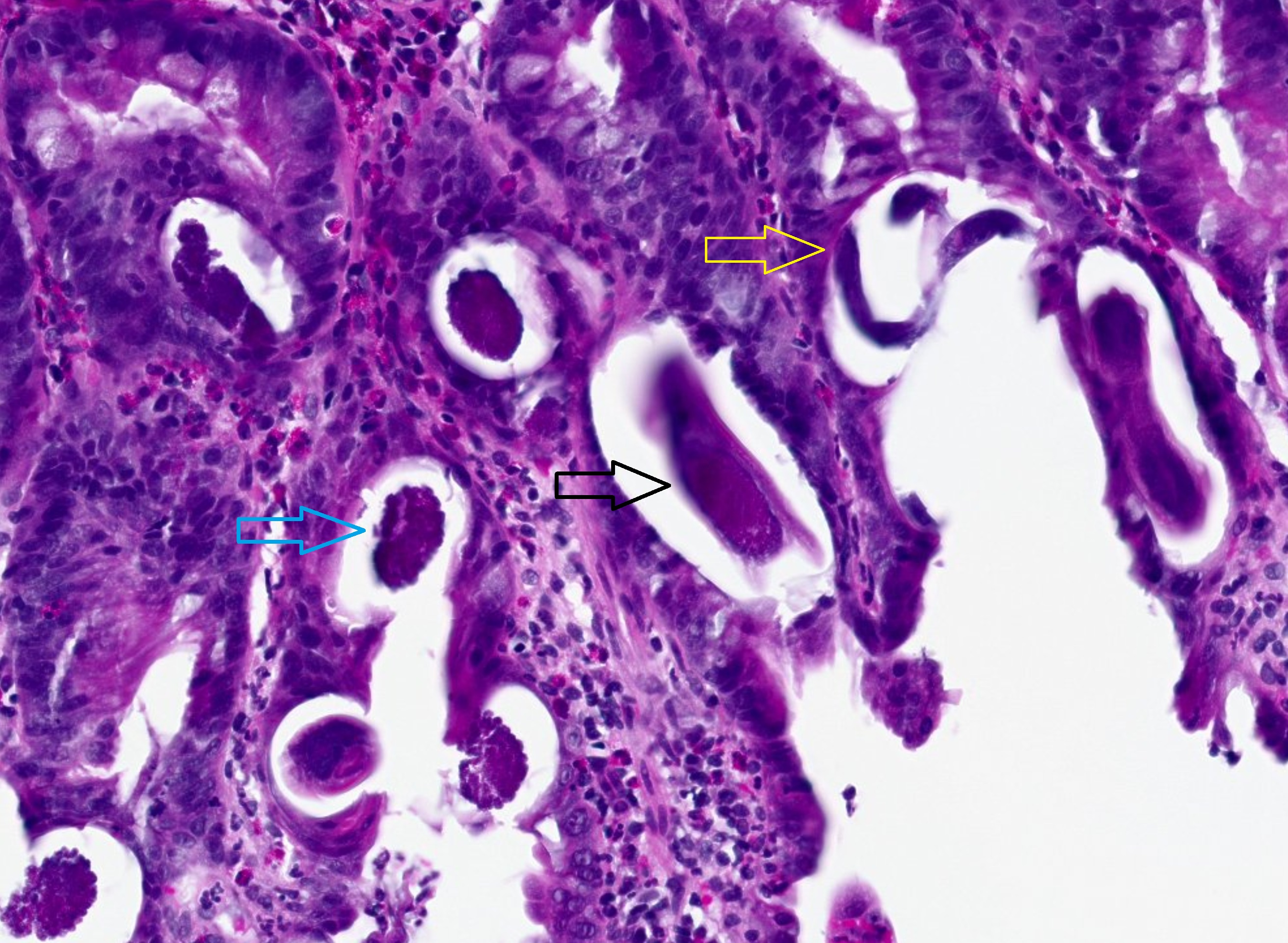 Strongyloides hyperinfection syndrome.
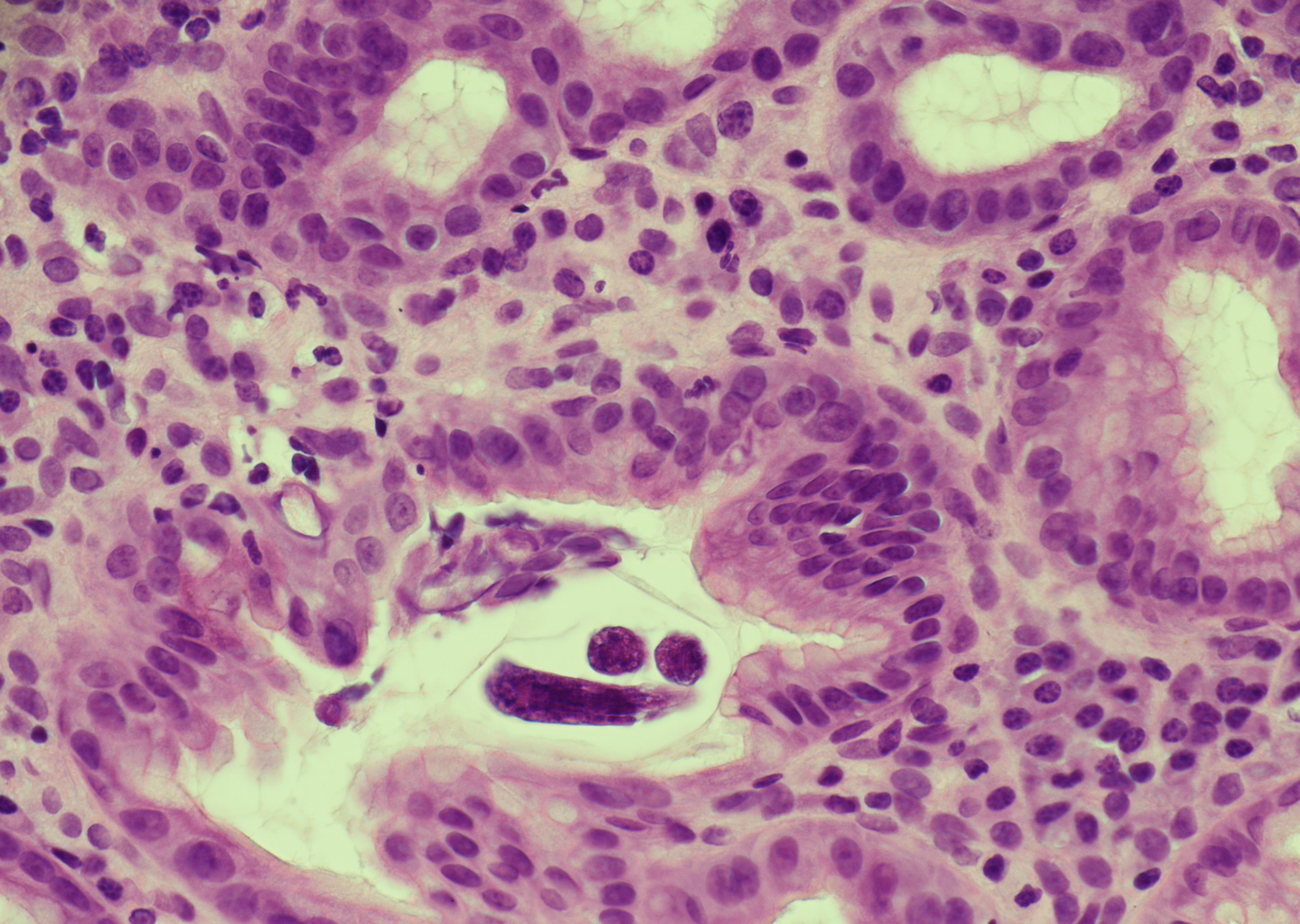
 Duodenal biopsy with a rhabditiform larva in…
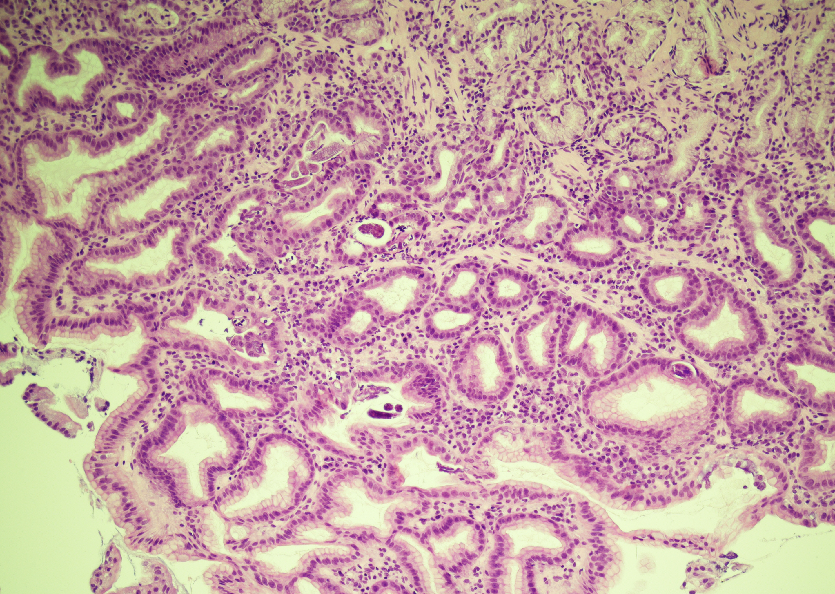
 Gastric antrum biopsy with intraepithelial…
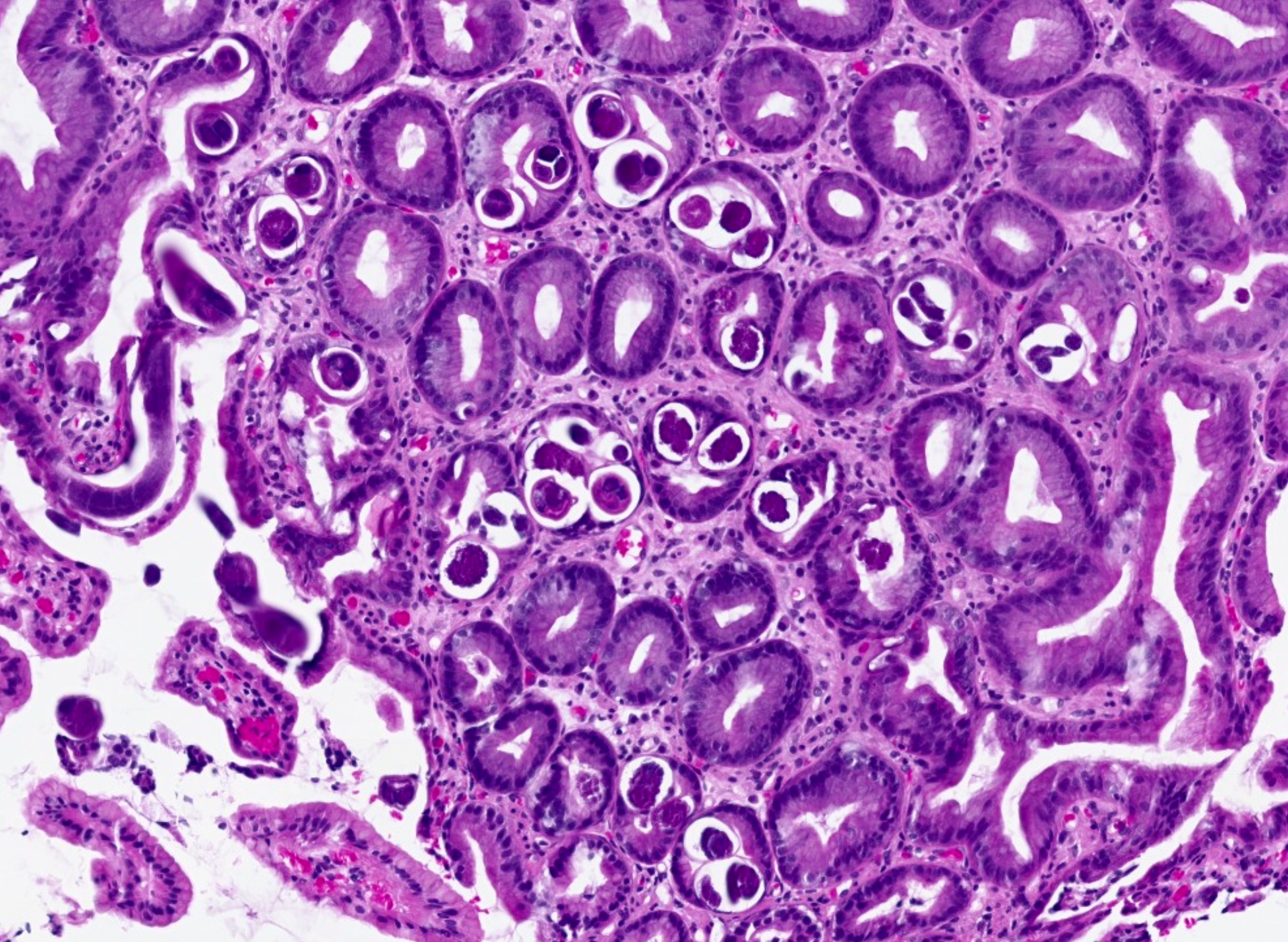
 Gastric antrum biopsy with intraluminal and…
 Gastric mucosa with intraluminal…
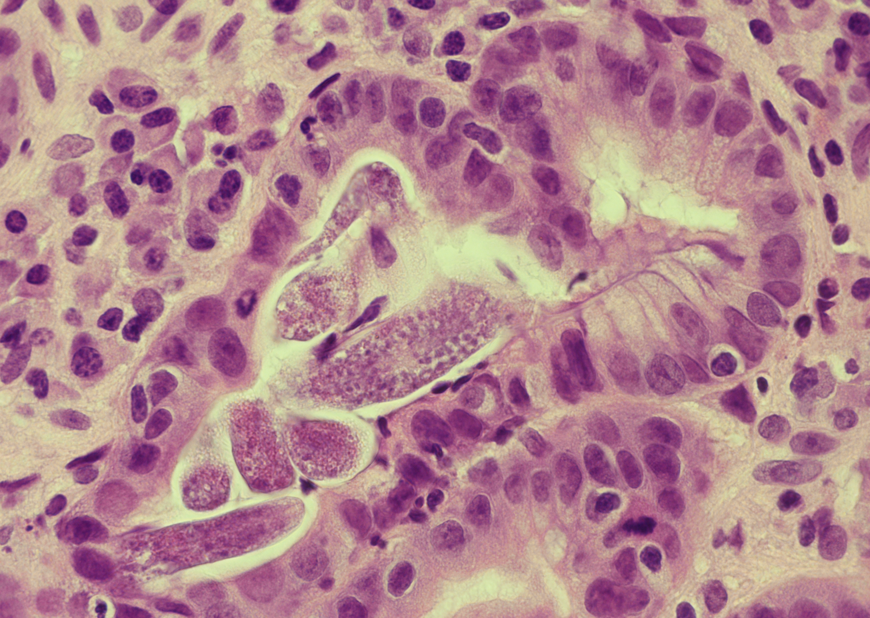 Gastric mucosa with intraluminal sections of…
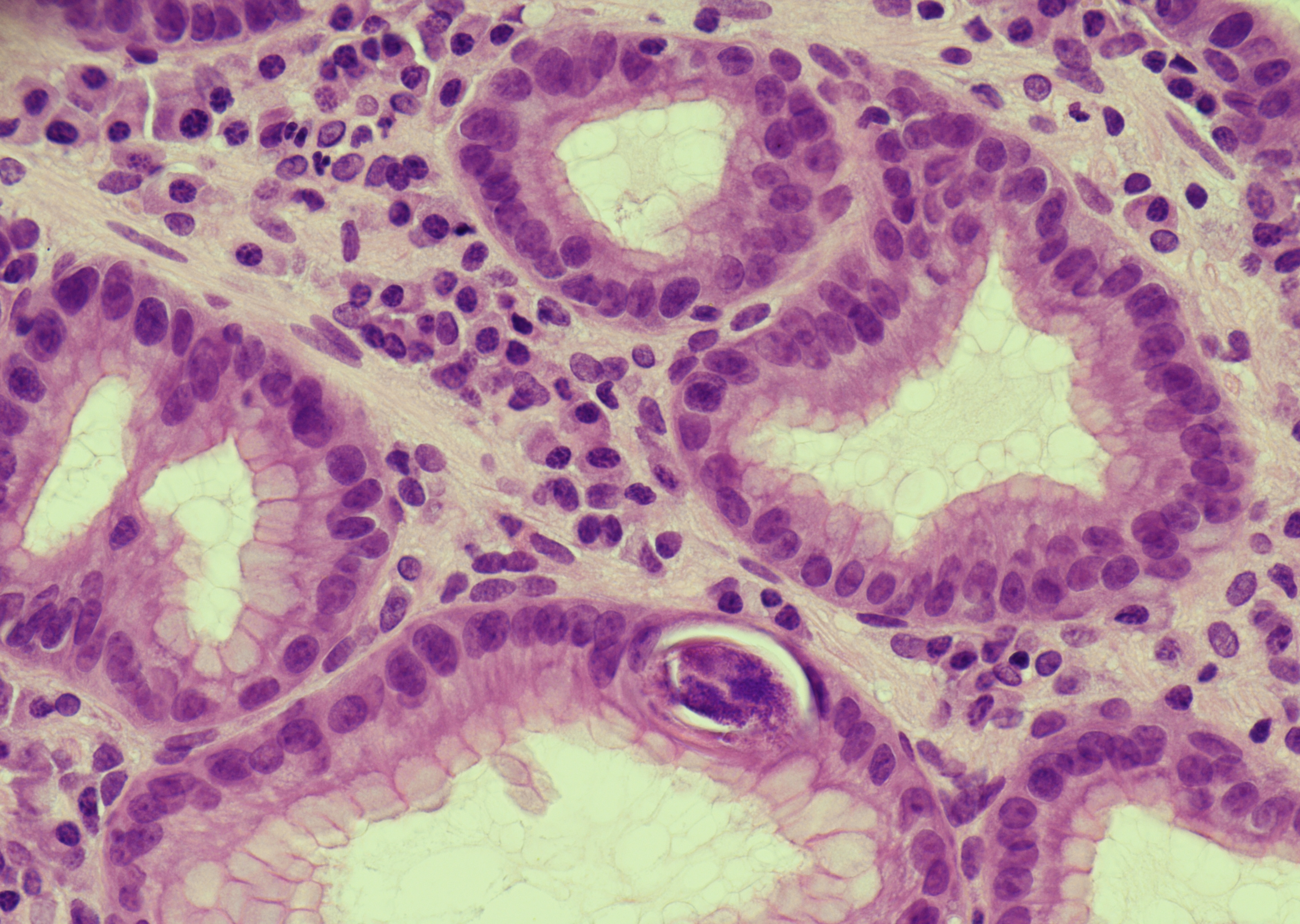 Gastric mucosa with an intraepithelial…
Morphology
- Adult female worm: 2-3 mm long & 30-40 μm in diameter, cylindrical, pointed tail, rounded anterior end
- Rhabditiform (juvenile) larvae: 180-400 μm long & 15-20 μm in diameter
- Filariform larvae: 500-600 μm long & 15-20 μm in diameter, notched tail
- Egg: 50-60 μm x 30-40 μm; rarely seen in stool
Cytology:
- Eosinophils may be seen in the background
- Larvae may be identified in duodenal fluid, sputum, bronchoalveolar lavage, bronchial washings, and, in disseminated disease, occasionally in pleural or peritoneal fluid
- Cytologic recognition relies on slender curved larvae; filariform forms show a notched tail
Histology:
- Biopsies, especially from the duodenum/small intestine, may show adult females and larvae within crypts, glands, or mucosa/submucosa
- Associated findings include eosinophil-rich lamina propria inflammation, active duodenitis/cryptitis, villous blunting or villous atrophy, and crypt hyperplasia
- In severe disease, eggs, eosinophilic crypt abscesses, granulomatous inflammation, or colonic involvement may also be seen
Differential Diagnosis:
- Schistosomiasis
- Amebiasis
- Balantidiasis
- Ancylostomiasis/Necatoriasis
Clinical notes
Mostly asymptomatic, with possible symptoms if larvae migrate to gastrointestinal (abdominal pain, diarrhoea, vomiting) or respiratory tract (dyspnea, wheeze).
Hyperinfection syndrome or disseminated strongyloidiasis in immunocompromised patients, if left untreated, can be fatal.
Ancillary Testing
Serial stool examination, serology, stool PCR/qPCRAdditional Information
Filariform larvae in contaminated soil can penetrate human skin and migrate via the lungs to the gastrointestinal tract, where matured adult females reproduce by parthenogenesis. Eggs hatch in the intestinal mucosa, releasing rhabditiform larvae that are either passed in stool or develop into filariform larvae causing autoinfection.
Differential Diagnosis
References
Copyright ® 2025 Österreichische Gesellschaft für Zytologie