
- EN
- DE

Parasites
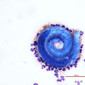
Differential Diagnosis of Strongyloides stercoralis:
Schistosoma Species

- Organ System
- Genitourinary Tract, Digestive Tract
- Diagnostic Method
- Cytology, Histology

- Geographical Region
- Islands,
- Tropical and Subtropical Region
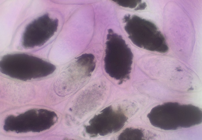
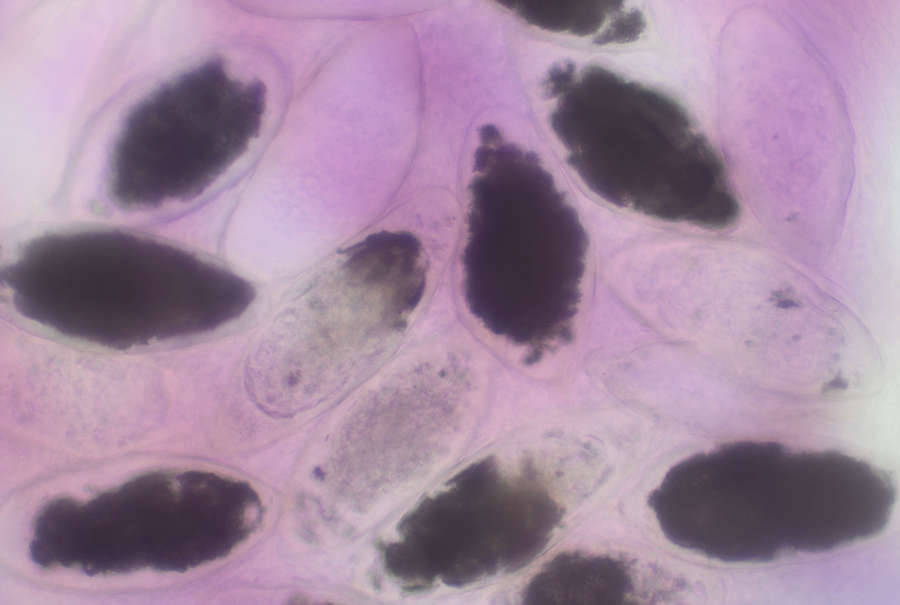 Non-viable Schistosoma haematobium eggs with…
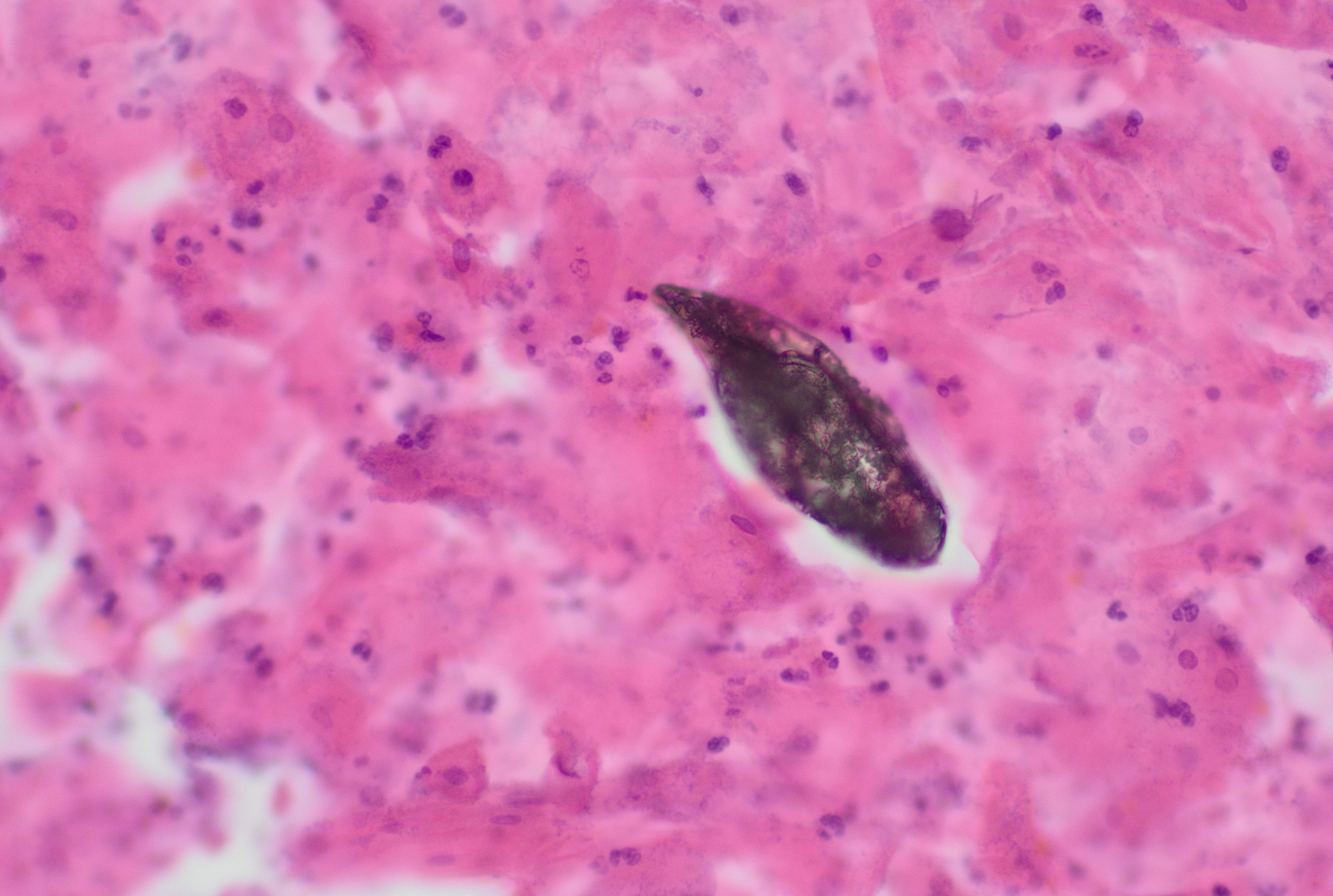 Conventional Pap smear, calcified, opaque…
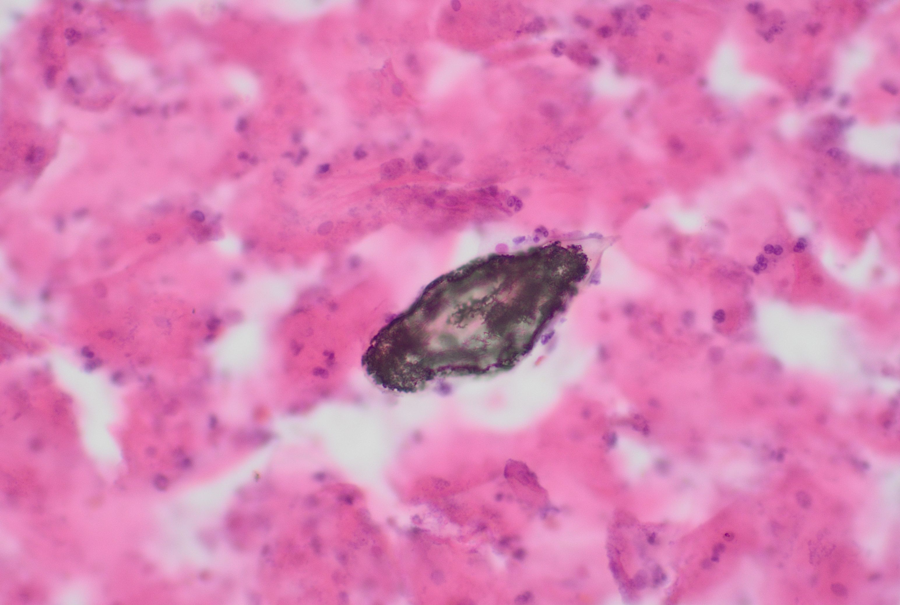 Conventional Pap smear, calcified, opaque…
 Conventional Pap smear, Schistosoma in the…
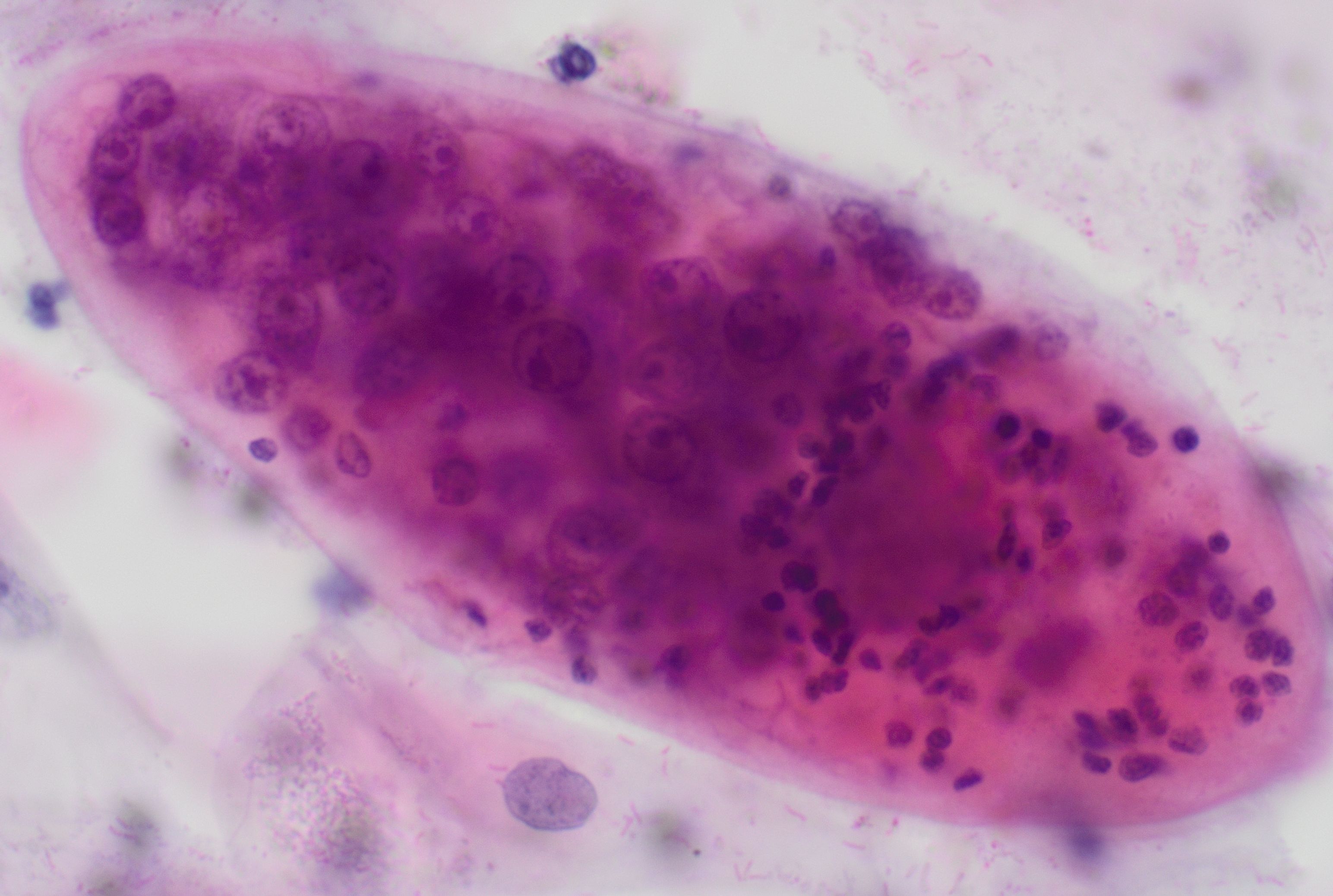 Conventional Pap smear, Schistosoma…
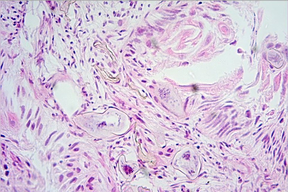 Bladder, squamous cell carcinoma with…
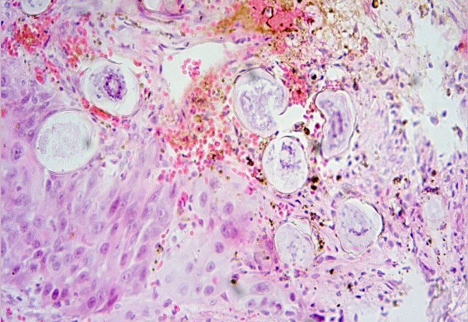 Bladder, squamous cell carcinoma with…
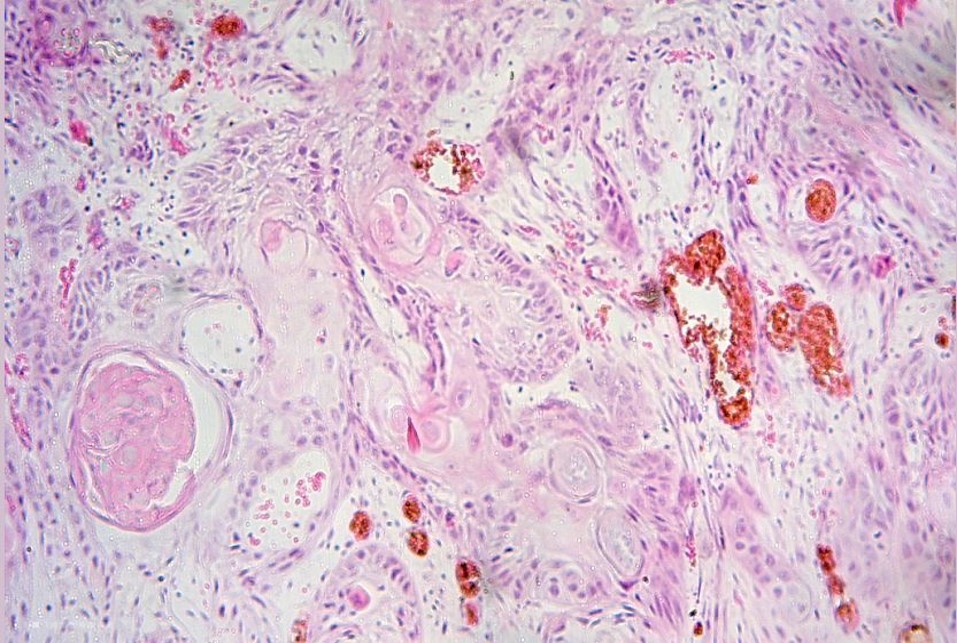 Bladder, squamous cell carcinoma with…
Morphology
Cytology:
- Detection of characteristic Schistosoma eggs in urine or stool specimens, less commonly also in cervical smears
- Neutrophils, eosinophils, granulomas, and fibrosis may be seen in the background
- The eggs, measuring 110 to 170 µm × 50 to 70 µm, are oval to elongated
- Viable ova contain a miracidium composed of eosinophilic cells that differentiate into various larval structures
- Non-viable ova appear empty and may present as blackened, shrunken, or calcified
- Species assignment is based on egg morphology and the type of specimen:
- S. haematobium with a terminal spine, usually in urine
- S. mansoni with a prominent lateral spine, usually in stool
- S. japonicum with a small, often inconspicuous lateral spine
Histology:
- Usually a granulomatous inflammatory reaction to retained eggs, often with fibrosis and calcification
- In intestinal or hepatosplenic schistosomiasis, eggs are found in the bowel wall, liver, and periportal tissue, with possible ulceration, hyperplasia, polyposis, periportal fibrosis, and portal hypertension
- In urogenital schistosomiasis, eggs are found in the urinary tract and are accompanied by chronic inflammation, fibrosis, and calcification
- In histological sections, the egg shell of S. mansoni stains usually positive (red), whereas the egg shell of S. hematobium stains negative (blue) with ZN (1)
Clinical notes
Schistosomiasis (syn. bilharzia) is a parasitic intravascular infection in which chronicity and severity are largely determined by the host's immune response to the trematode worms and to egg deposition in the tissues.
Cercarial dermatitis (urticarial rash) and Katayama syndrome (systemic hypersensitivity reaction) are early manifestations. Following freshwater exposure, cercarial dermatitis may initially occur; Katayama syndrome typically develops weeks later, with fever, rash, headache, myalgia, respiratory symptoms, and eosinophilia.
Chronic/advanced manifestations are caused by local inflammatory response to Schistosoma eggs trapped in host tissues and are species-dependent: S. mansoni and S. japonicum more often cause intestinal and hepatosplenic disease, with abdominal pain, diarrhea, constipation, and blood in the stool; severe cases may be associated with liver fibrosis, splenomegaly, and portal hypertension. Urogenital disease is characterized by dysuria and hematuria. Chronic S. haematobium infection is also associated with bladder fibrosis, renal damage, and an increased risk of bladder carcinoma. CNS manifestations due to ectopically deposited eggs are rare.
Ancillary Testing
Urine filtration, Kato-Katz technique for stool examination, serology, antigen detection, PCR, radiology (CXR, abdominal/pelvic US), shells of ovum can stain positively with a Ziehl-Neelsen stainAdditional Information
Infection occurs after contact with freshwater through skin penetration by cercariae; the snail serves as the intermediate host. Eggs typically appear in urine or stool 1 to 3 months after exposure.
After freshwater exposure in regions where S. mansoni and S. haematobium are co-endemic, both stool and urine samples should be examined.
Differential Diagnosis
References
CDC - DPDx - Schistosomiasis Infection
Gray DJ, Ross AG, Li YS, McManus DP. Diagnosis and management of schistosomiasis. BMJ. 2011 May 17;342:d2651. doi: 10.1136/bmj.d2651. PMID: 21586478; PMCID: PMC3230106.
- Vieira S, Belo S, Hänscheid T. Ziehl-Neelsen in Schistosomiasis: Much More Than Staining the Shell and Species Identification. Am J Trop Med Hyg. 2016;94(4):699-700. doi:10.4269/ajtmh.15-0798