
- EN
- DE

Virus
Human Polyomavirus (HPyV)

- Organ System
- Genitourinary Tract, CNS and Sensory Organs
- Diagnostic Method
- Cytology, Histology

- Geographical Region
- Dry Areas,
- Islands,
- Tropical and Subtropical Region,
- Polar and Subpolar Zone,
- Temperate Zone
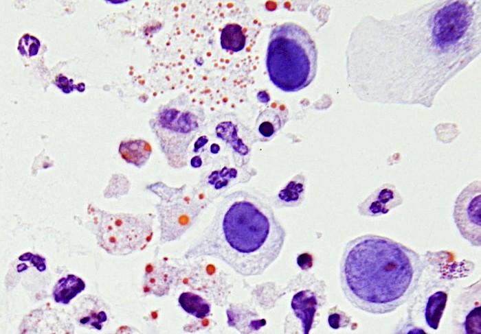
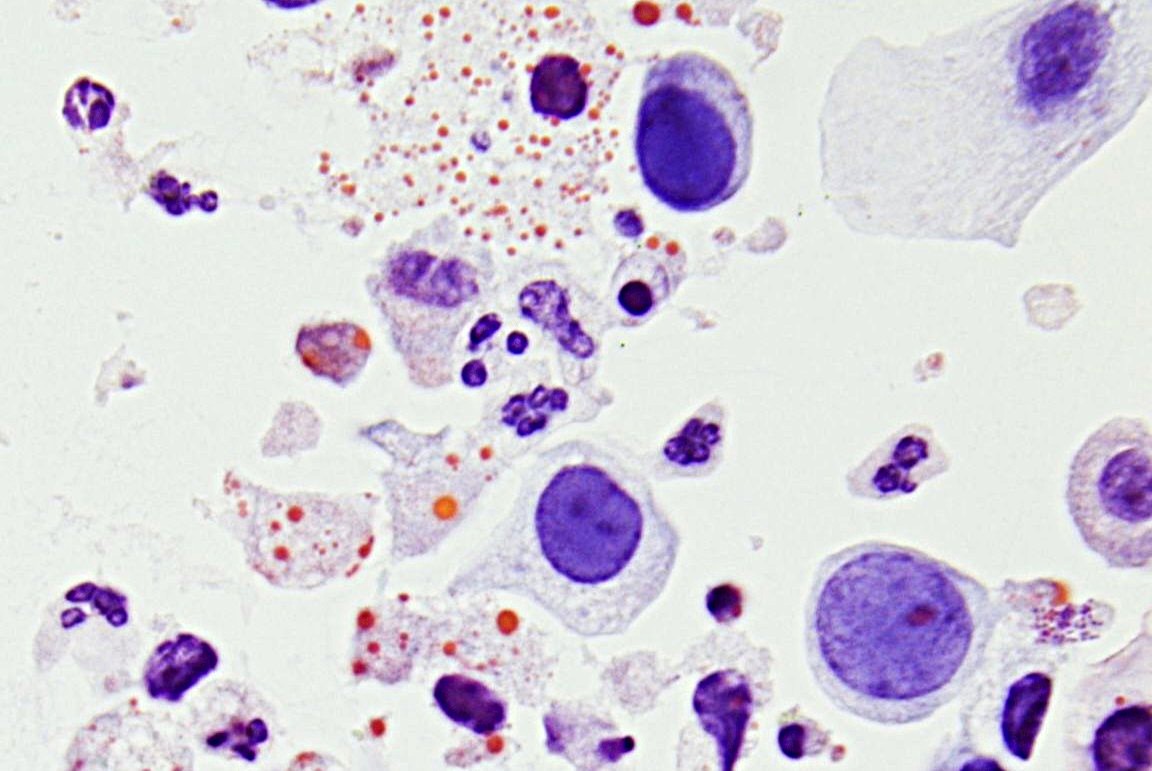 Urine specimen showing granulocytes and…
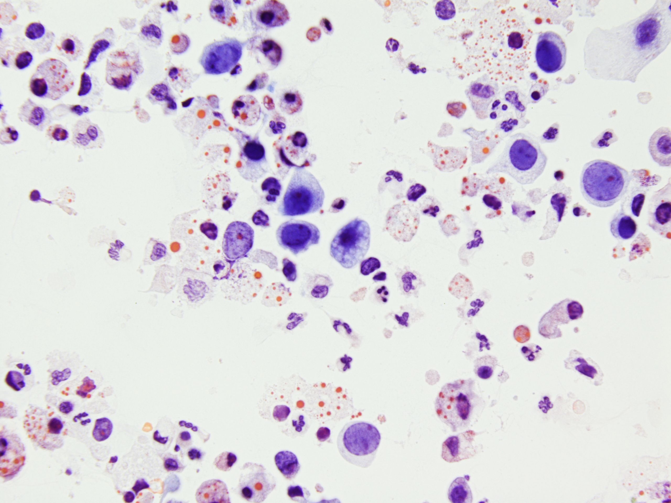 Urine specimen with degenerative urothelial…
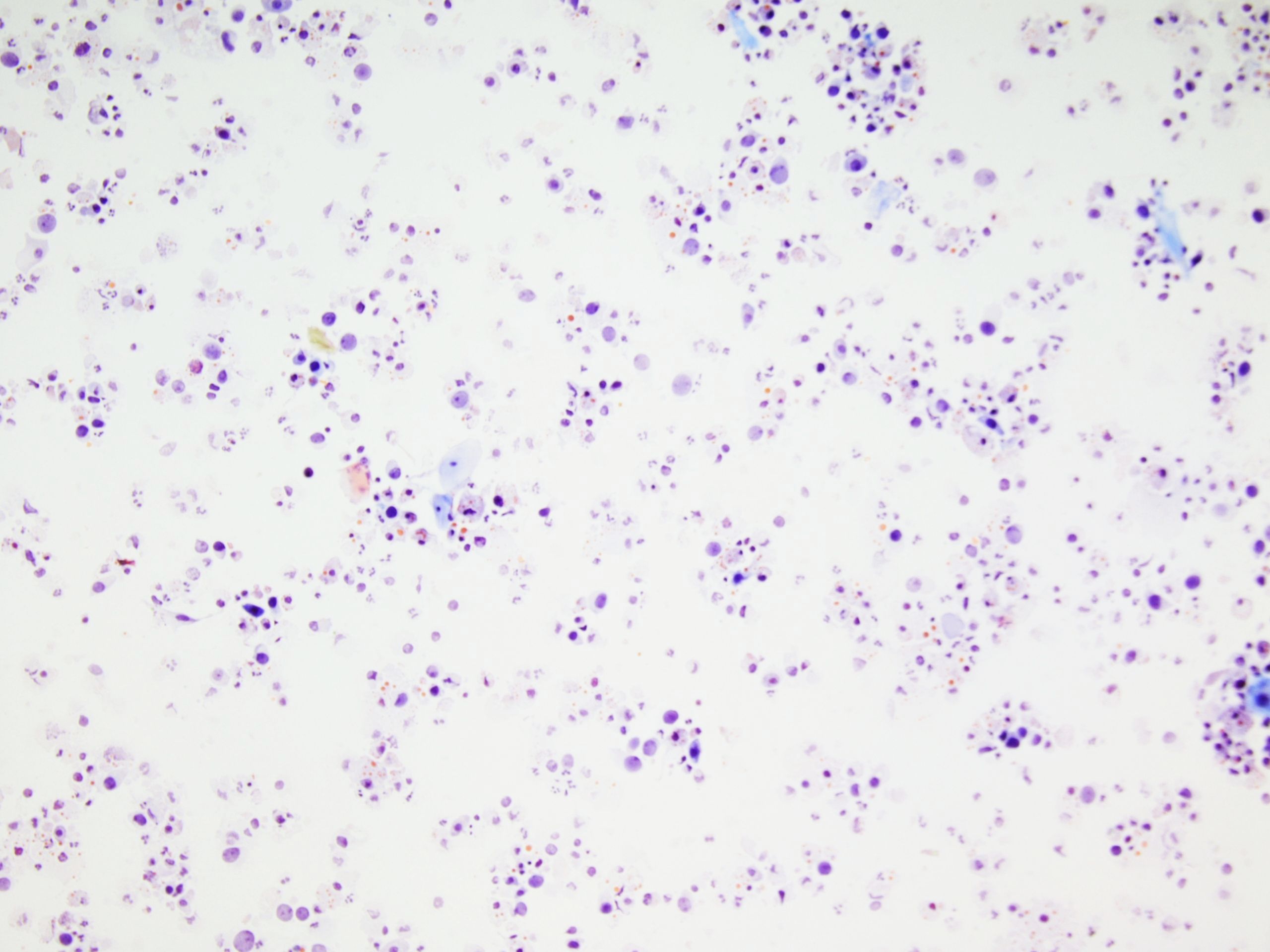 Urine, overview with a variety of different…
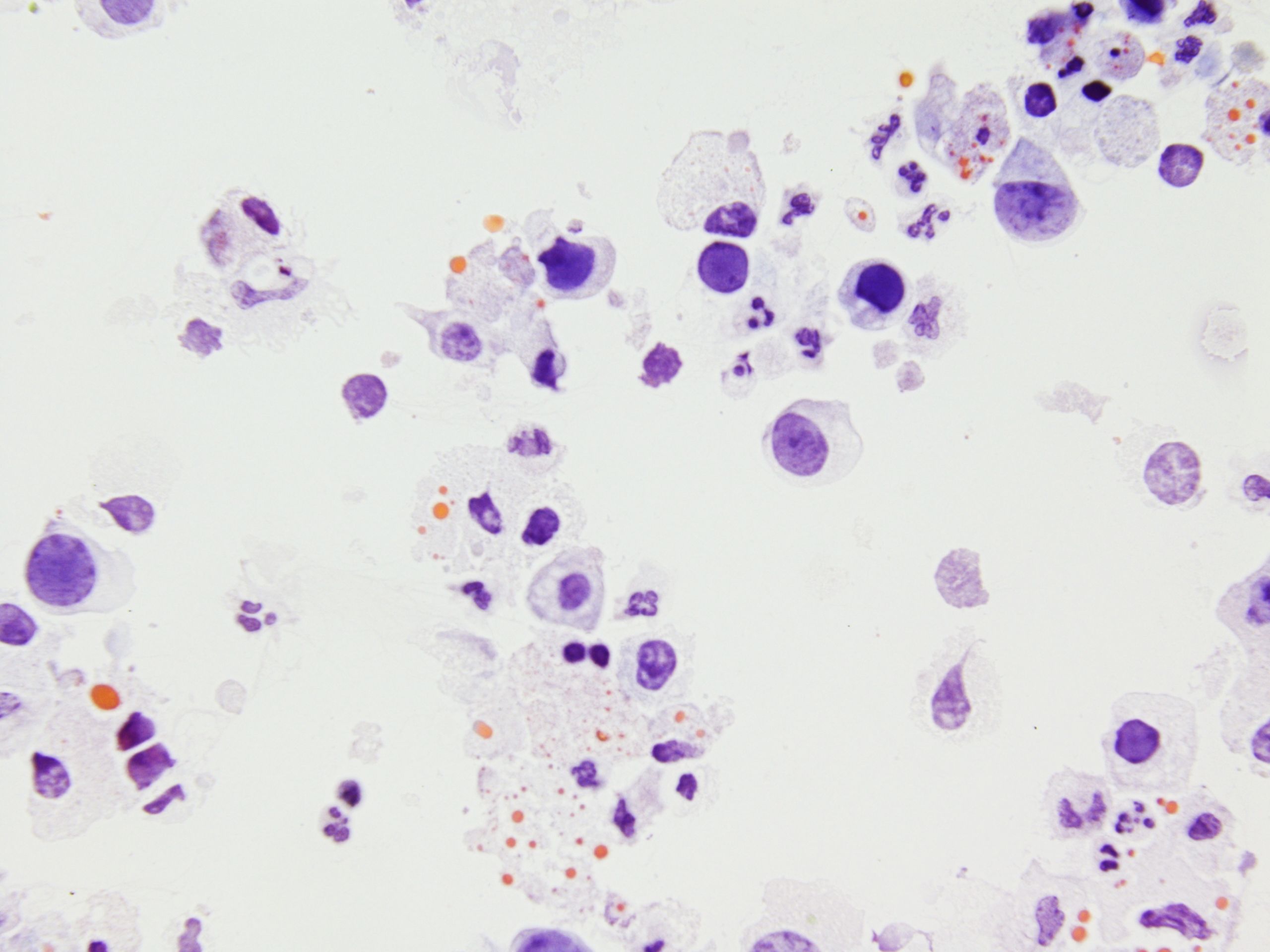 Urine specimen, decoy cells with comet…
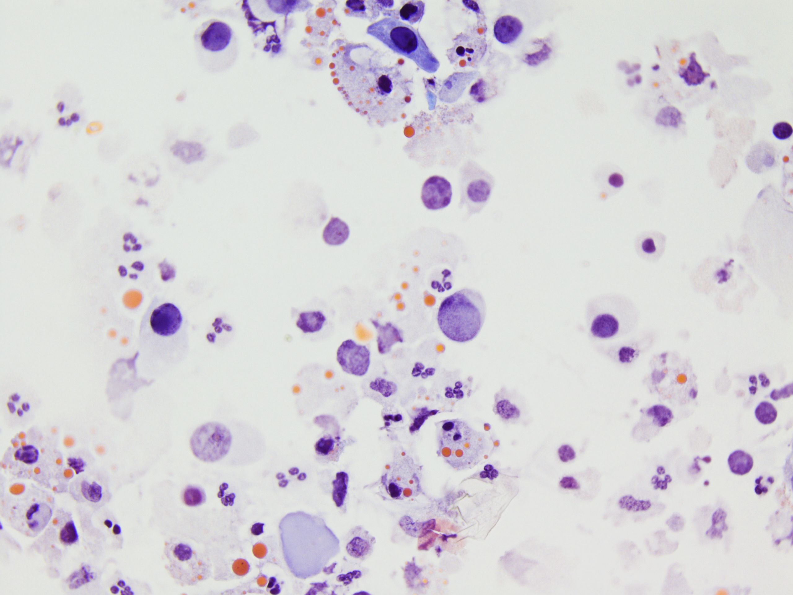 Urine specimen, multiple cells showing viral…
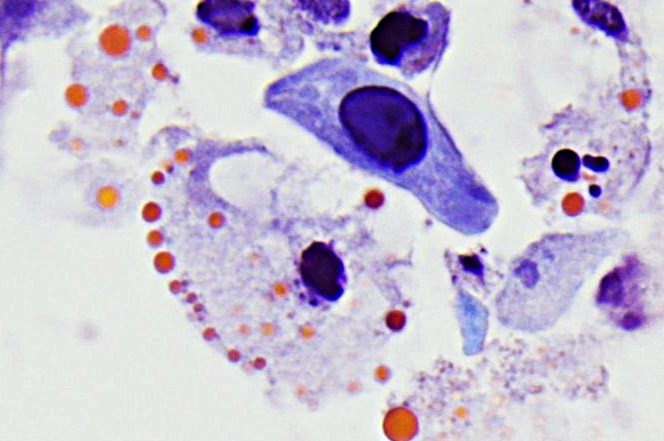 Urine specimen, conspicuous cell with a…
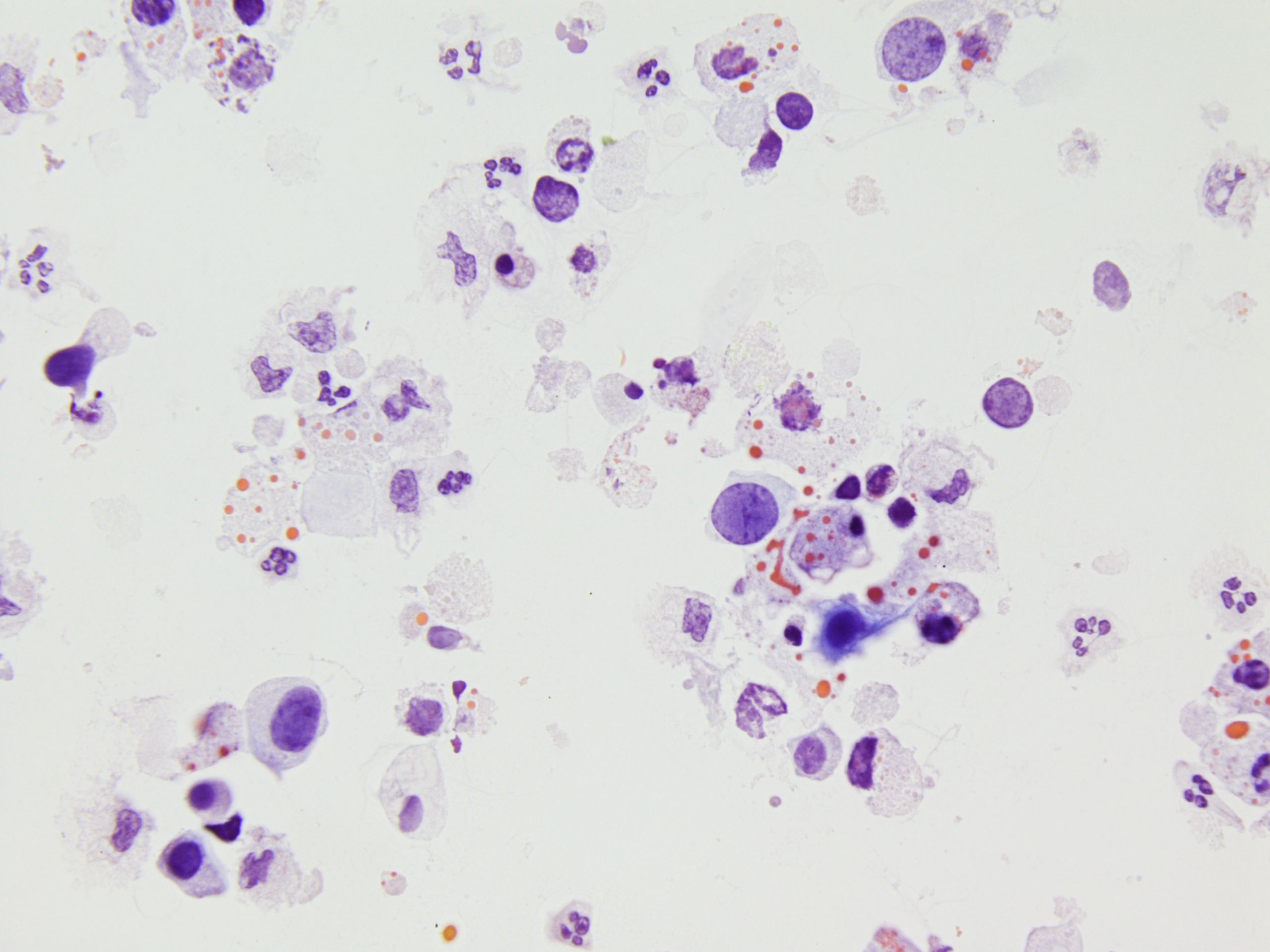 Urine specimen, variable nuclear size…
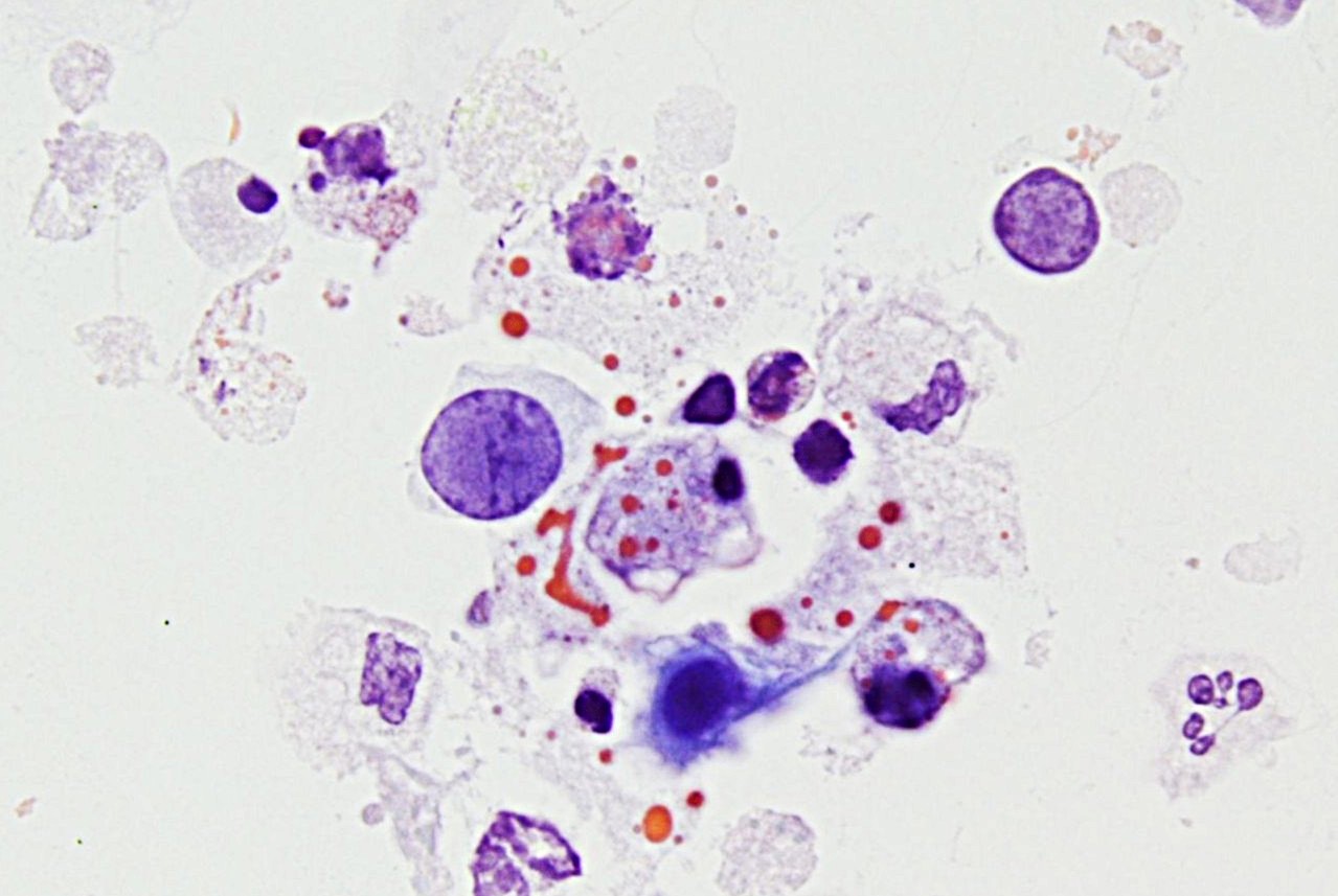 Urine specimen, classic type of decoy cells…
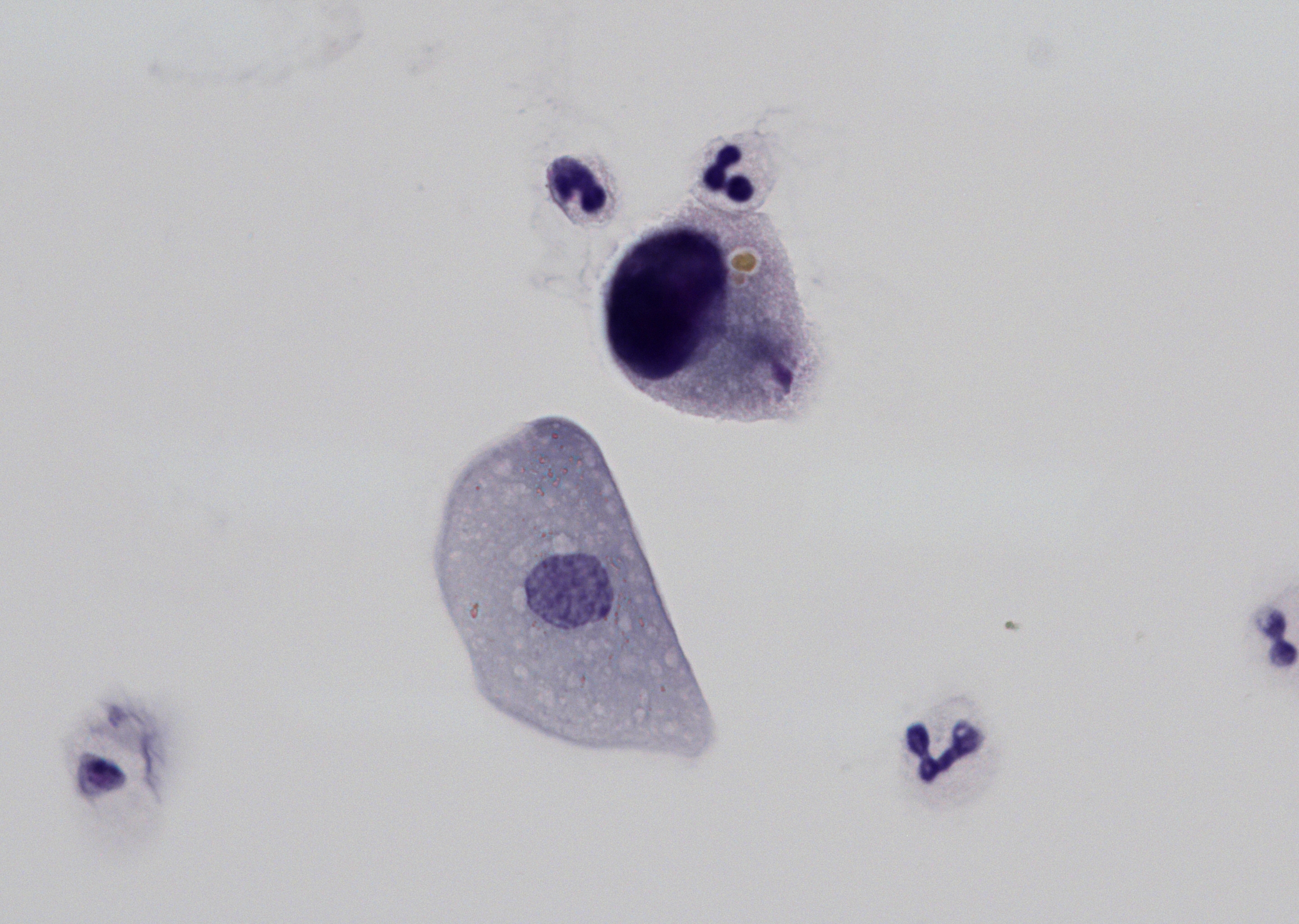 Urine, a prominent single cell with a round…
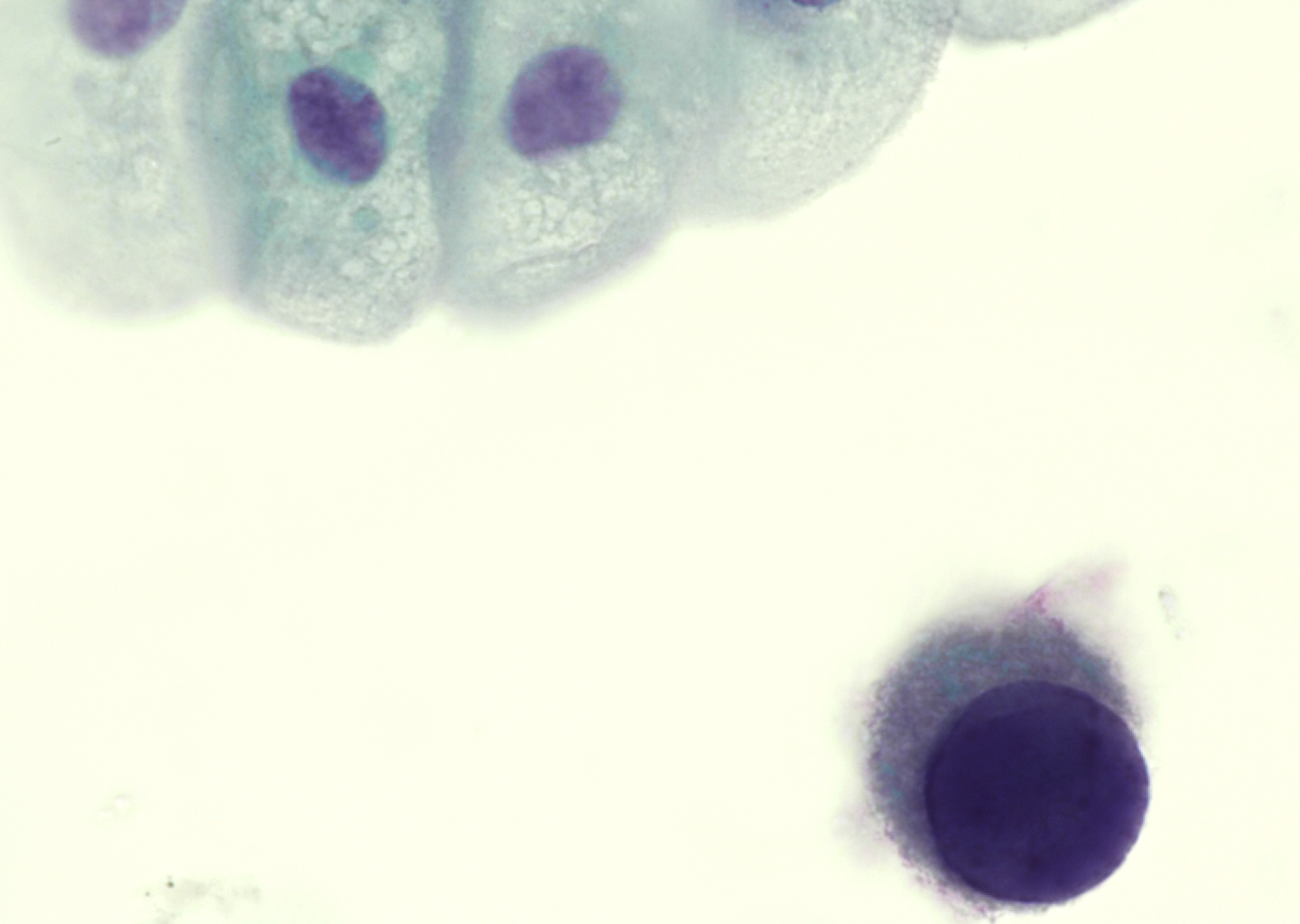 Urine specimen, a conspicuous isolated cell…
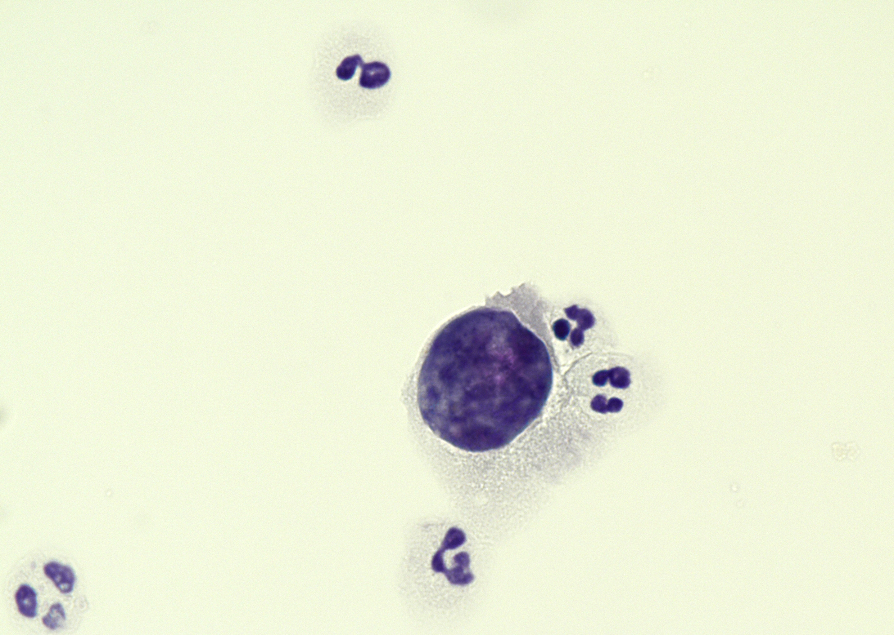 Urine specimen, conspicuous isolated cell…
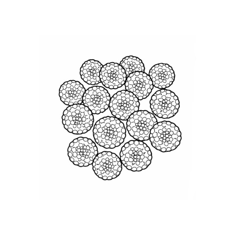 Schematic line drawing based on TEM…
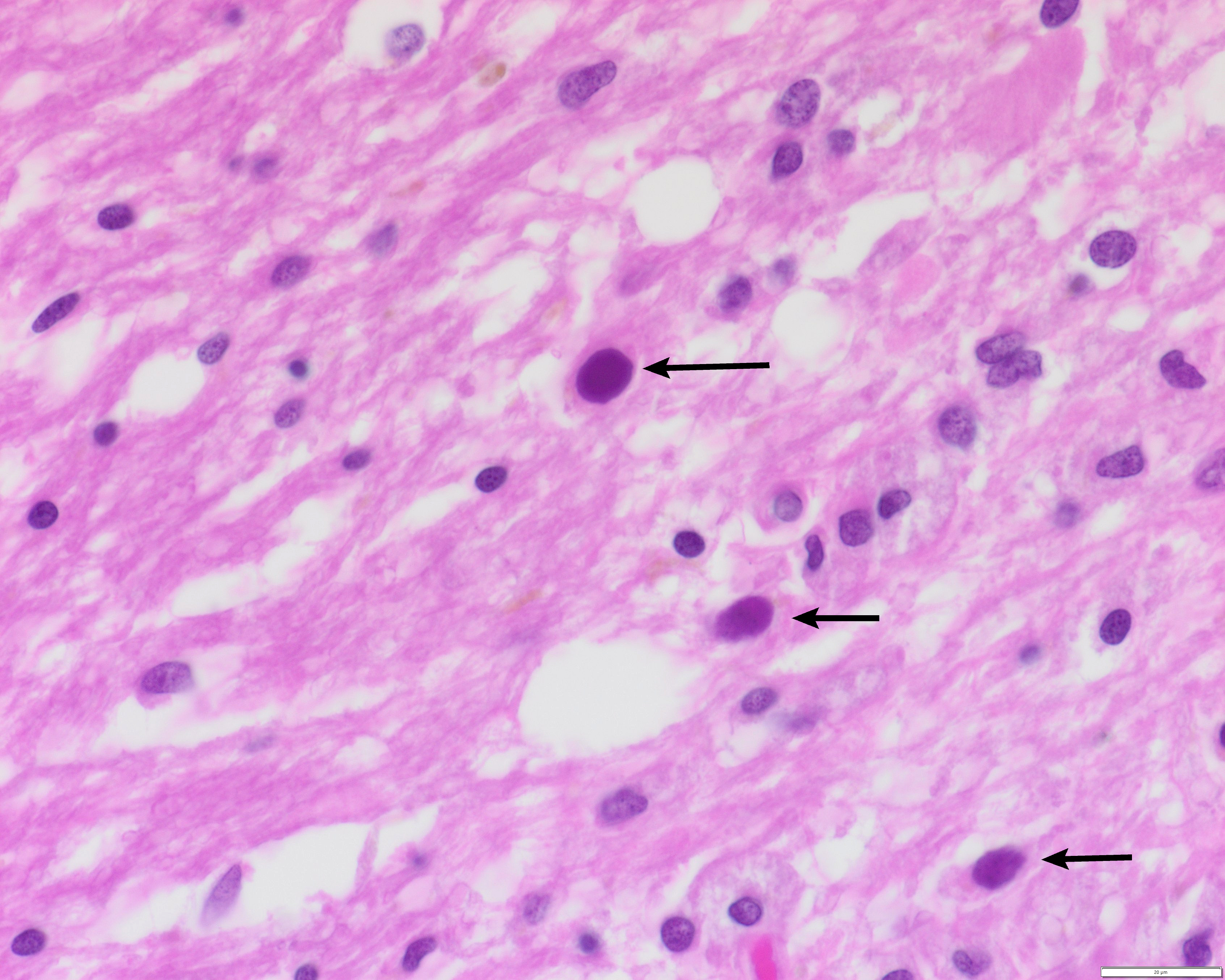 Brain tissue, autopsy case, CLL patient with…
Morphology
Cytology:
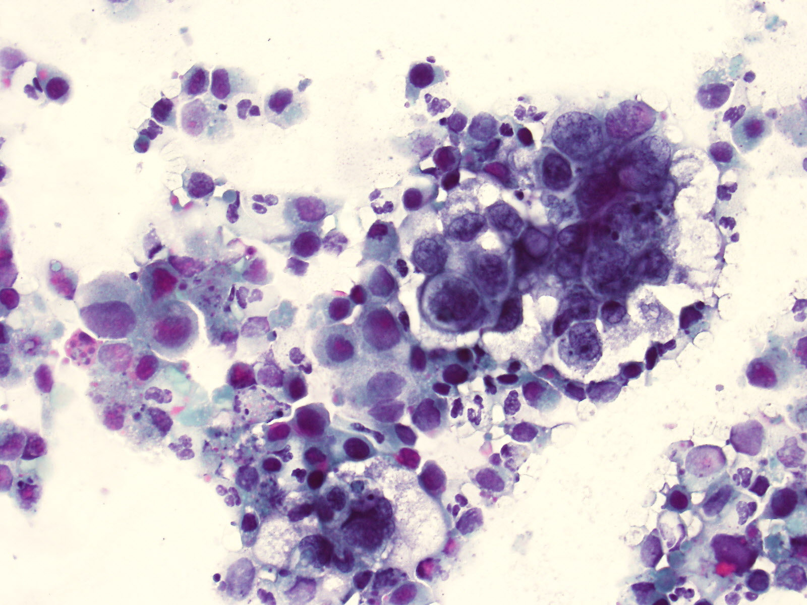
Decoy cells are the cytological hallmark of polyomavirus infection, in particular, BKV.
Four main morphological types of decoy cells are described (1, 2):
- Type 1: classic type with large, uniform amorphous ground-glass-like intranuclear inclusion bodies and a condensed rim of chromatin at the nuclear edge
- Type 2: CMV-like features with granular intranuclear inclusions surrounded by a clear halo
- Type 3: multinucleated cells with granular chromatin
- Type 4: vesicular nuclei with coarsely clumped chromatin, with or without intracytoplasmic inclusions
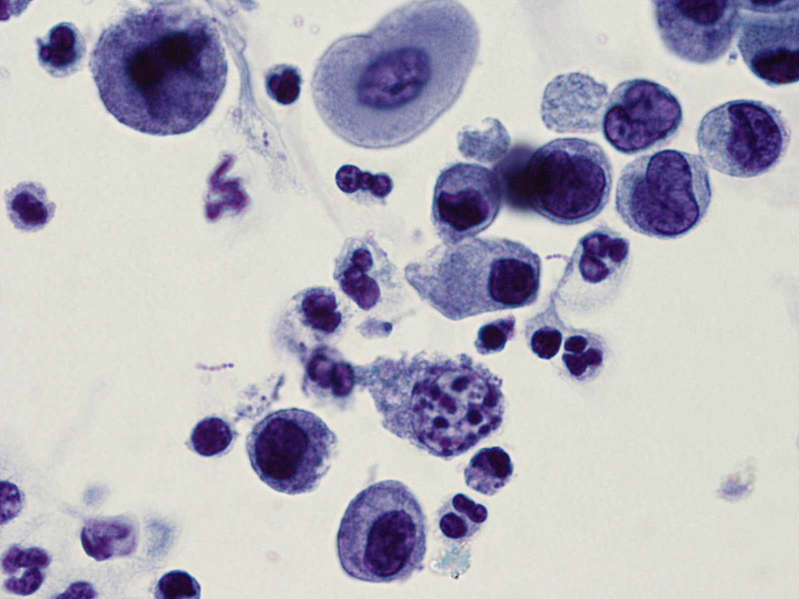
Decoy cells can mimic high-grade urothelial carcinoma cells. Still, decoy cells generally have a more finely dispersed, homogenous chromatin, whereas high-grade urothelial carcinoma cells have a more distinctive, coarse, and inhomogeneous chromatin. In routine urine cytology, they are just called "decoy cells".
Histology:
Infected cells show enlarged, round, hyperchromatic nuclei with an intranuclear inclusion body and a "ground glass" appearance, peripheral chromatin condensation, and scant cytoplasm.
Clinical notes
The primary infection usually occurs in childhood and is generally asymptomatic or mild, with respiratory symptoms and fever. After infection, the virus remains latent in different human cells, depending on the virus type. In immunosuppressed patients, the virus can reactivate.
BKV remains latent in the urothelial and renal tubular epithelial cells, with periodic shedding into the urine; reactivation due to immunosuppression can lead to polyomavirus-associated nephropathy (PVN) or hemorrhagic cystitis and is associated with an increased risk of polyomavirus-associated urothelial carcinoma. Especially in kidney transplant patients, this has significant clinical implications and can lead to graft loss.
Those shed infected epithelial cells show altered nuclei with viral cytopathic effects and are called "Decoy cells", and can be observed in urine cytology, especially in immunosuppressed patients with PVN, most commonly in the post-transplant setting.
Urine cytology may suggest polyomavirus replication by demonstrating decoy cells; however, cytology alone is not sufficient to diagnose BKPyV-associated nephropathy/PVN (3, 4).
Ancillary Testing
BKV (PCR plasma, urine), JCV (PCR plasma, urine, CSF, brain tissue); IHC (SV40)Additional Information
Ubiquitous, non-enveloped dsDNA virus. There are 14 human pathogenic polyomavirus types, with the most common and clinically relevant representatives being BK-, JC-, and MC-virus.
A wide range of seropositivity across the global population, depending on specific virus types and populations; for example, BKV has a seropositivity of over 90% in the general population.
JCV exhibits strong neurotropism; reactivation leads to infection of oligodendrocytes and astrocytes, resulting in progressive multifocal leukoencephalopathy (PML), a severe demyelinating disease of the central nervous system. PML occurs primarily in patients with advanced HIV infection, hematologic malignancies, organ transplantation, or under immunomodulatory therapies, and is associated with high morbidity and mortality.
The SV40 (Simian Polyomavirus 40) immunohistochemical antibody shows cross-reactivity with BKV and JCV (5).
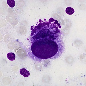
Differential Diagnosis
References
- Singh HK, Bubendorf L, Mihatsch MJ, et al. Urine Cytology Findings of Polyomavirus Infections. In: Madame Curie Bioscience Database [Internet]. Austin (TX): Landes Bioscience; 2000-2013. Available from: https://www.ncbi.nlm.nih.gov/books/NBK6541/
- Geetha V, Rao L, Monappa V, et al. Decoy cells in urine cytology: A useful clue to post-transplant polyoma virus infection. J Cytol. 2012;29(2):133-134. https://doi.org/10.4103/0970-9371.97157
- BTS BK Guideline Working Group. (2024, December 4). UK guideline on management of BK polyomavirus (BKPyV) infection and disease following kidney transplantation. British Transplantation Society.
- Kotton, C. N., et al. (2024). The second international consensus guidelines on the management of BK polyomavirus in kidney transplantation. Transplantation, 108(9), 1834–1866. https://doi.org/10.1097/TP.0000000000004976
- Costa, C., & Cavallo, R. (2012). Polyomavirus-associated nephropathy. World Journal of Transplantation, 2(6), 84–94. https://doi.org/10.5500/wjt.v2.i6.84