
- EN
- DE

Virus
Differential Diagnosis of Human Polyomavirus (HPyV):
Cytomegalovirus (CMV)

- Organ System
- Respiratory Tract, Digestive Tract, Lymphatic System, Blood & Bone Marrow, CNS and Sensory Organs, Genitourinary Tract
- Diagnostic Method
- Cytology, Histology

- Geographical Region
- Dry Areas,
- Islands,
- Tropical and Subtropical Region,
- Polar and Subpolar Zone,
- Temperate Zone
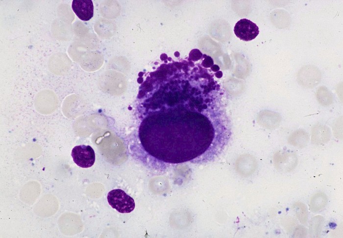
 BAL, mononuclear cell with CMV-like…
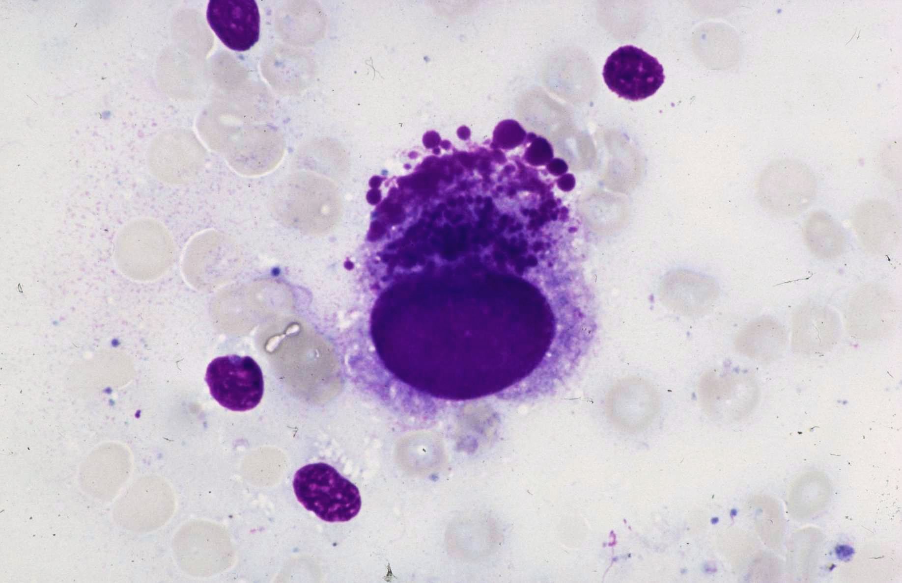
 BAL, mononuclear cell with enlarged dark…
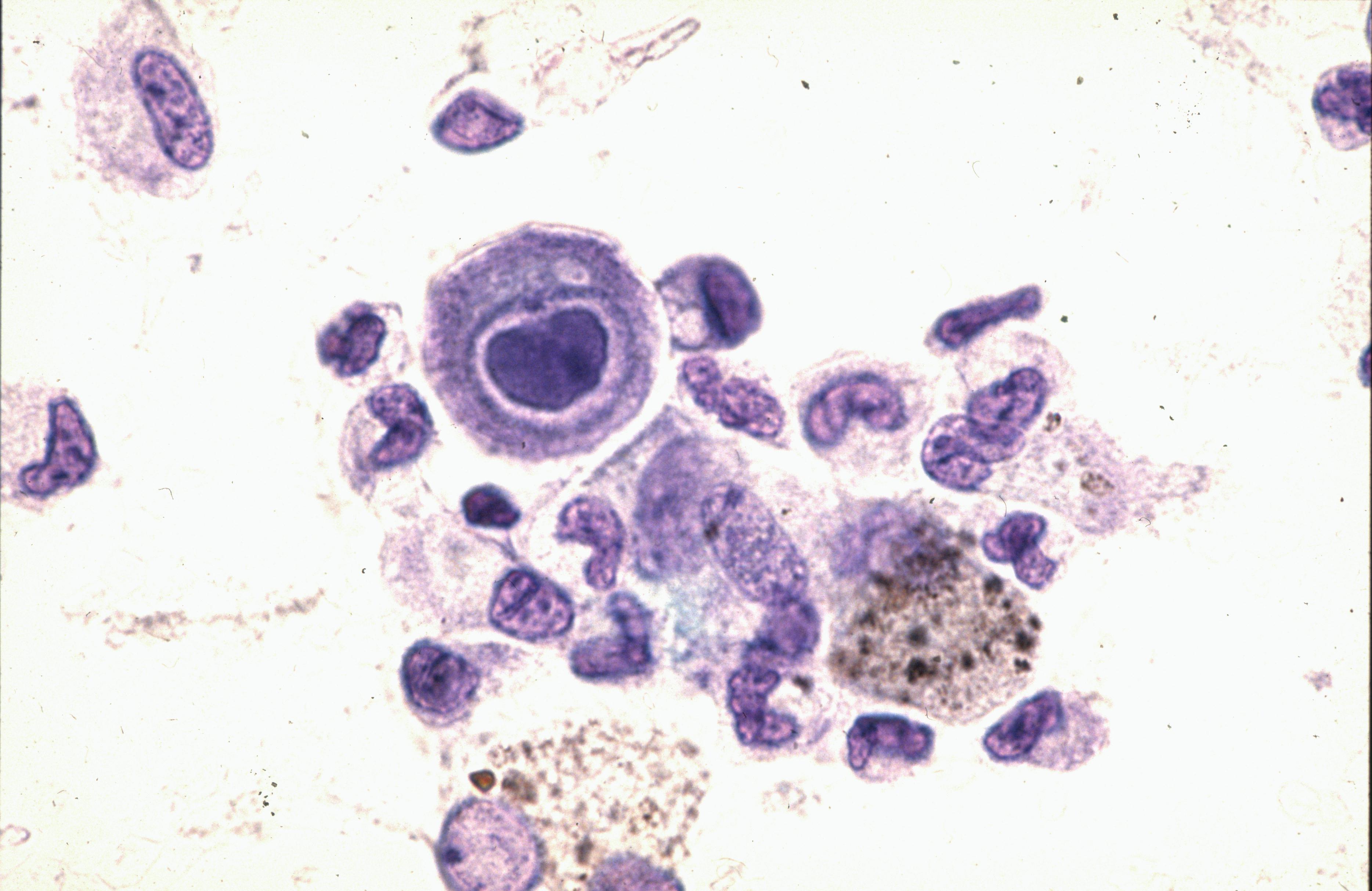
 BAL, in the setting of known HIV infection…
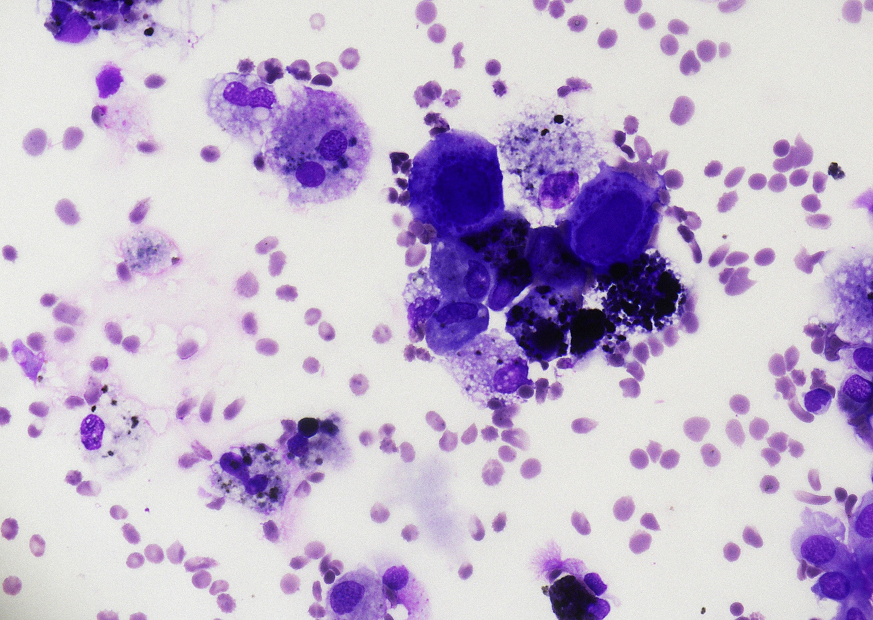 BAL, HIV patient, cell with dark, rather…
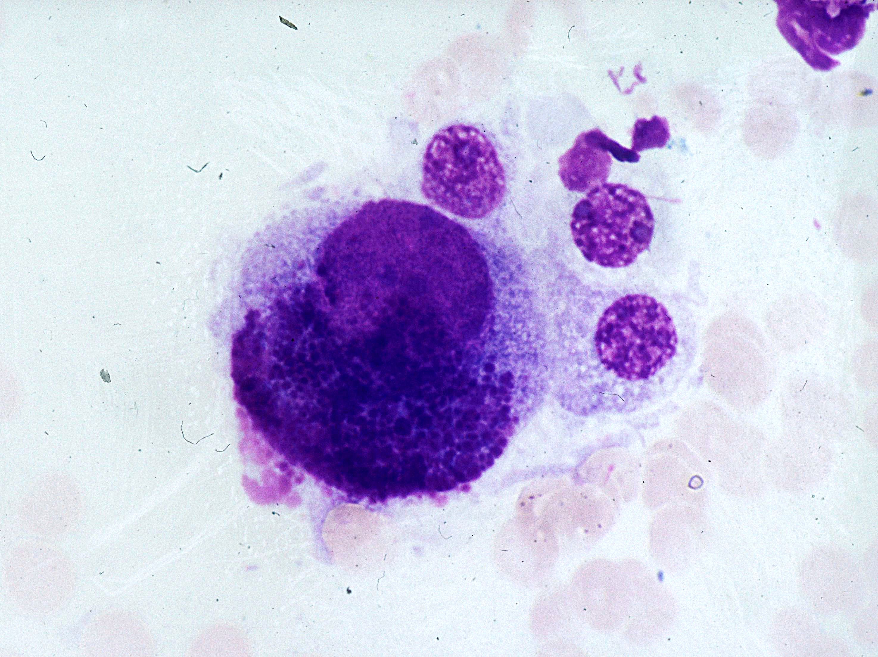 BAL, large cell with an enlarged excentric…
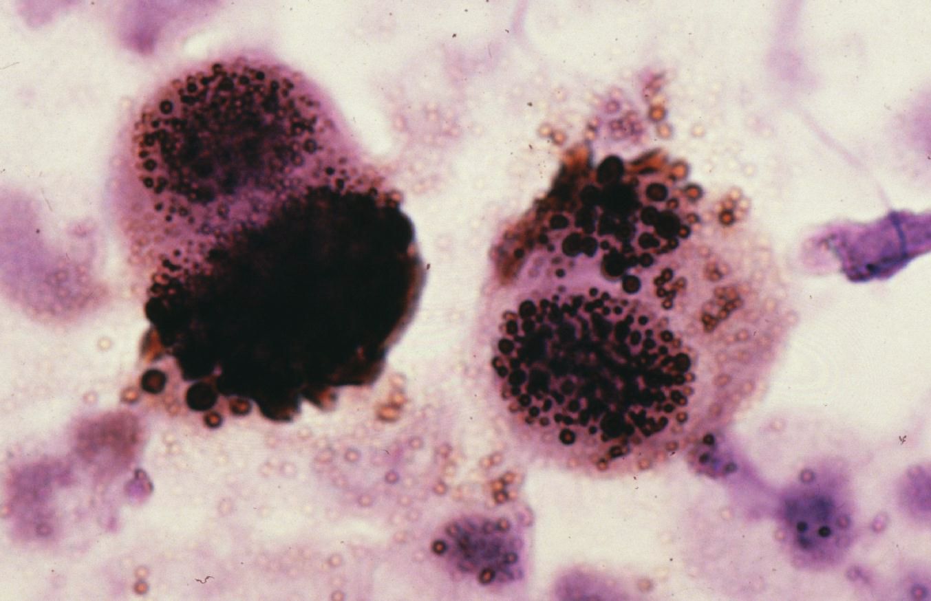 BAL, positive brown chromogenic signal…
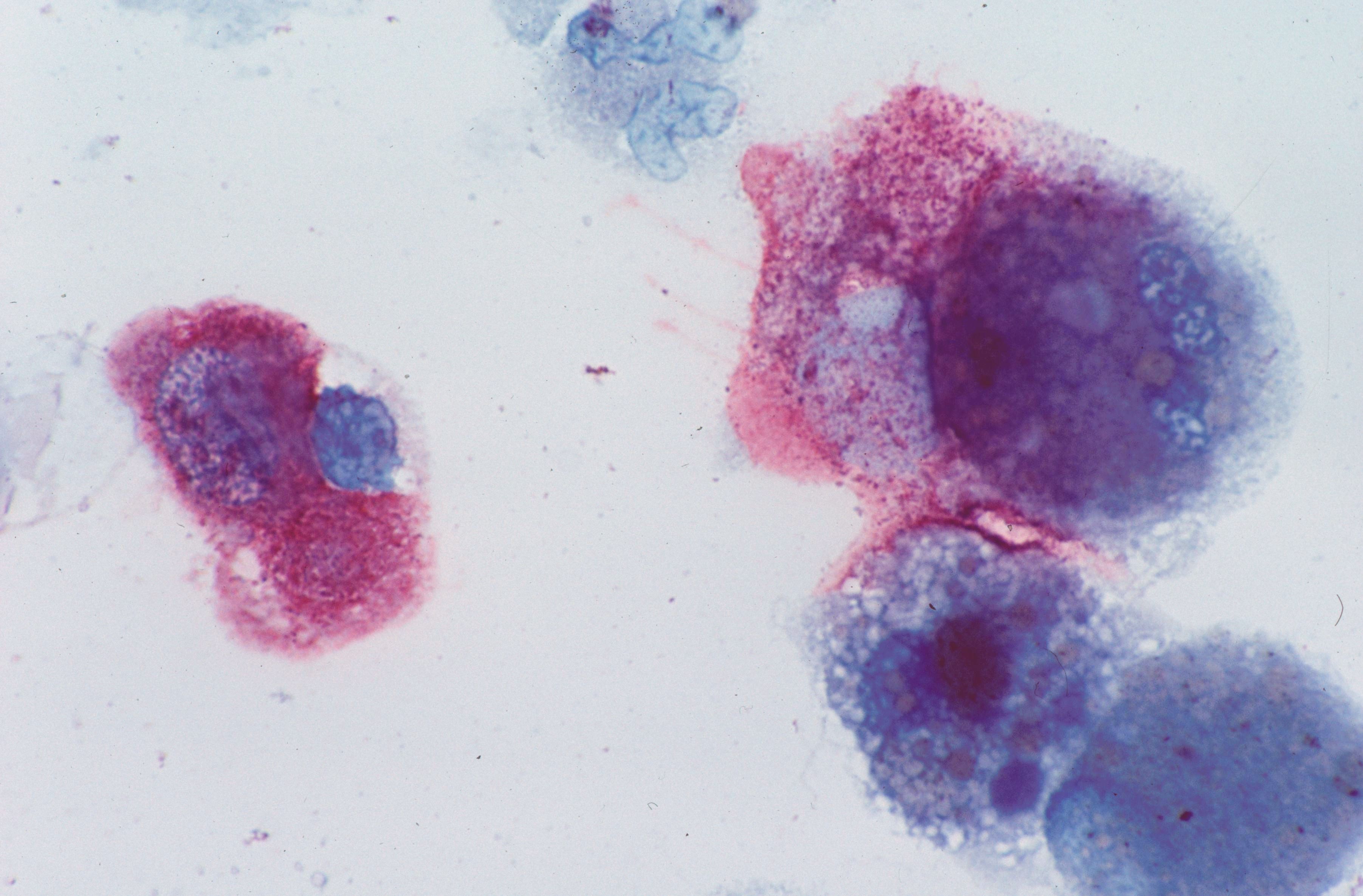 BAL, positive signal indicating…
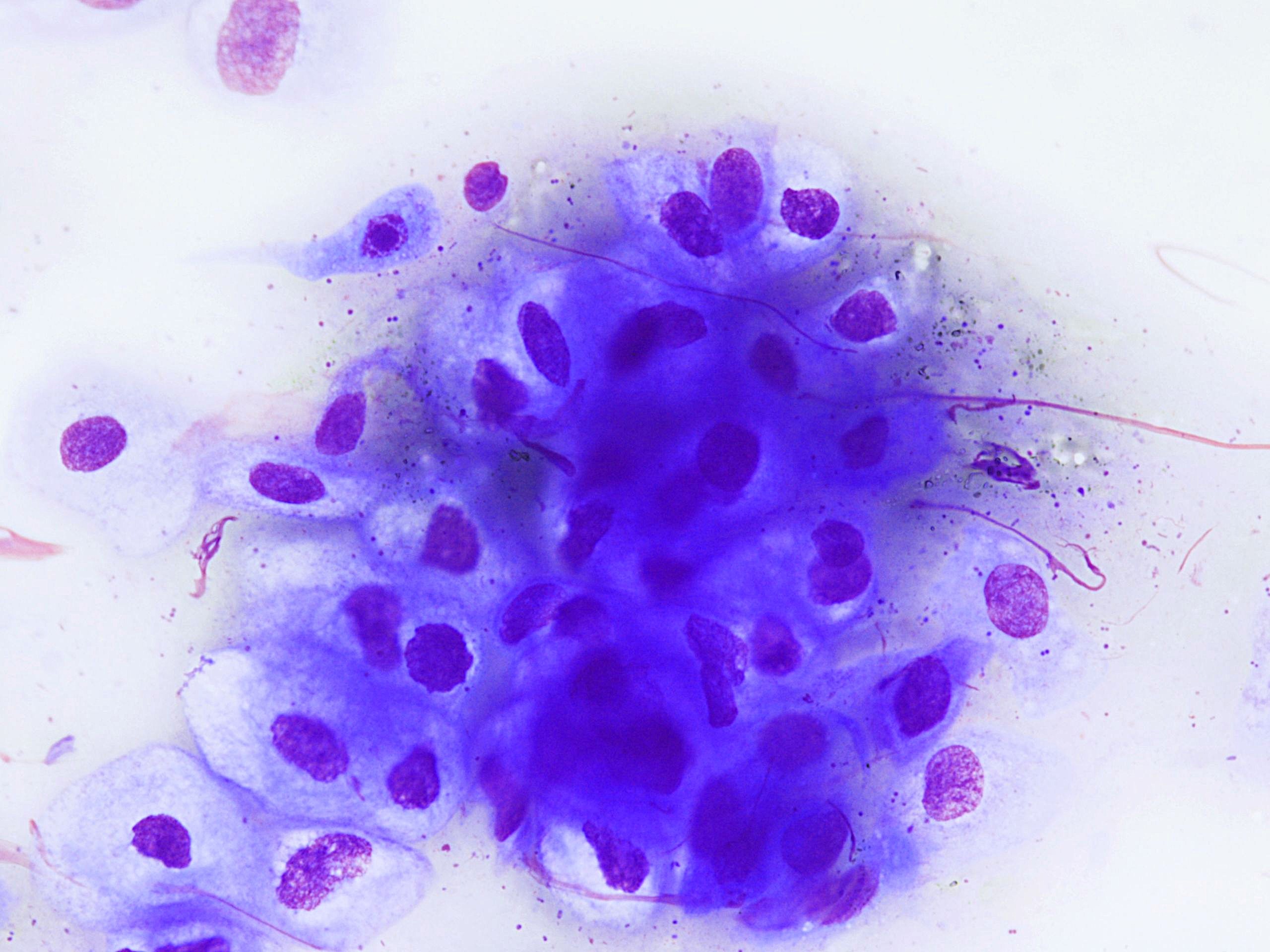
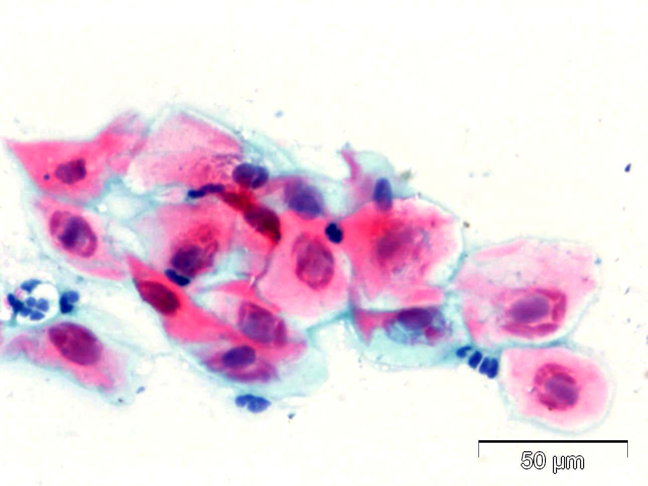 Cervical smear, CMV infected squamous…
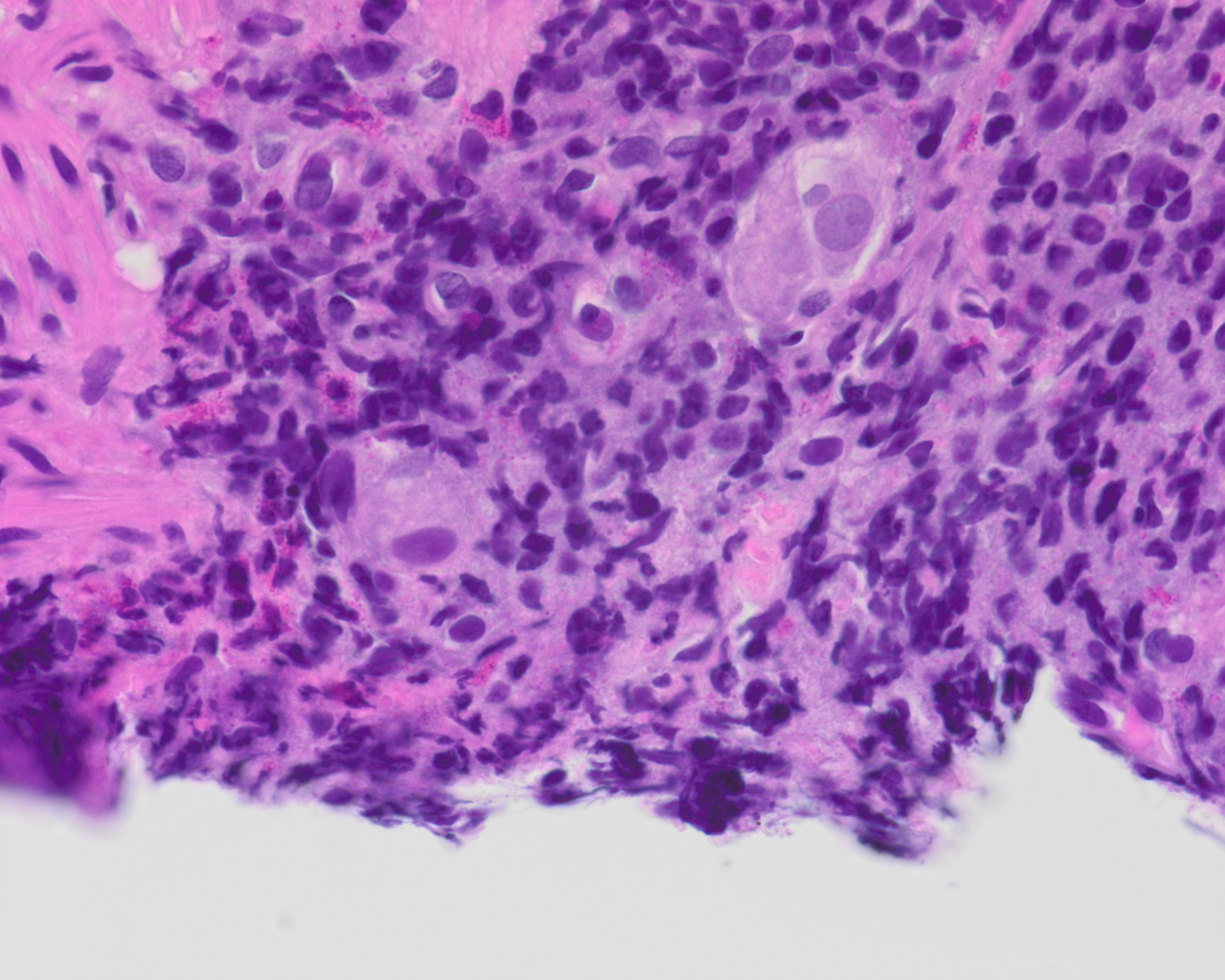 Colon biopsy, a few greatly enlarged…
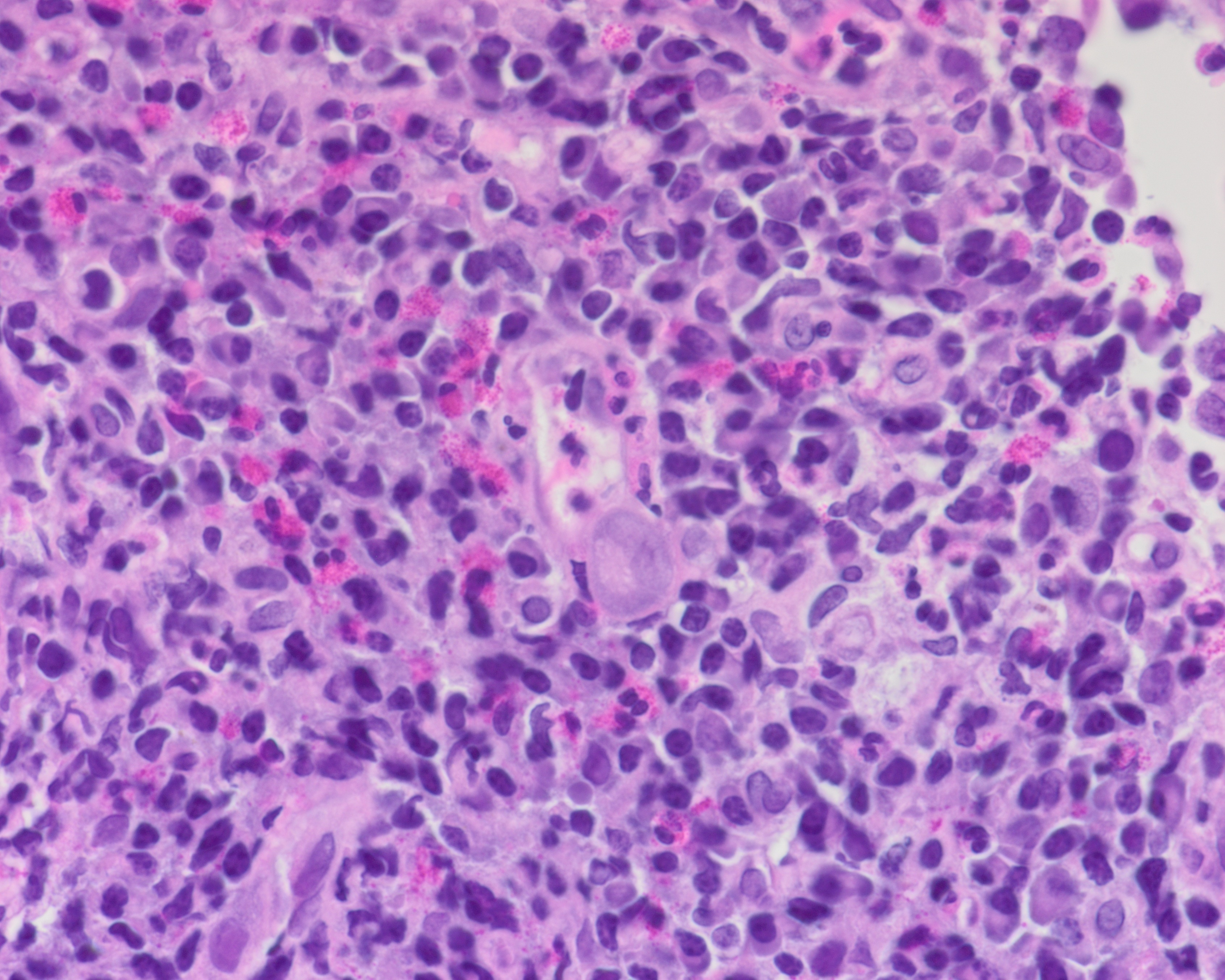 Colon biopsy, enlarged infected endothelial…
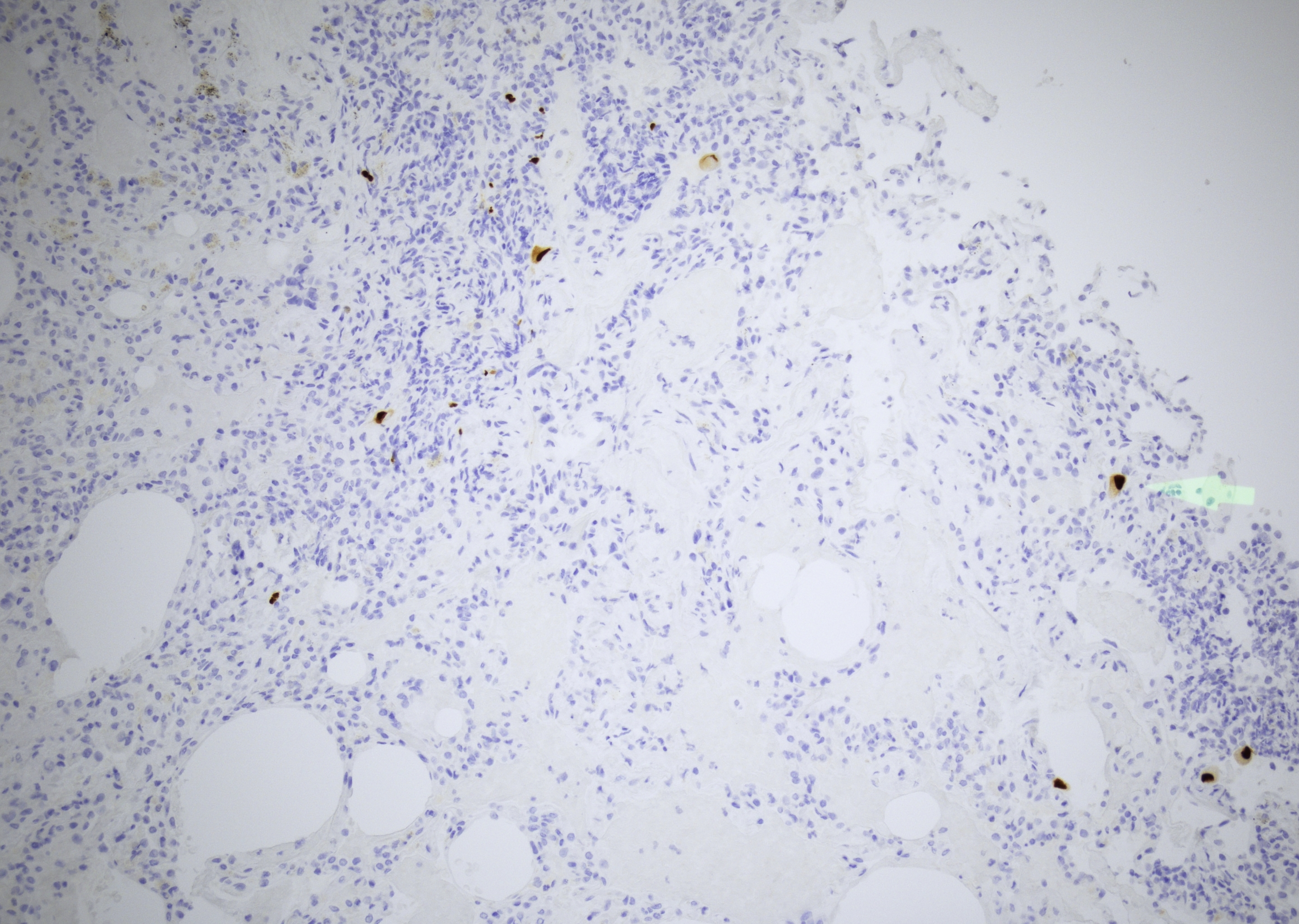 Colon biopsy, multiple CMV-infected cells…
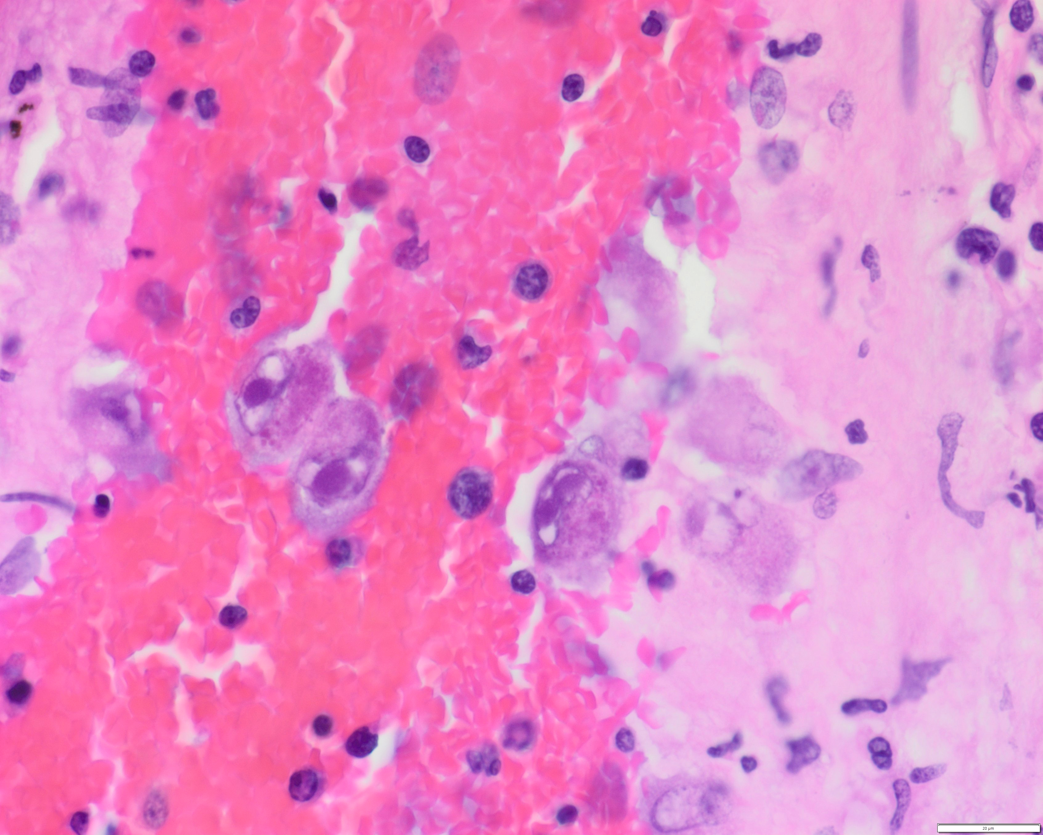 Lung section, autopsy case…
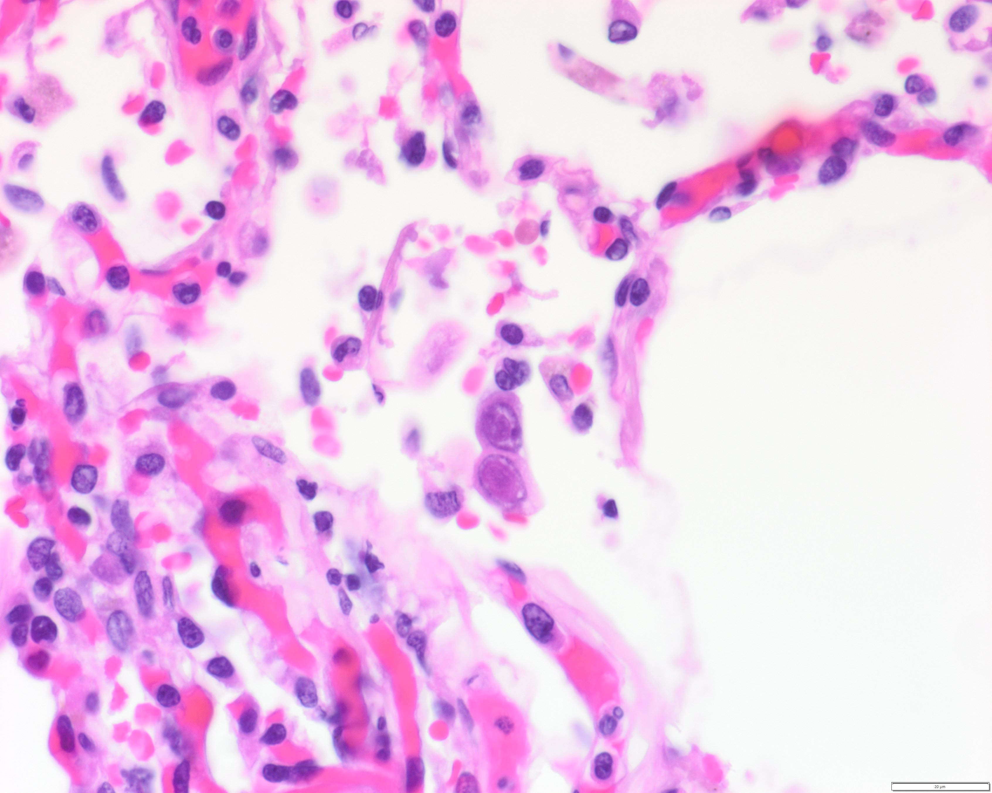 Lung section, autopsy case…
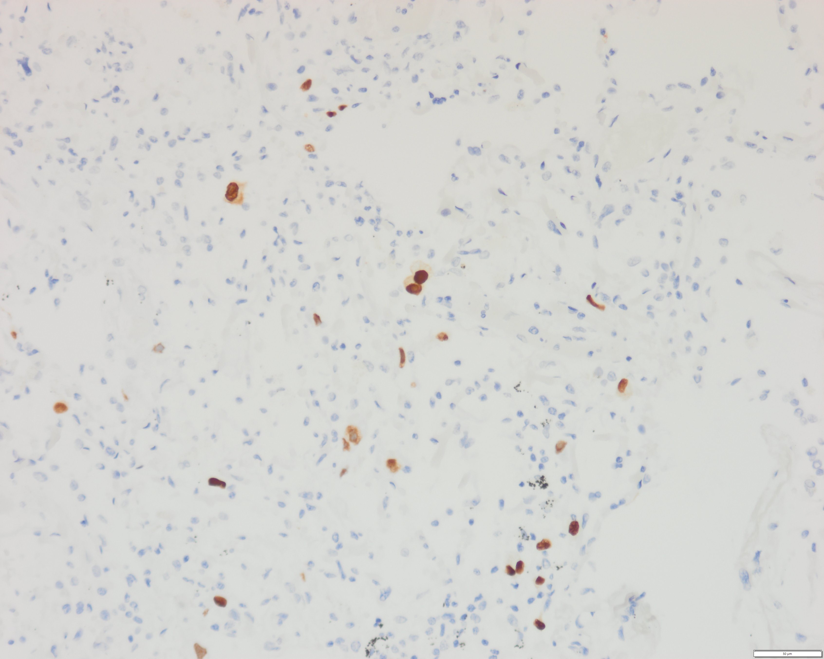 Lung section, autopsy case…
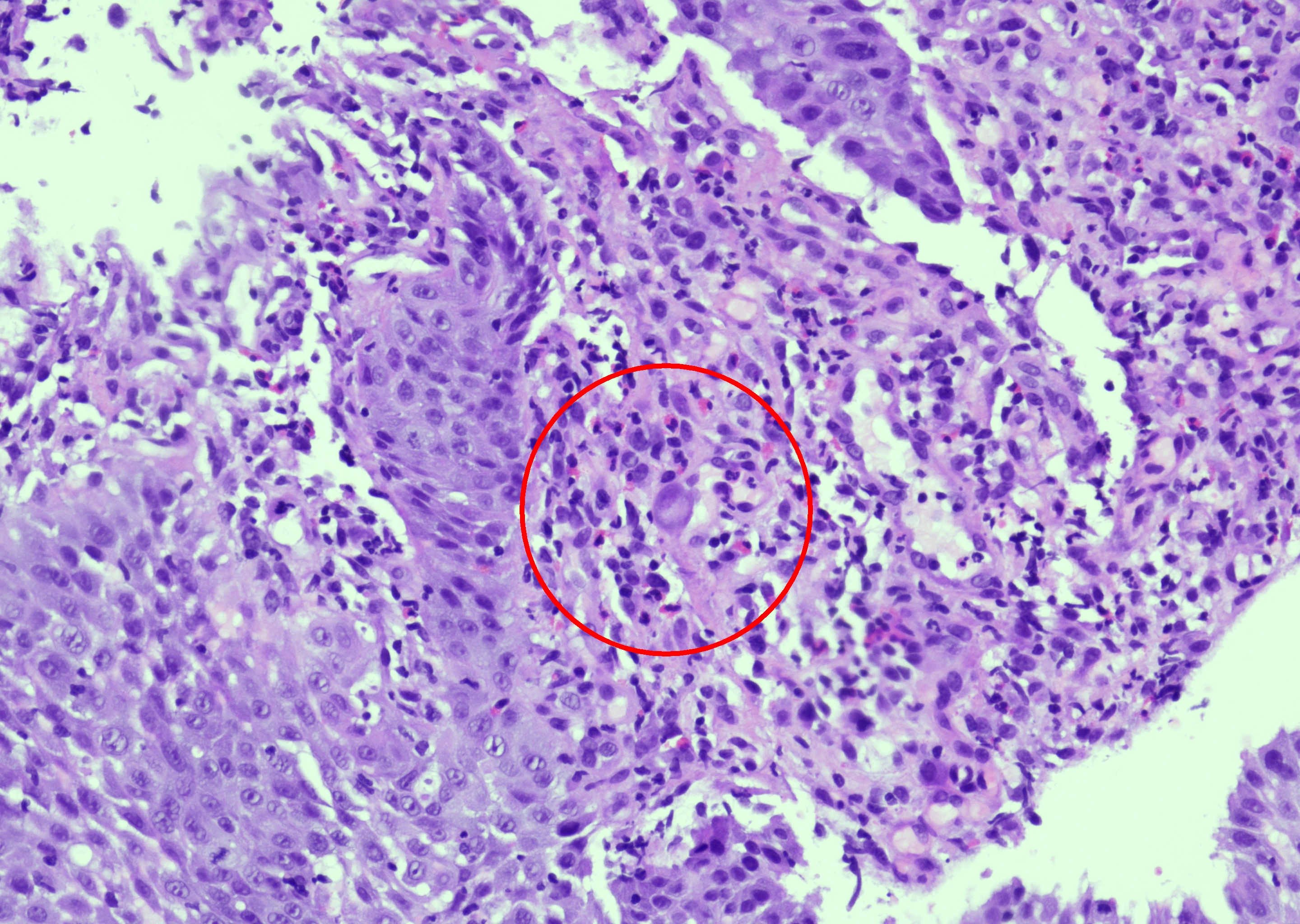 Gastroesophageal junction, biopsy with mixed…
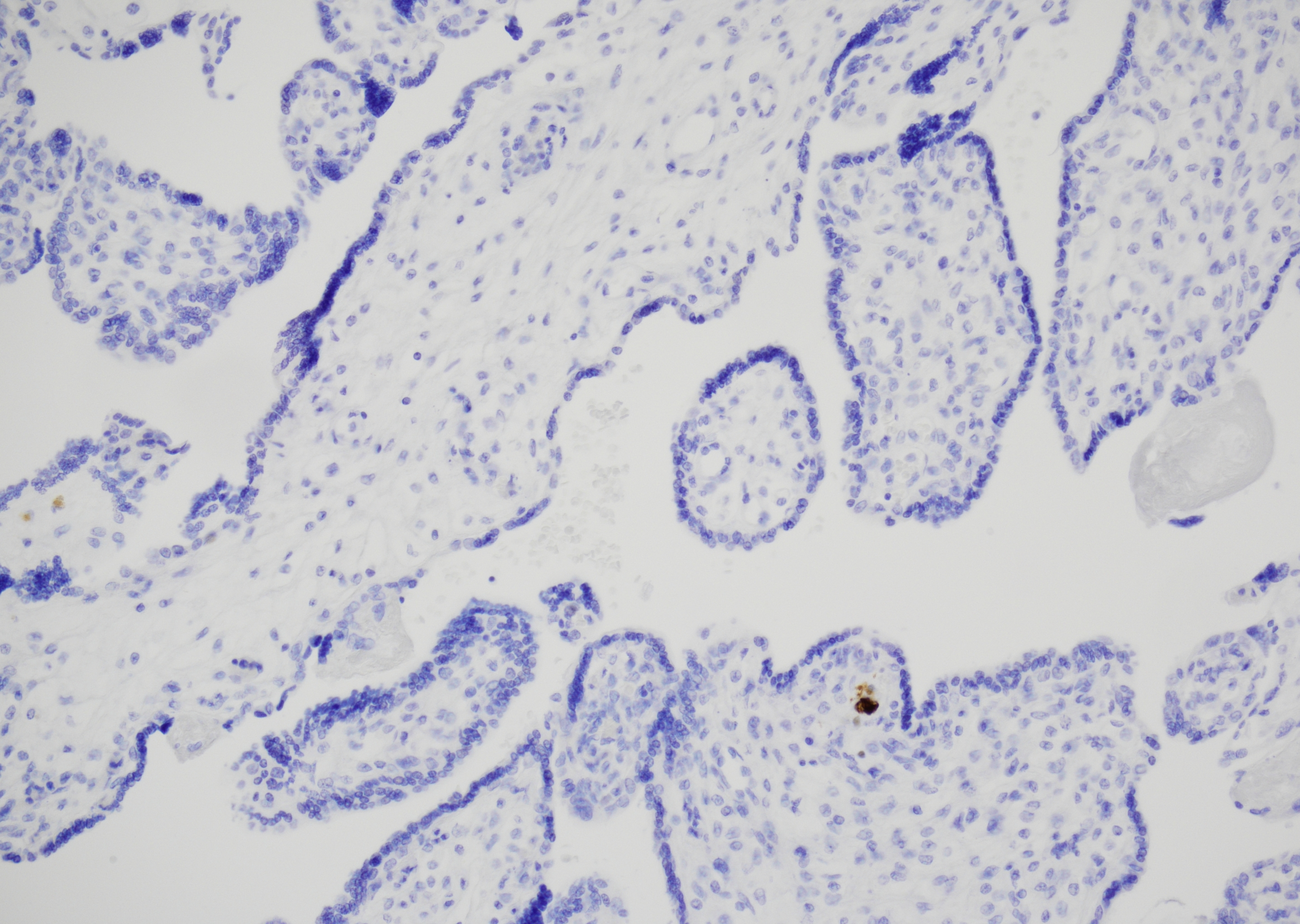 Placental chorionic villi, CMV-infected…
Morphology
Cytology:
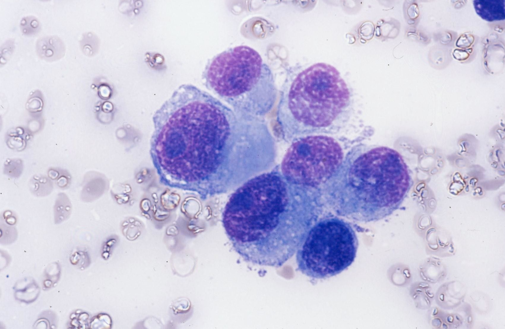
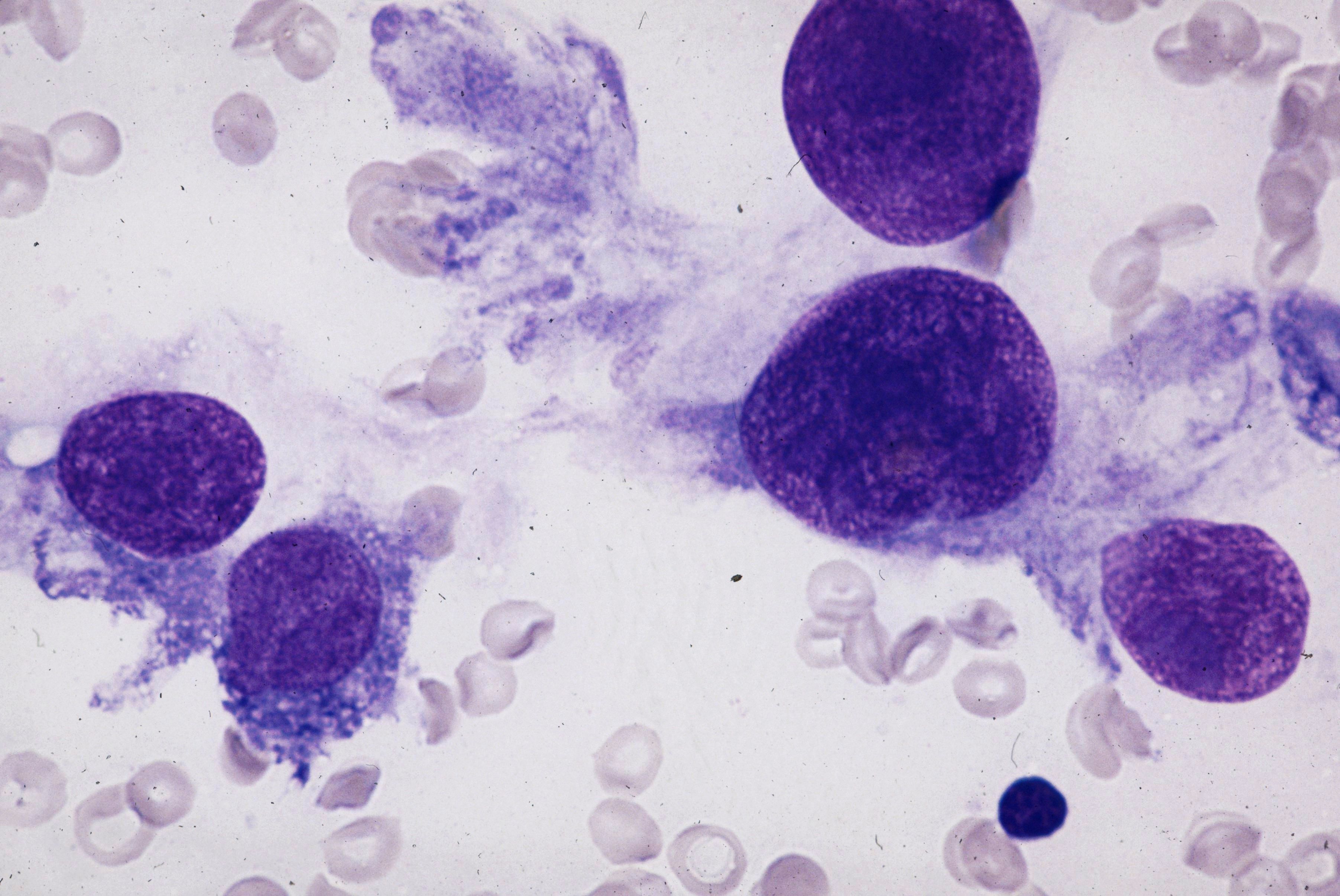
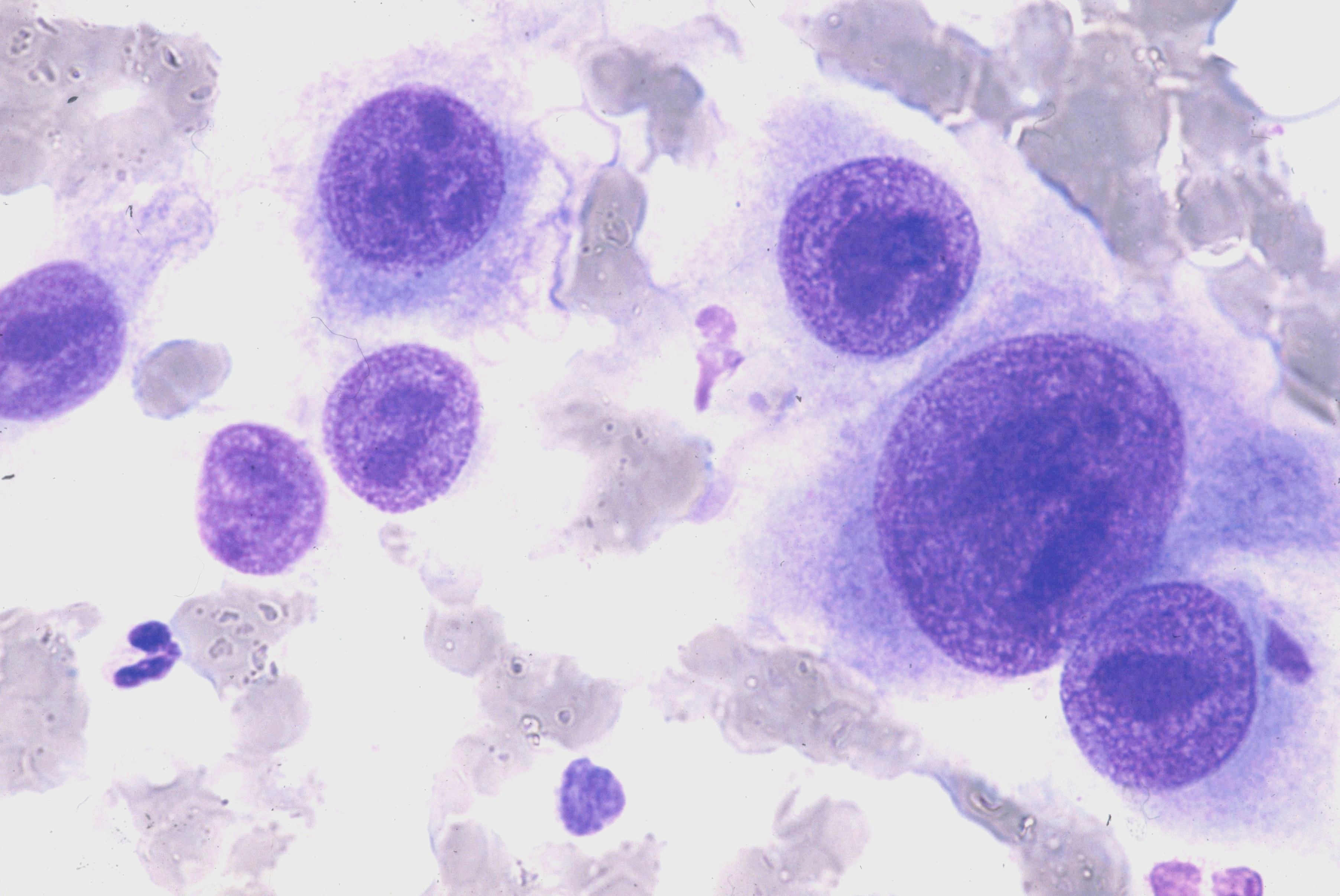
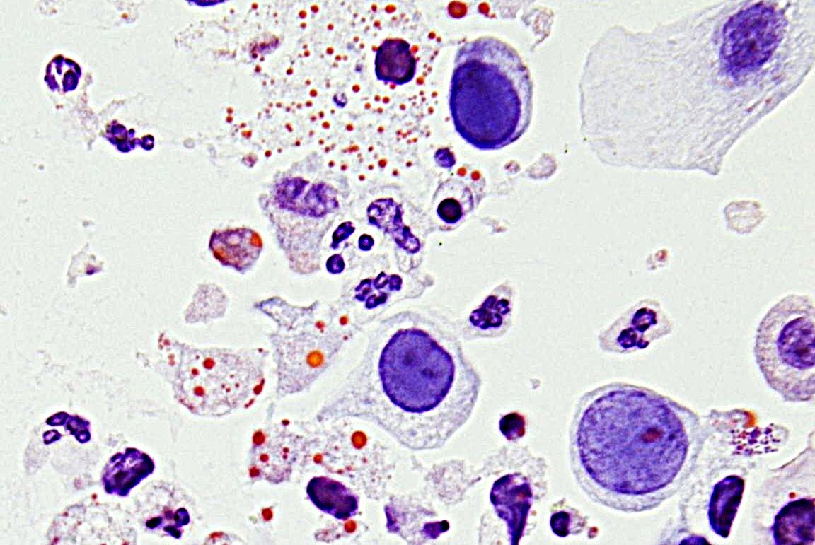
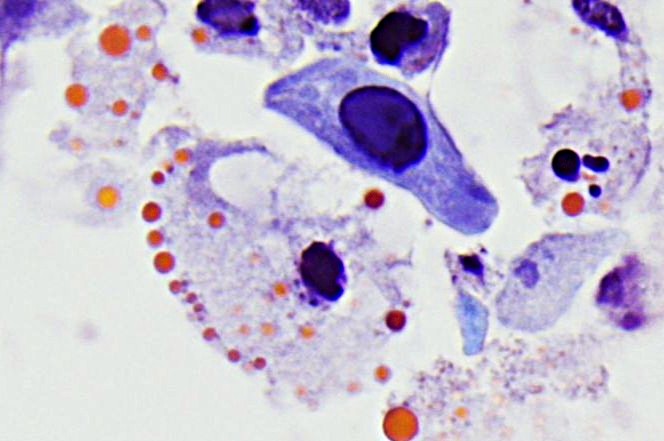
- Single mononuclear cells (around 3-4x size of macrophages) with enlarged, hyperchromatic nuclei and homogeneous, blue-violet intracytoplasmic inclusions on MGG stain and/or classical intranuclear inclusions
- "Owl's eye cells" - enlarged uninucleated cells with a large basophilic intranuclear inclusion, perinuclear clearing, and a sharply outlined nuclear membrane
Histology:
- The most commonly infected cells are macrophages, endothelial, and stromal cells
- Epithelial cells are less frequently conspicuous
- Immunohistochemistry against anti-CMV antibodies highlights CMV-infected cells, even those lacking morphologically evident viral cytopathic changes.
Surrounding Reaction:
- Unspecific pattern with strong reactive epithelial changes
- Often ymphocytic aggregates
- Ischemic pattern with ulceration and/or necrosis, particularly in the gastrointestinal tract in histology
Clinical notes
Primary CMV infection is usually self-limited and asymptomatic in immunocompetent individuals. Adults may develop nonspecific symptoms, such as a cold or the flu, during the initial CMV infection; children and young adults most commonly present with mononucleosis-like symptoms.
CMV infection may be dangerous for people with a weakened immune system, with highest risk of severe disease in solid organ transplant recipients, hematopoietic stem cell transplant recipients, and patients with HIV infection and low CD4 count. For newborns or individuals with immunodeficiency or undergoing immunosuppressive therapy, the infection can affect multiple organ systems (e.g., lung, liver, eyes, brain, gastrointestinal tract). In neonates infected in utero, growth delays, hearing loss, and even long-term neurological damage may occur. Immunosuppression may lead to viral reactivation.
In patients with inflammatory bowel disease (IBD), particularly ulcerative colitis (UC), CMV colitis constitutes a notable complication that presents diagnostic and management challenges due to clinical features resembling disease flares (1). Evidence suggests that CMV worsens the severity of colitis in IBD and increases the risk of treatment refractoriness and need for colectomy, particularly in patients refractory to corticosteroid therapy. The preferred diagnostic methods for CMV infection in IBD patients include immunohistochemistry, tissue PCR, or rapid viral culture methods, as histological examination with HE staining alone has poor sensitivity for detecting CMV disease (2).
Ancillary Testing
CMV DNA detection by PCR or ISH: FFPE tissue, BAL in suspected CMV pneumonia, blood, cerebrospinal fluid (CSF) in case of CNS involvement, possibly amniotic fluid from the 21st week of pregnancy, urine or saliva within the first 3 weeks of life in a newborn. Serology: CMV IgM antibodies, IgG antibodies, and IgG avidity during pregnancy. Clinical examinations: fetal ultrasound, and in newborns, ophthalmological examination, hearing screening, and cranial ultrasound.Additional Information
CMV is a member of the human herpes virus family, known as herpes virus type 5 (HHV-5).
After the initial infection, the virus remains latent in many different cell types.
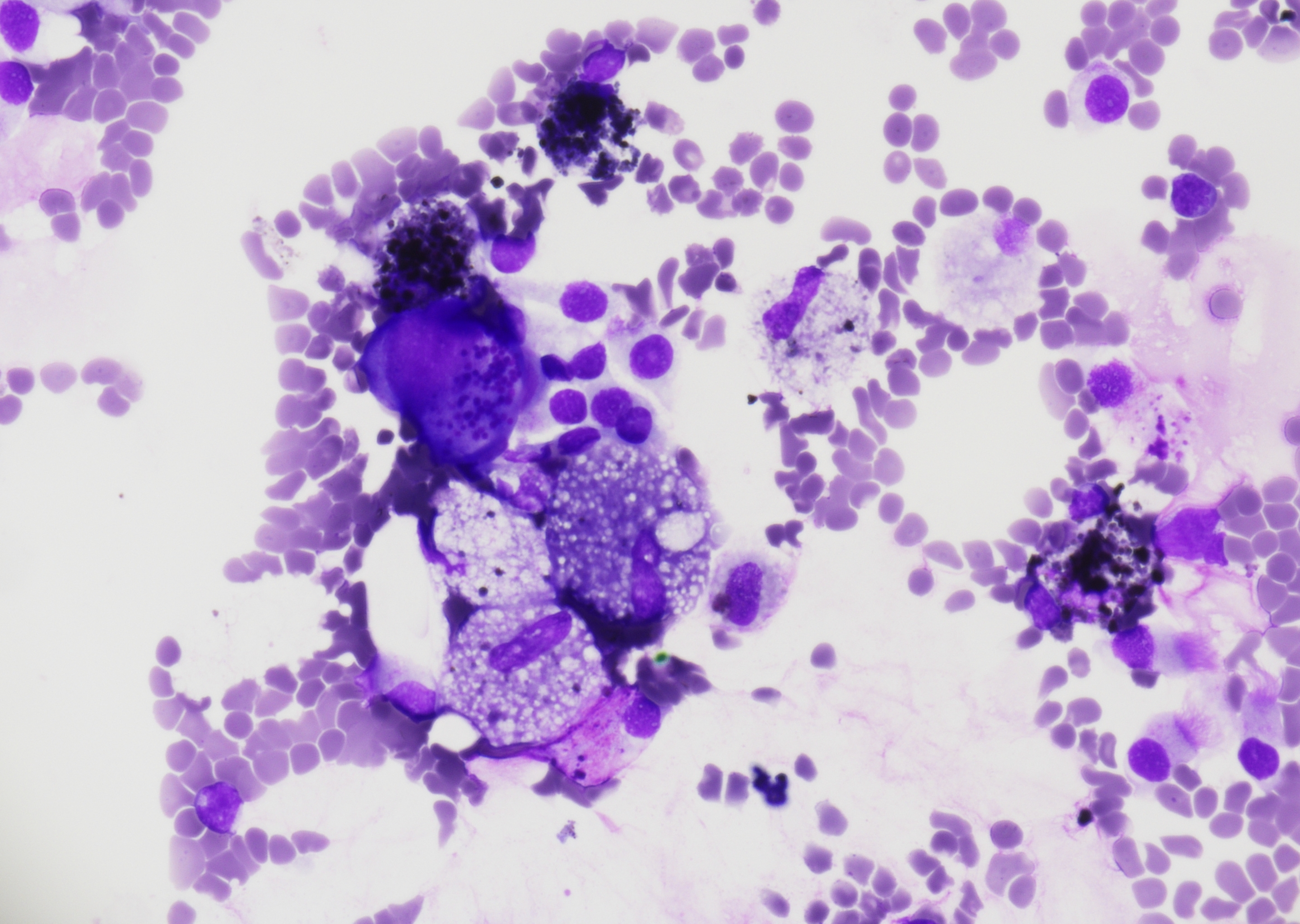
CMV-induced changes in the lung are mainly seen in the BAL.
Differential Diagnosis
References
- Soni K, Puing A. Cytomegalovirus Colitis in Adult Patients with Inflammatory Bowel Disease. Viruses. 2025 May 24;17(6):752. doi: 10.3390/v17060752. PMID: 40573343; PMCID: PMC12197619.
- Rubin, David T. MD, FACG1; Ananthakrishnan, Ashwin N. MBBS, MPH, FACG2; Siegel, Corey A. MD, MS3; Barnes, Edward L. MD, MPH, FACG4; Long, Millie D. MD, MPH, FACG4. ACG Clinical Guideline Update: Ulcerative Colitis in Adults. The American Journal of Gastroenterology 120(6):p 1187-1224, June 2025. | DOI: 10.14309/ajg.0000000000003463