
- EN
- DE

Bacteria
Differential Diagnosis of Helicobacter heilmannii sensu lato:
Helicobacter pylori

- Organ System
- Digestive Tract
- Diagnostic Method
- Cytology, Histology

- Geographical Region
- Dry Areas,
- Islands,
- Tropical and Subtropical Region,
- Polar and Subpolar Zone,
- Temperate Zone
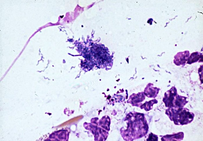
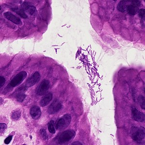
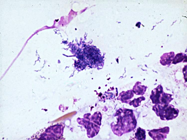 Stomach, brush smear, curved, S-shaped or…
 Elongated, slightly S-shaped, curved rod…
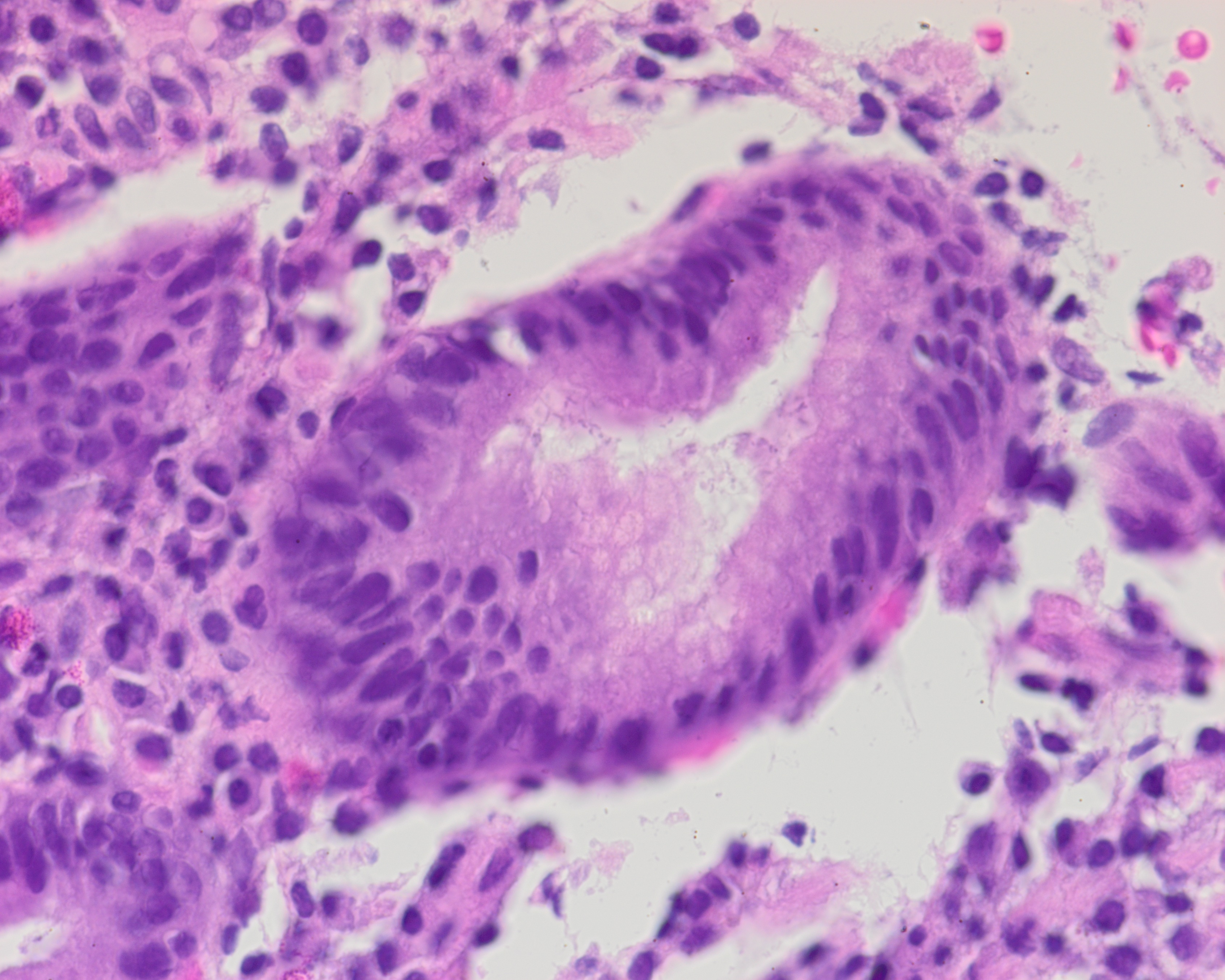 Gastric mucosa with H. pylori infection…
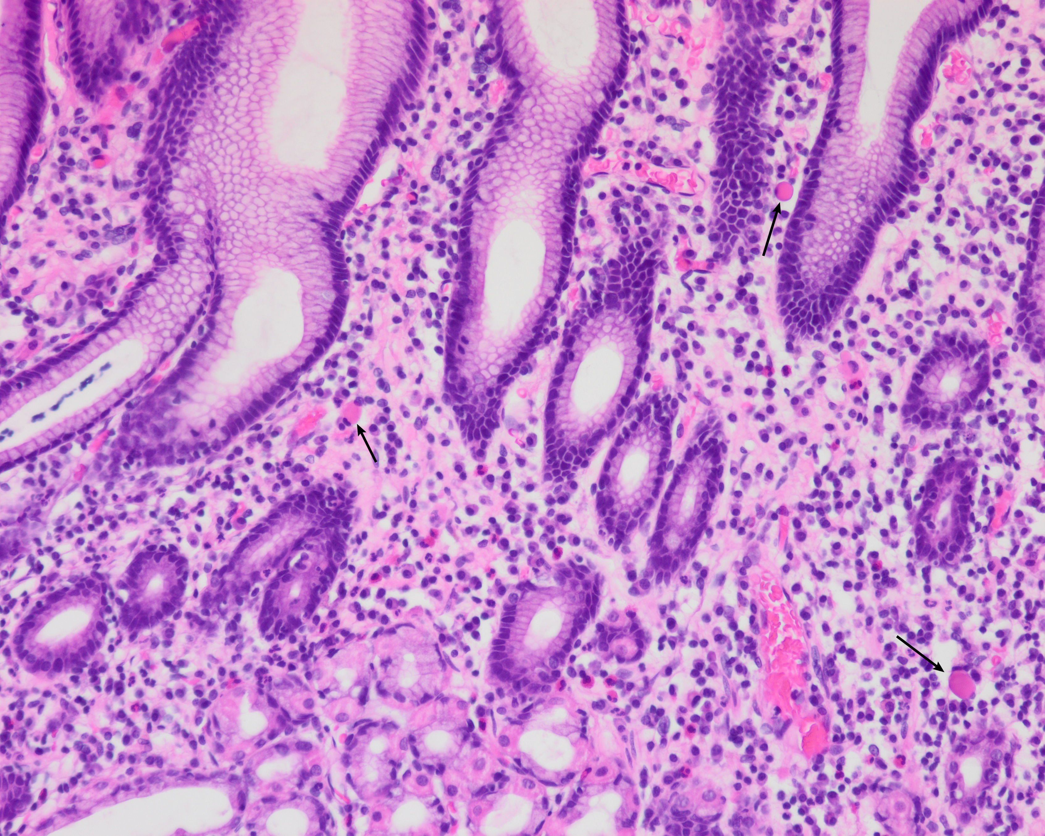 Gastric corpus mucosa with superficial…
 Gastric antrum mucosa with an acute…
 Gastric antrum mucosa. The typical…
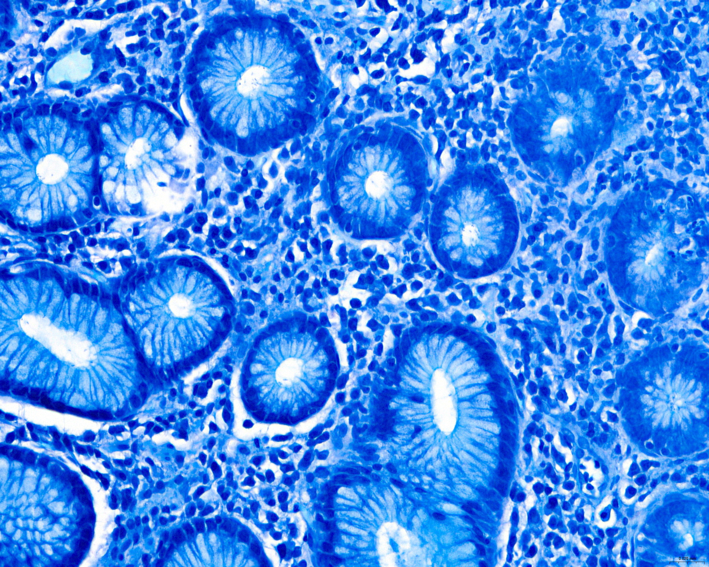 Modified Giemsa highlights Helicobacter-like…
Morphology
Cytology:
- Curved, S-or comma-shaped rods about 2-5 µm
- Lining the mucus layer and foveolar surface, clustering in pits
- Flagella not seen on routine stains
- Mucin and oft neutrophil-rich inflammation in background
Histology:
- Organisms adherent to foveolar epithelial cells, in the surface mucin and within pits
- Distribution often focal and patchy
- Chronic active gastritis with usually antral predominance
- Surface epithelial damage
- Lymphoplasmacytic cellular infiltrate with superficial ("top-heavy") distribution, pit abscesses and lymphoid aggregates
- Intestinal metaplasia and Russell bodies may be present
- Density reduced in atrophic mucosa and after acid suppression
- Rarely seen in areas of intestinal metaplasia
- Use of IHC or special stains (Giemsa, Diff-Quick, toluidine blue, Alcian yellow, Romanowsky) increases sensitivity
Differential Diagnosis:
- Helicobacter heilmannii - larger, thicker, and more tightly helical ("corkscrew") rods with lower bacterial density, and associated with milder inflammation. Co-infection with H. pylori is possible.
- Campylobacter-like curved bacteria and oral flora in contaminated samples.
Clinical notes
H. pylori is a Gram-negative, microaerophilic, flagellate, spiral bacterium with catalase, oxidase, and urease activity. Urease converts urea into ammonia, which neutralizes gastric acid and promotes bacterial protein synthesis, aiding bacterial survival in the stomach.
Most patients are asymptomatic, but may present with dyspepsia, epigastric pain, nausea, vomiting, and gastrointestinal bleeding. Disease associations include chronic gastritis, peptic ulcer disease, and atrophic gastritis, as well as a risk factor for intestinal-type gastric adenocarcinoma and mucosa-associated lymphoid tissue (MALT)-type gastric lymphomas.
H. pylori selectively colonizes the gastric mucosa and can colonize ectopic gastric mucosa at almost any site.
Transmission via fecal-oral or oral-oral routes and a worldwide distribution.
If eradication therapy is planned, molecular resistance testing by PCR/sequencing may be considered, particularly for clarithromycin resistance and, if applicable, fluoroquinolone resistance. (1)
Ancillary Testing
Urea breath test and stool antigen test for noninvasive detection and post-treatment testing, rapid urease test (RUT) from gastric biopsies as an invasive point-of-care test during endoscopy, PCR from biopsy or stool for detection and macrolide resistance mutations, serology (detetcts exposure, not presence; not recommended for screening or post-treatment testing)Additional Information
Widespread use of PPIs, antibiotics, and bismuth compounds may decrease bacterial load, alter the intragastric distribution of H. pylori, and affect microscopic appearance, including coccoid forms. (2, 3)
Differential Diagnosis
References
- Chey, William D. MD, FACG1; Howden, Colin W. MD, FACG2; Moss, Steven F. MD, FACG3; Morgan, Douglas R. MD, MPH, FACG4; Greer, Katarina B. MD, MSEpi5; Grover, Shilpa MD, MPH6; Shah, Shailja C. MD, MPH7. ACG Clinical Guideline: Treatment of Helicobacter pylori Infection. The American Journal of Gastroenterology 119(9):p 1730-1753, September 2024. | DOI: 10.14309/ajg.0000000000002968
- Garcés-Duran, R., et al. (2023). Belgian consensus for Helicobacter pylori management 2023. Acta Gastro-Enterologica Belgica, 86(1), 74–91. https://doi.org/10.51821/86.1.11327
- Ierardi, E., Losurdo, G., Mileti, A., Paolillo, R., Giorgio, F., Principi, M., & Di Leo, A. (2020). The Puzzle of Coccoid Forms of Helicobacter pylori: Beyond Basic Science. Antibiotics, 9(6), 293. https://doi.org/10.3390/antibiotics9060293