- EN
- DE

Bacteria
Differential Diagnosis of Helicobacter pylori:
Helicobacter heilmannii sensu lato

- Organ System
- Digestive Tract
- Diagnostic Method
- Histology

- Geographical Region
- Dry Areas,
- Islands,
- Tropical and Subtropical Region,
- Polar and Subpolar Zone,
- Temperate Zone
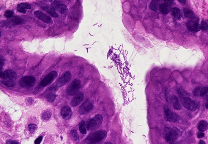
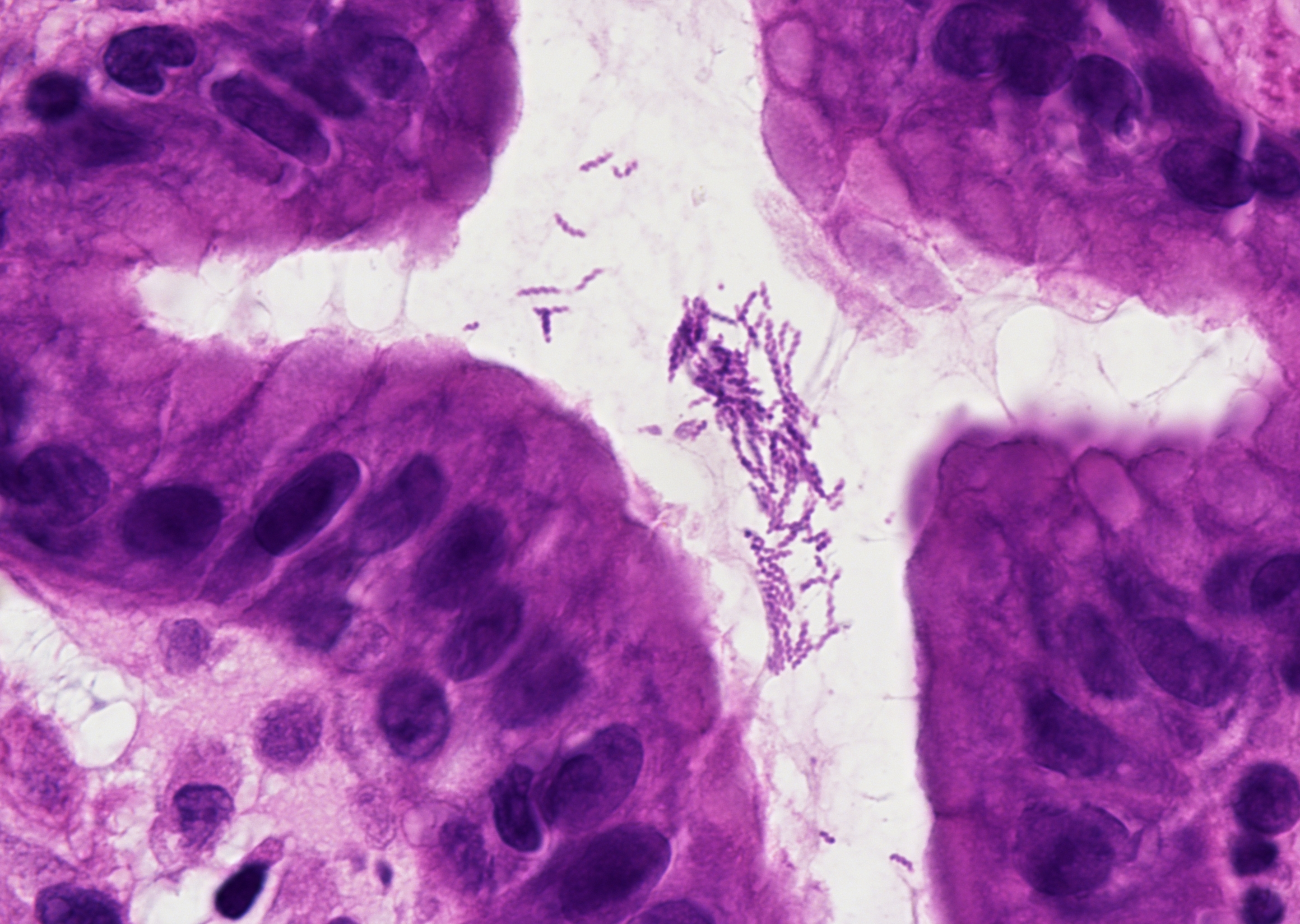 Gastric mucosa with spiral-shaped H.
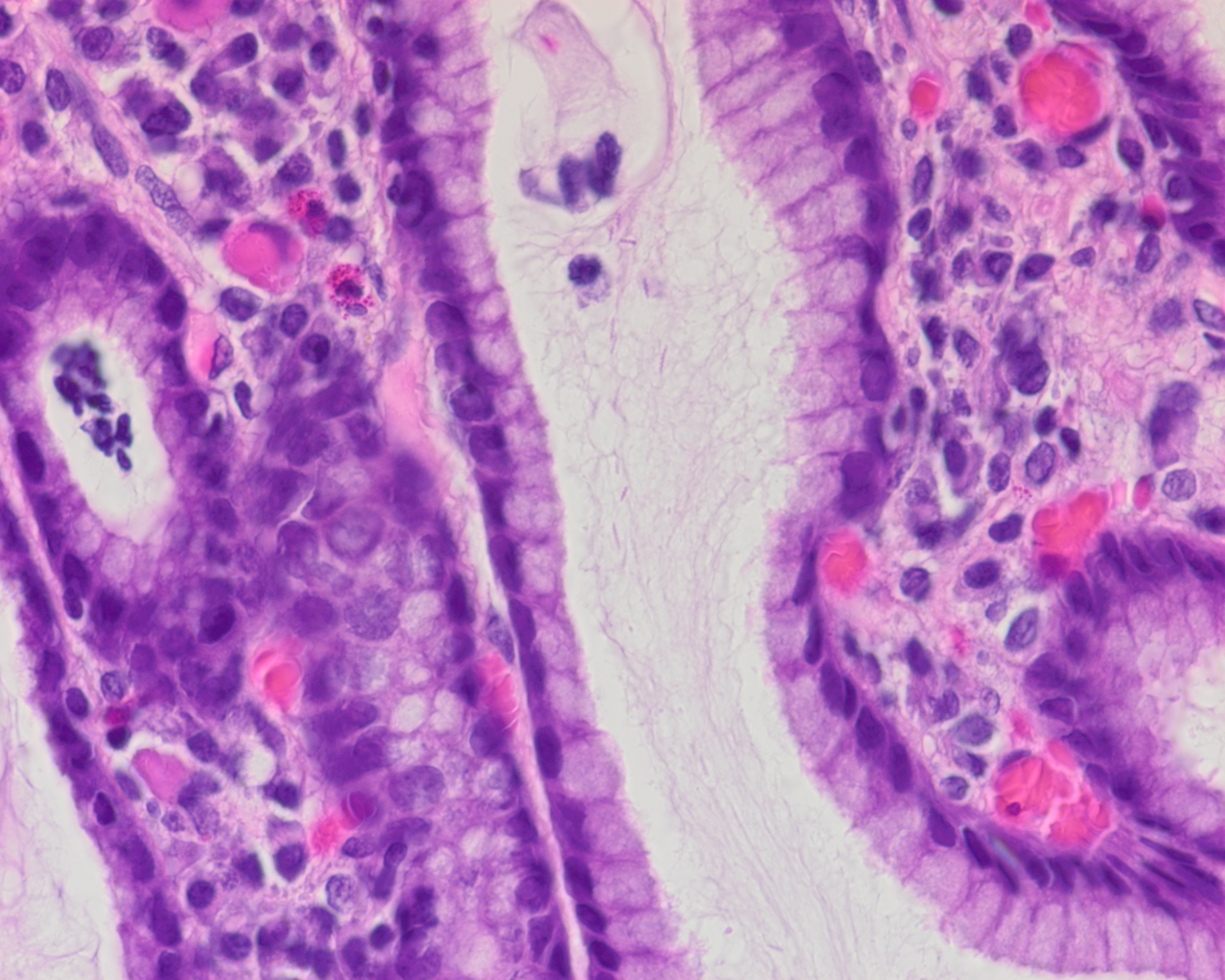 Gastric corpus mucosa with H. heilmannii…
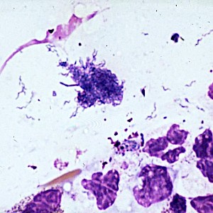
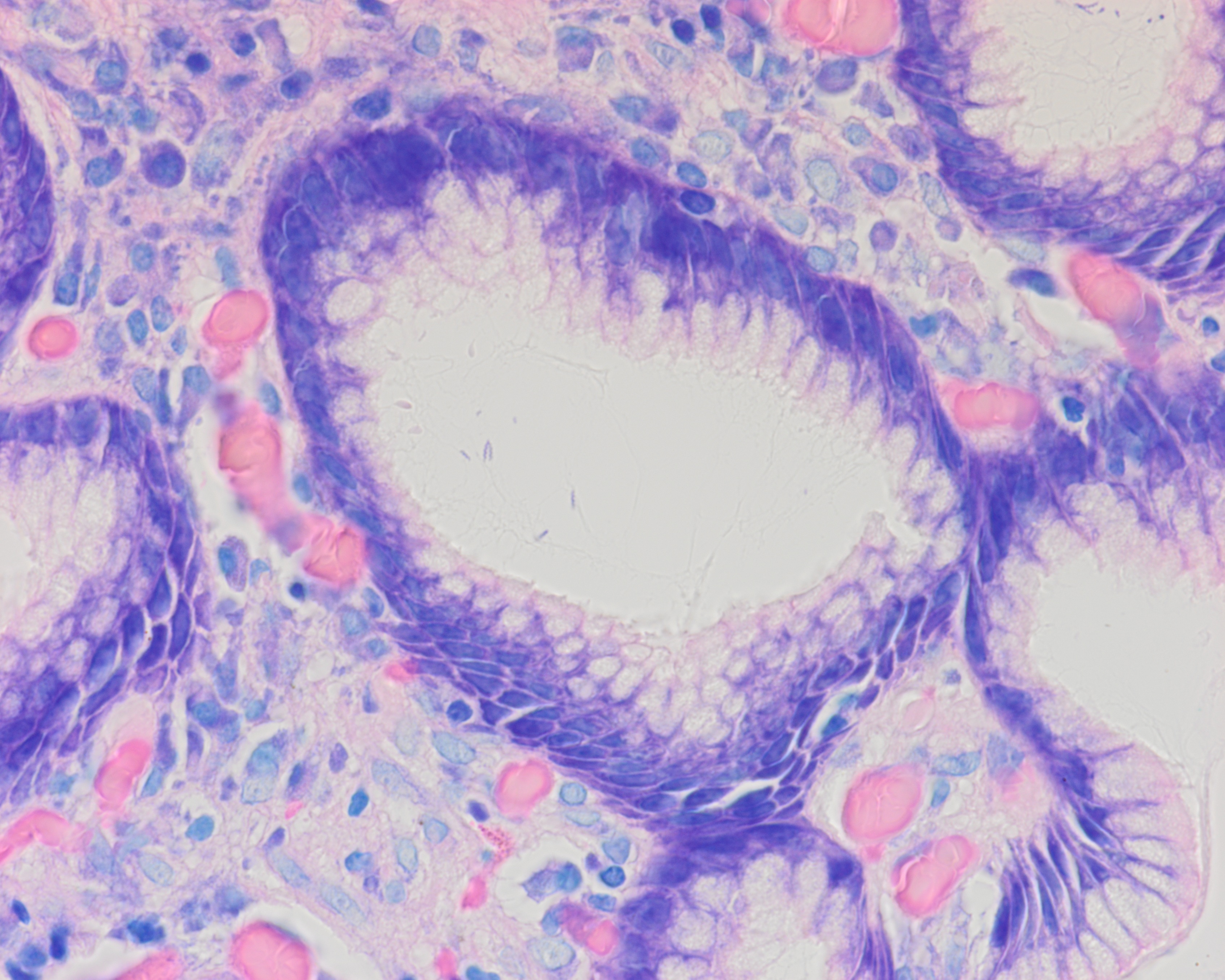 H. heilmannii highlighted by Giemsa stain…
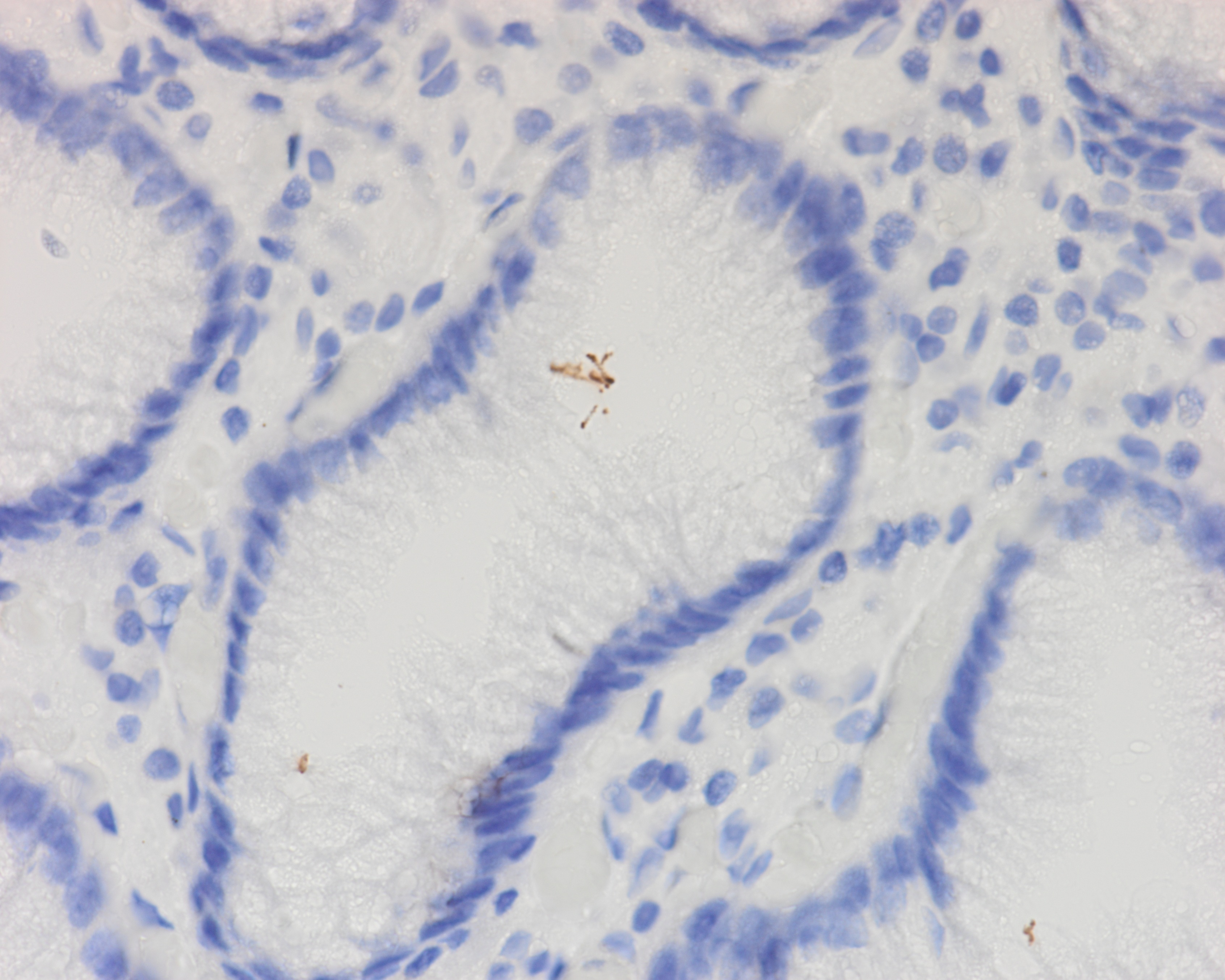 IHC-positive staining of H. heilmannii…
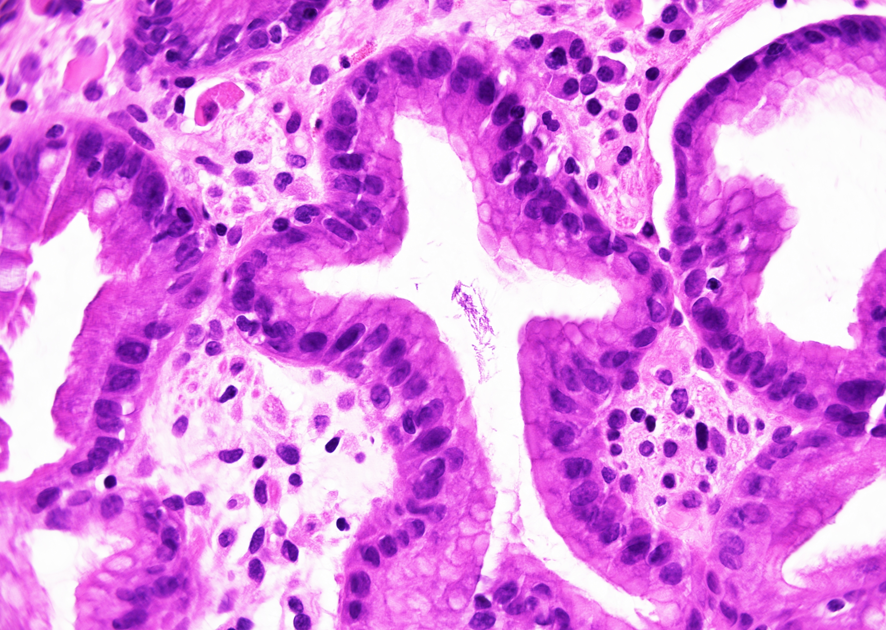 Gastric mucosa, H. heilmannii (HE, x400)
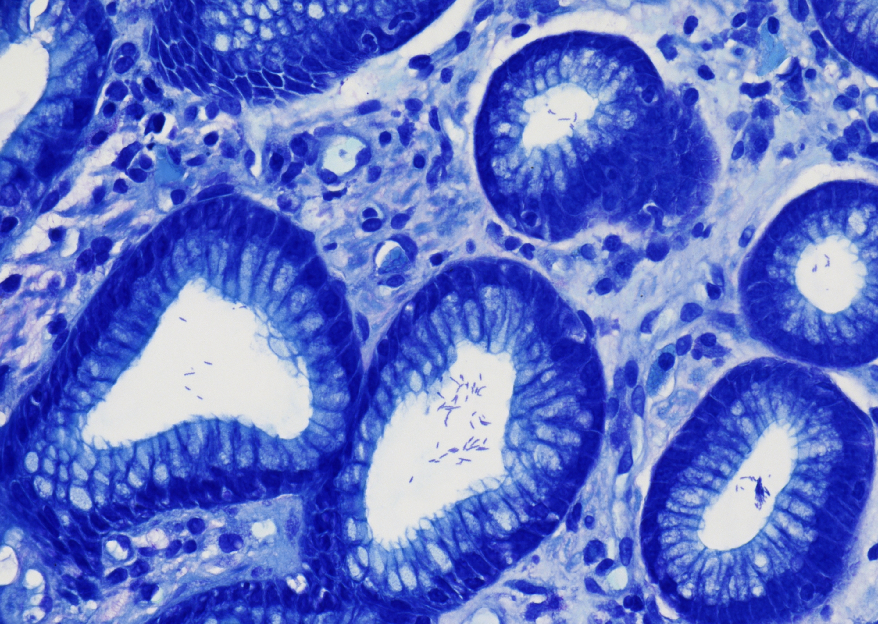 H. heilmannii in modified Giemsa stain (x400)
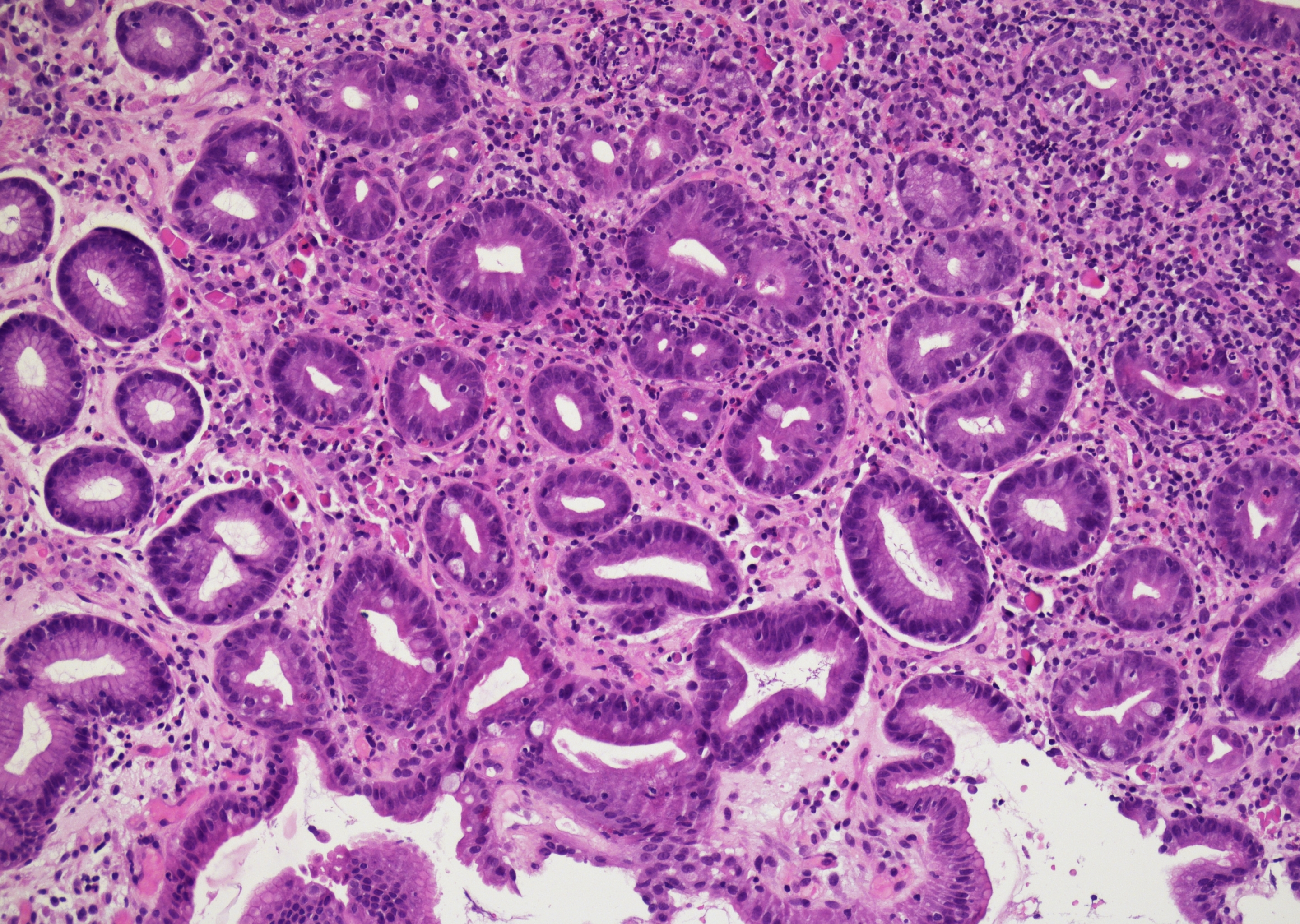 Chronically active inflamed gastric mucosa…
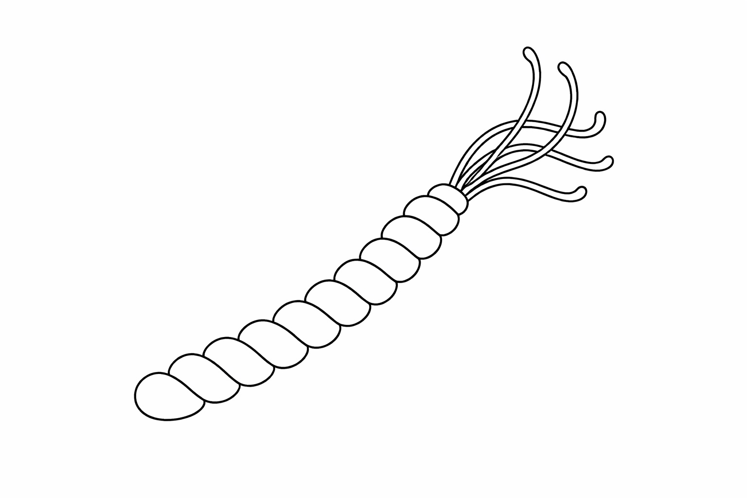 Long, tightly coiled spiral-shaped rod with…
Morphology
Cytology:
Large, tightly coiled, spiral-shaped bacteria, often with pronounced corkscrew morphology. Usually sparse, located along the mucus layer and surface epithelium. Background inflammation variable, often mild.
Histology:
Organisms located within the surface mucus layer and gastric pits, usually focal and patchy. The associated chronic gastritis is typically mild to moderate and often lacks significant activity. Neutrophils are usually sparse or absent. Lymphoid aggregates may be present, with possible association with MALT-type lesions. The bacterial density is generally lower than in Helicobacter pylori infection. Often underdiagnosed due to low organism density and patchy distribution.
Special stains (Giemsa, Warthin-Starry, Steiner) may increase detection rate, but sensitivity is limited due to low bacterial density. Immunohistochemistry for Helicobacter pylori often negative or only weakly reactive.
Differential Diagnosis:
- Helicobacter pylori - shorter, slightly coiled, S-shaped organisms, usually higher density.
- Other spiral or Campylobacter-like organisms in contaminated samples.
Clinical notes
Chronic gastritis, peptic ulcer disease. Reported association with gastric MALT lymphoma. Zoonotic origin suspected, commonly linked to contact with dogs, cats, or pigs. (1)
Ancillary Testing
Urease test, PCRAdditional Information
It is now recognized that H. heilmannii s.l. does not represent a single species, but rather a group of distinct gastric non-H. pylori Helicobacter species.