
- EN
- DE

Bacteria
Differential Diagnosis of Gram-negative rods:
Mycobacterium Species

- Organ System
- Respiratory Tract, Lymphatic System, Soft Tissue & Bone, Digestive Tract, CNS and Sensory Organs, Skin and Mucosa
- Diagnostic Method
- Cytology, Histology

- Geographical Region
- Dry Areas,
- Islands,
- Tropical and Subtropical Region,
- Polar and Subpolar Zone,
- Temperate Zone
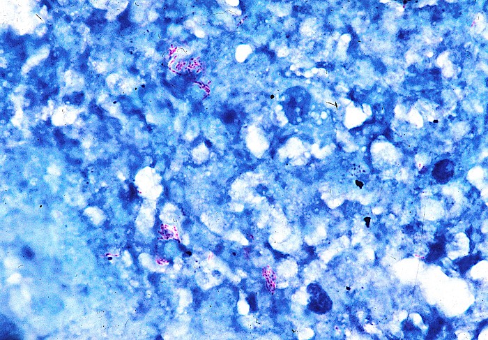
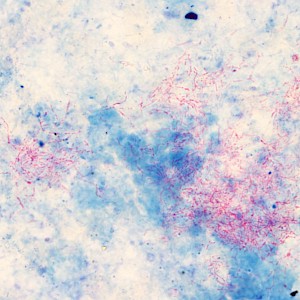
 Red-stained acid-fast rods consistent with…
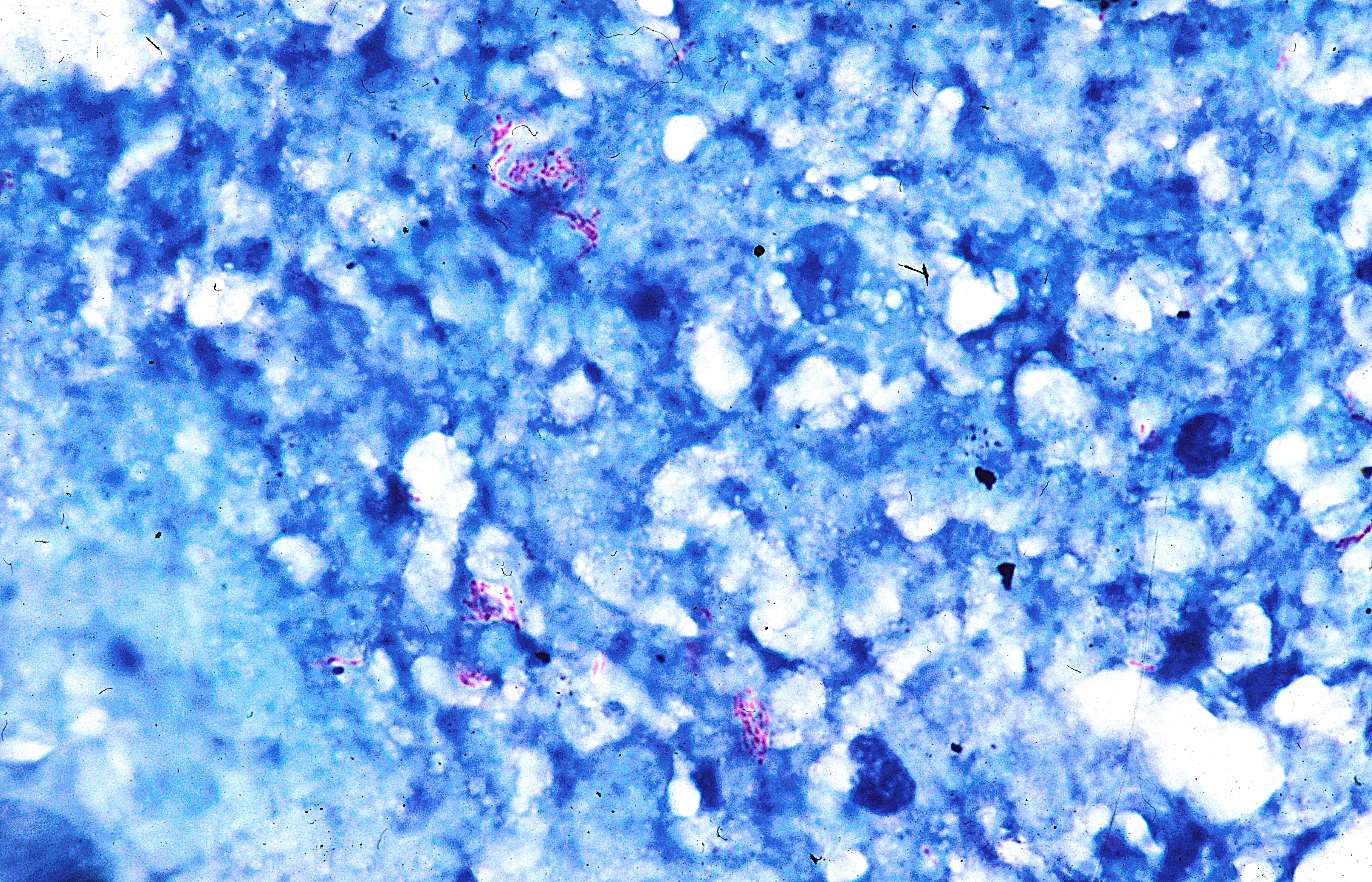
 Red-stained acid-fast rods consistent with…
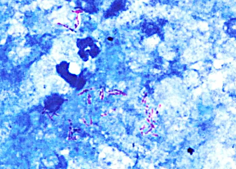
 FNA, axillary lymph node, acid-fast rods…
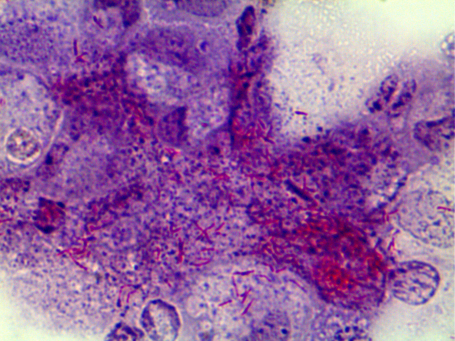 BAL, red-stained acid-fast rods (ZN)…
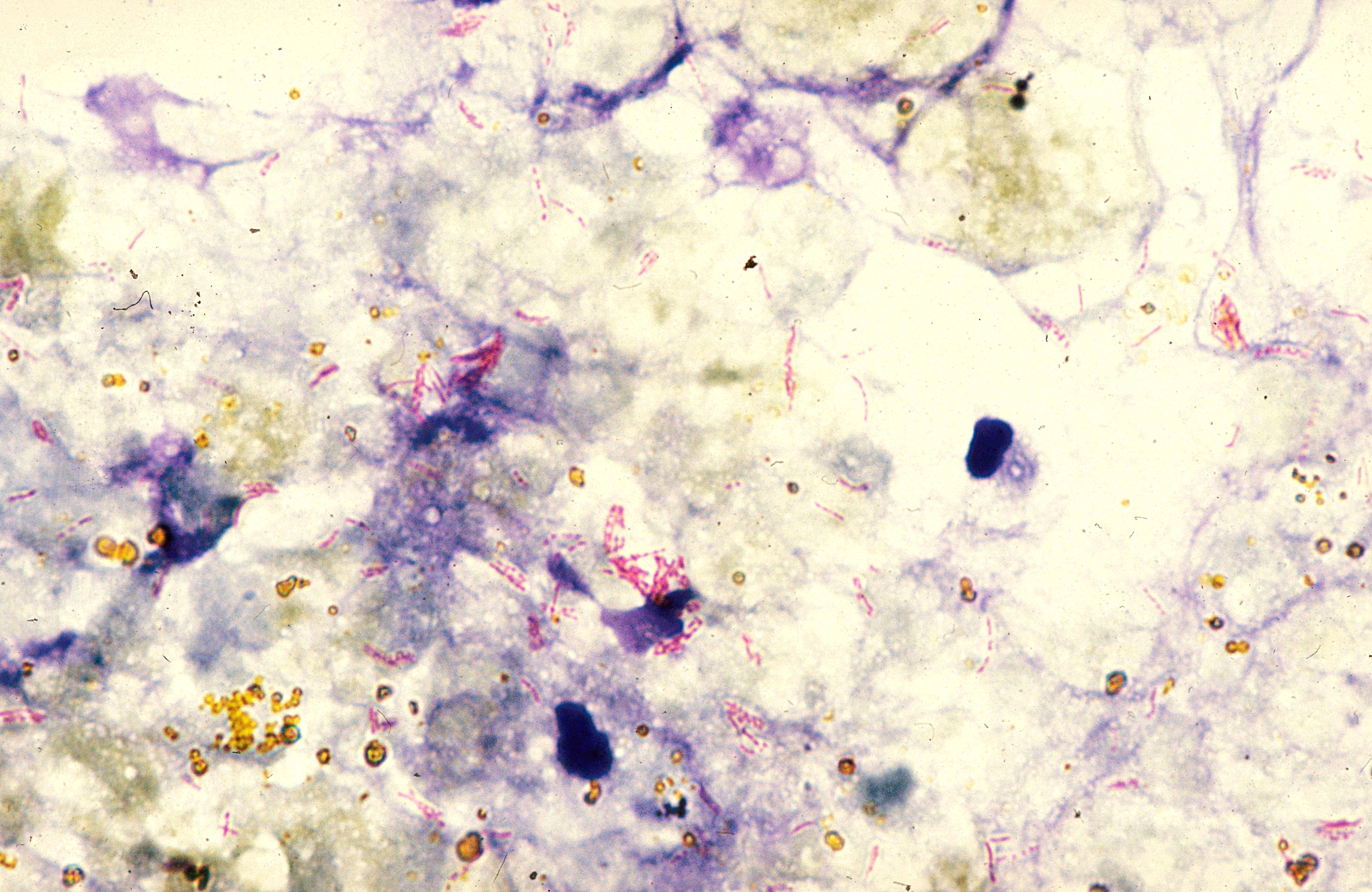 Acid-fast rods, amorphous detritus and some…
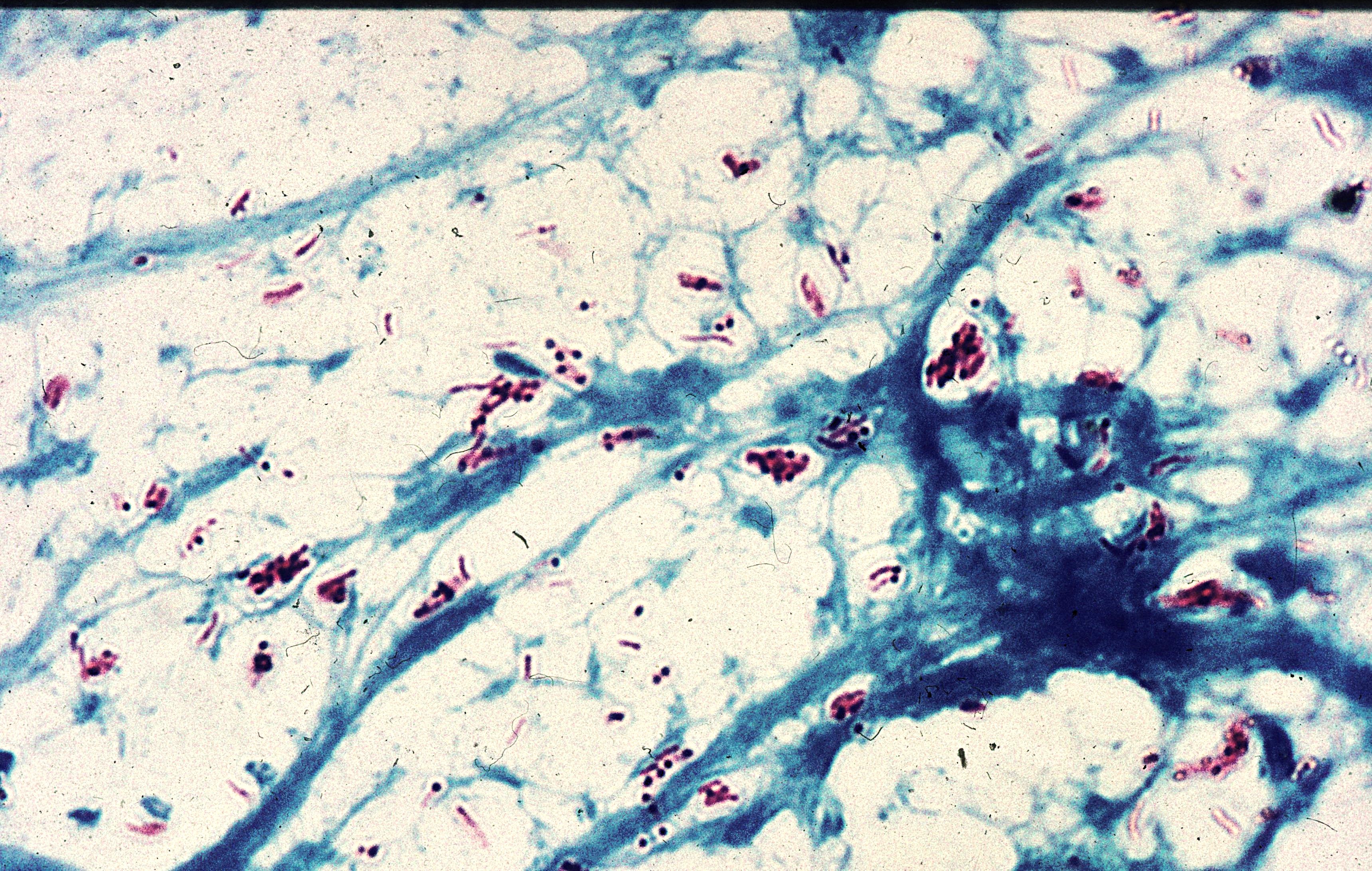 Acid-fast rods, amorphous detritus (ZN)
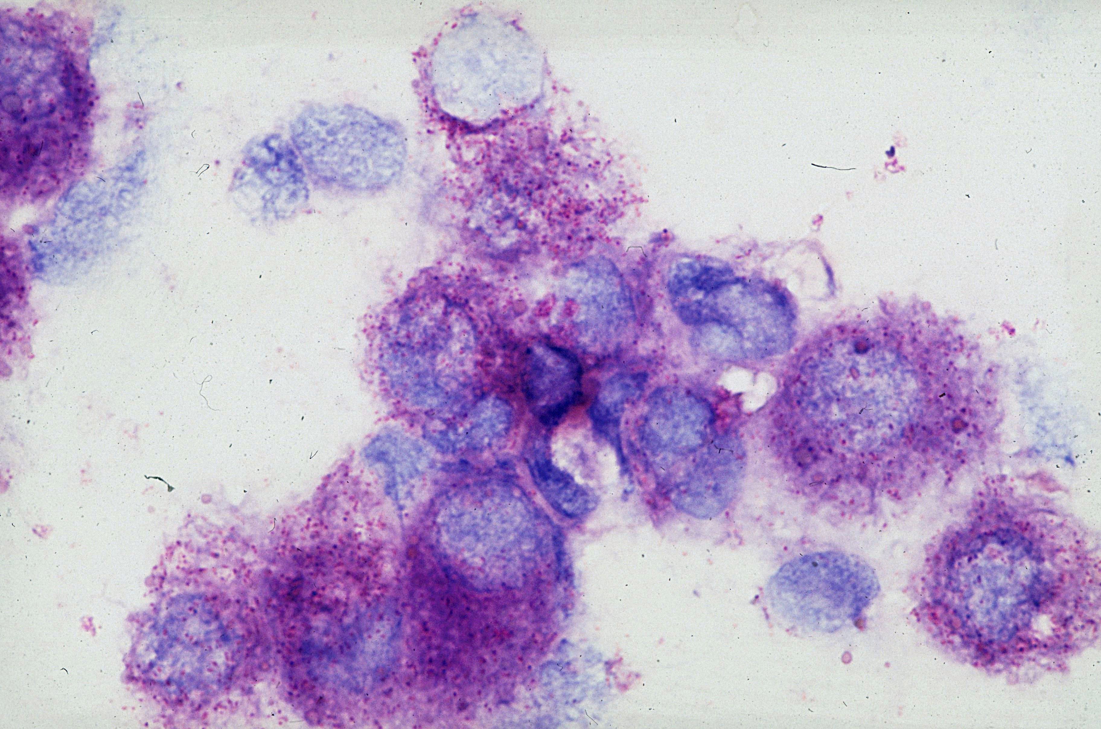 Red-stained acid-fast rods, consistent with…
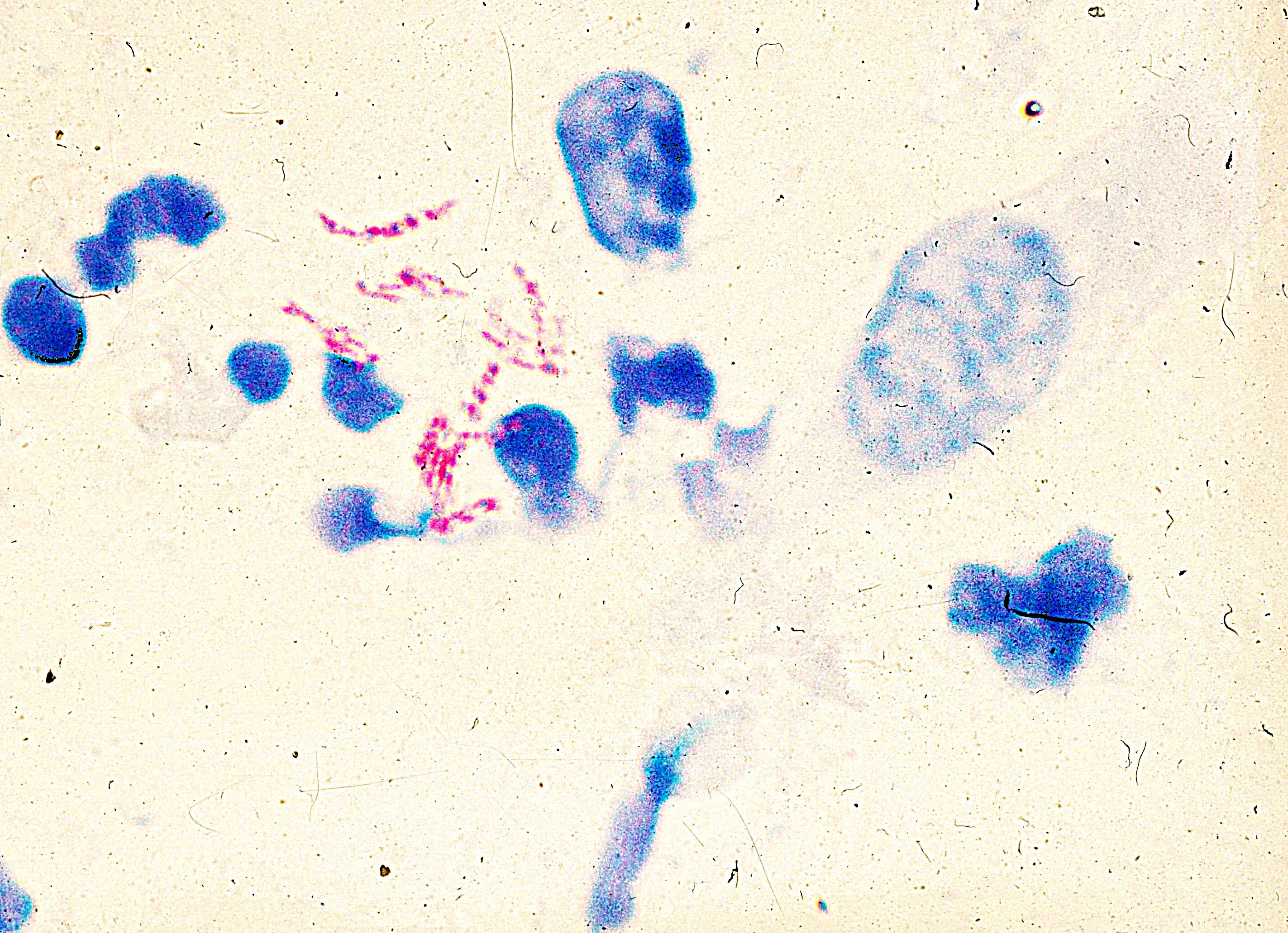 Red-stained acid-fast rods, consistent with…
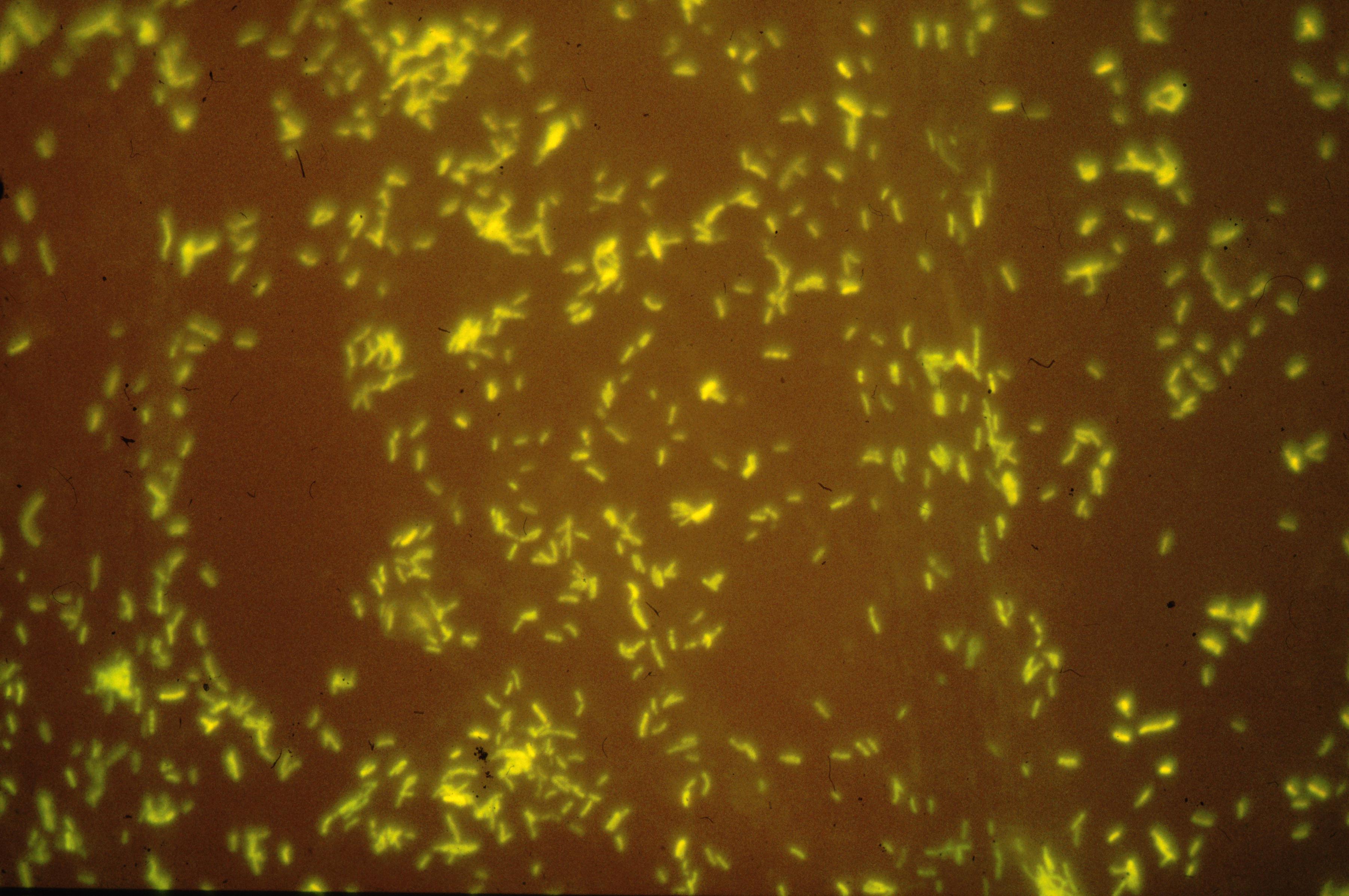 Yellow acid-fast rods, consistent with M.
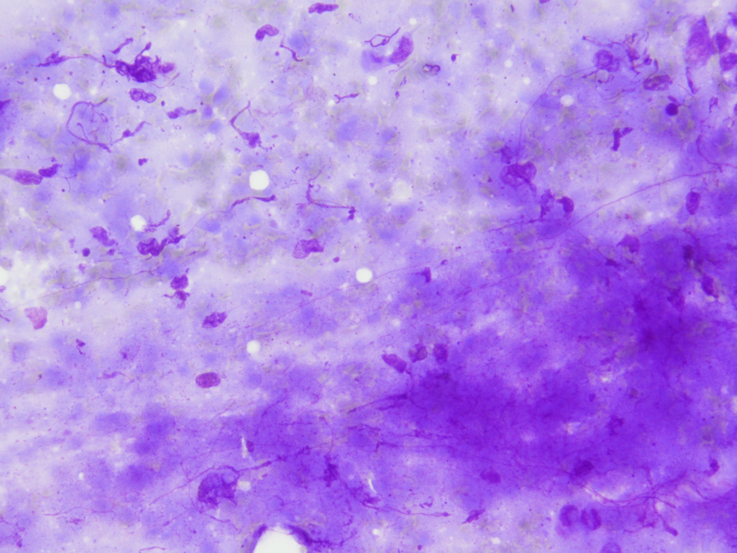 Amorphous detritus with few respiratory…
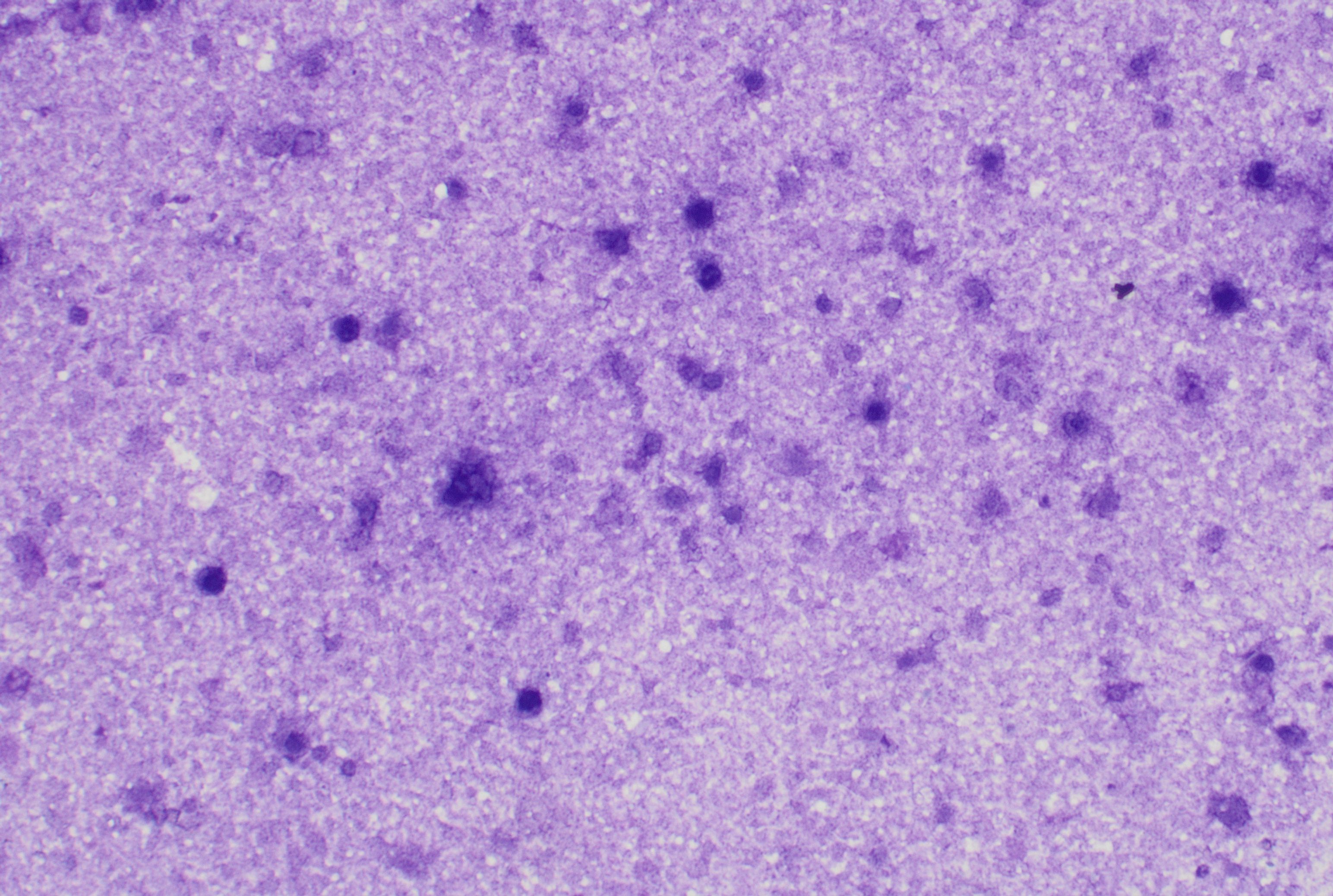 Lymph node FNA showing mainly necrosis in an…
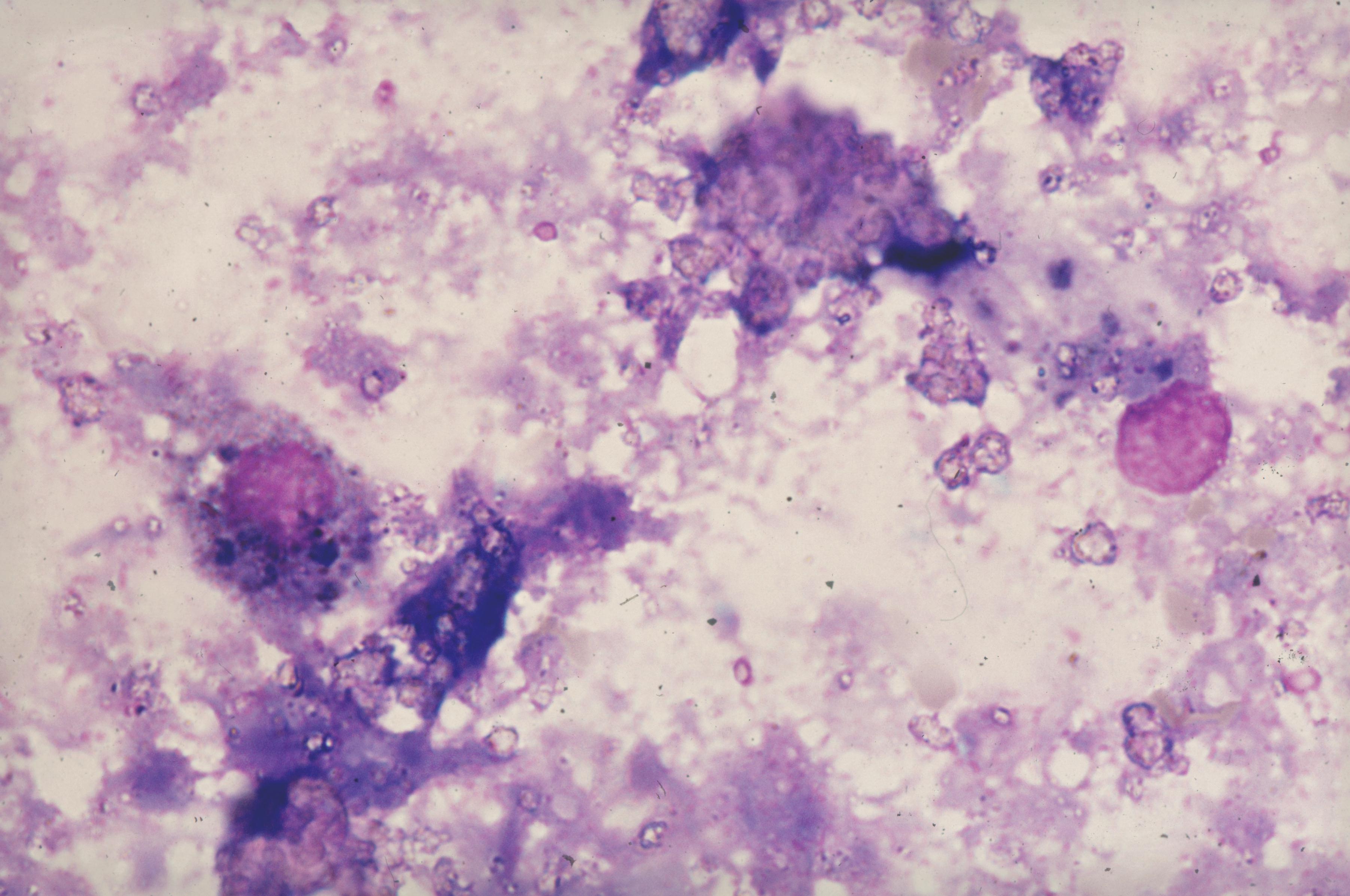 Amorphous detritus, few degeneratively…
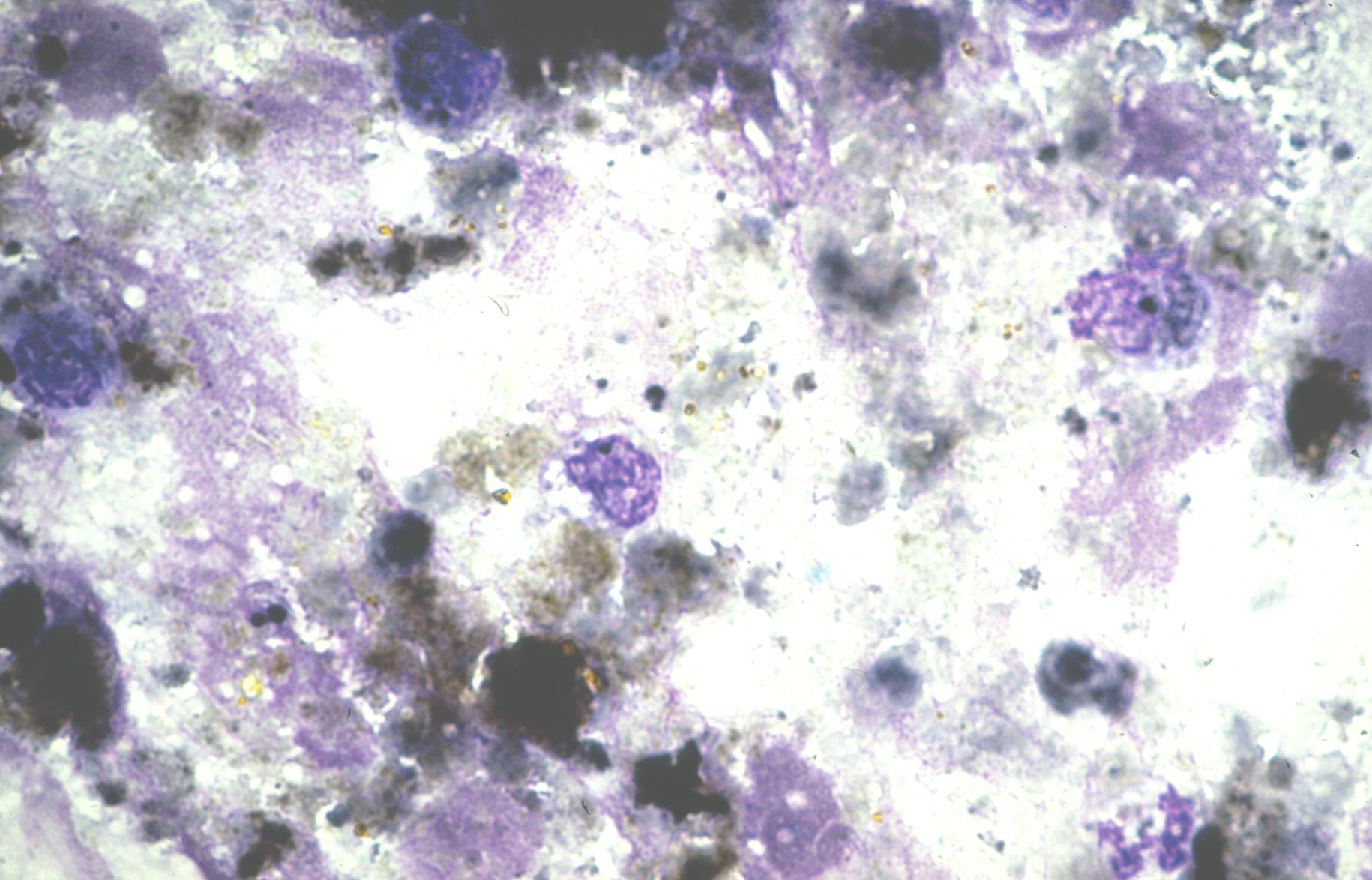 Amorphous detritus and some degeneratively…
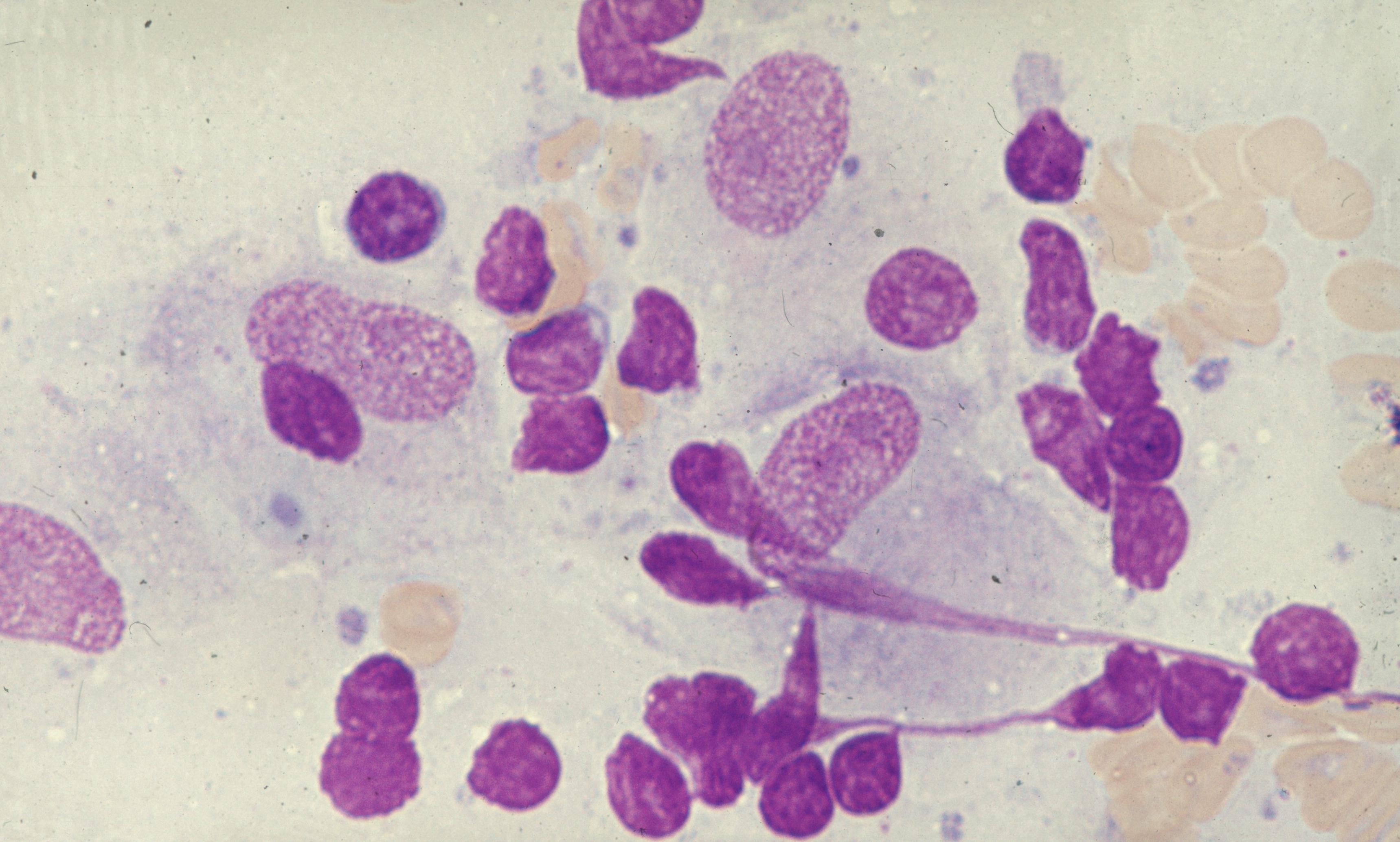
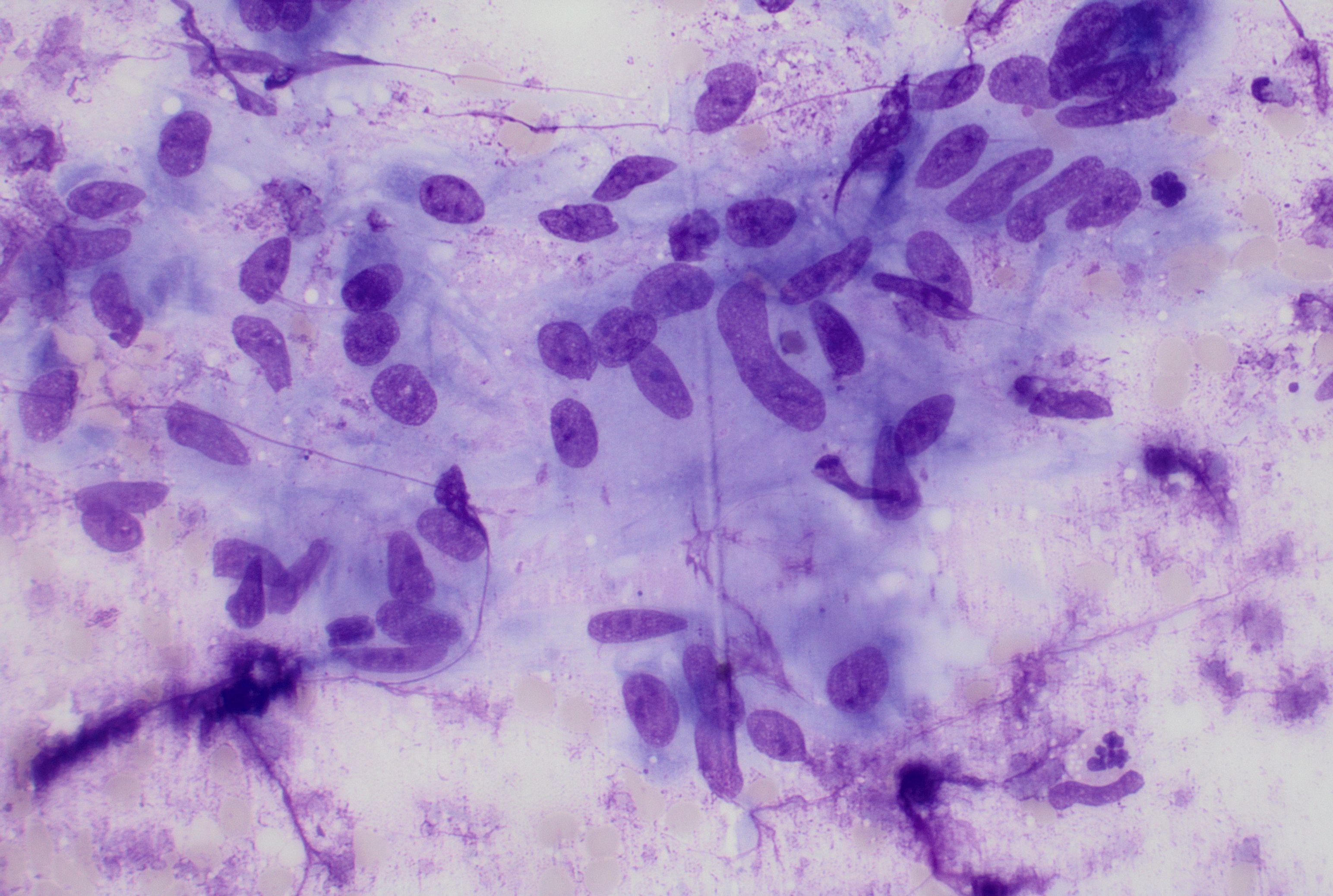 Lymph node, FNA with epitheloid cells in a…
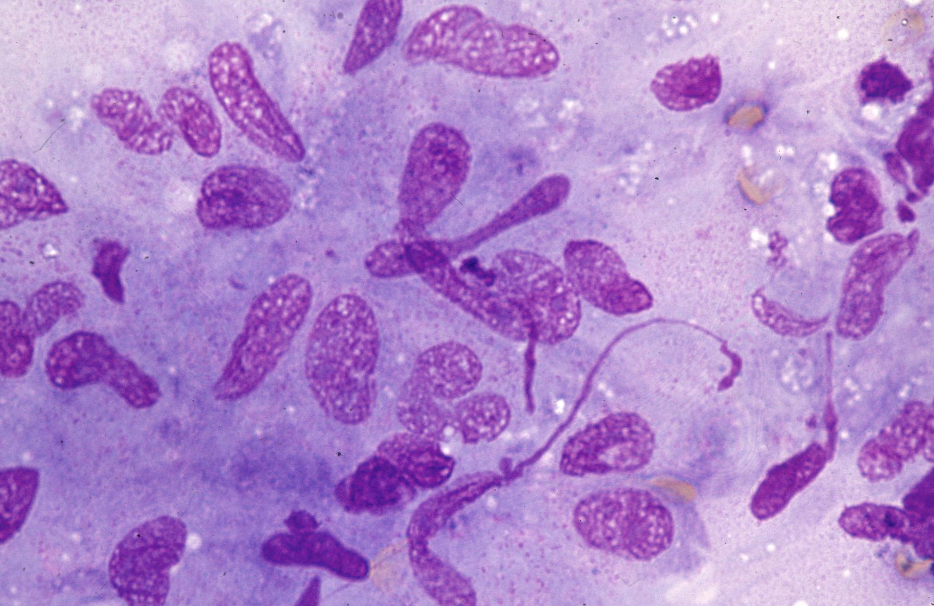 Epithelioid cells with elongated, narrow…
 Lymph node FNA with a poorly formed…
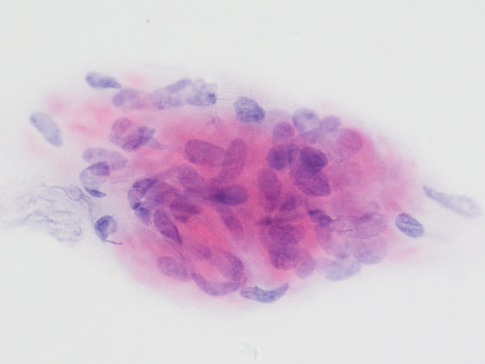 Epithelioid cells with elongated, narrow…
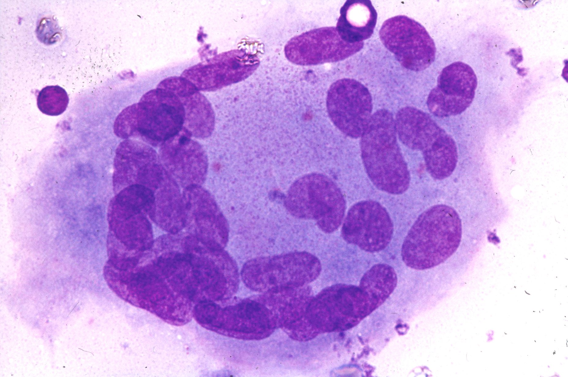 Langhans-type giant cell with peripherally…
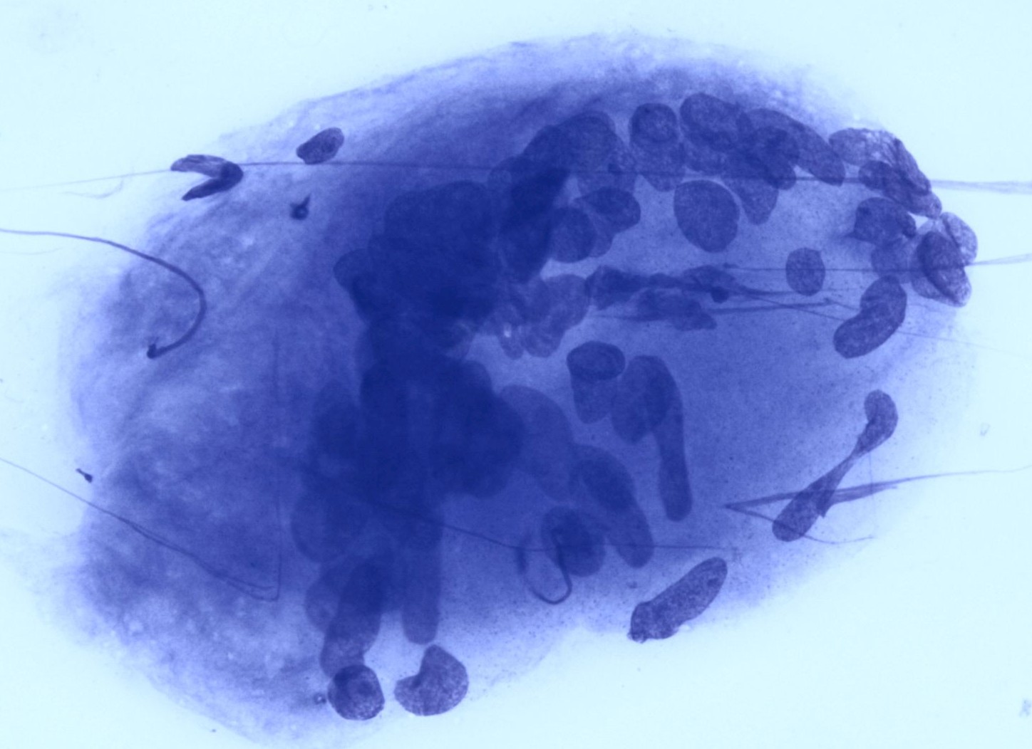 Langhans-type giant cell with peripherally…
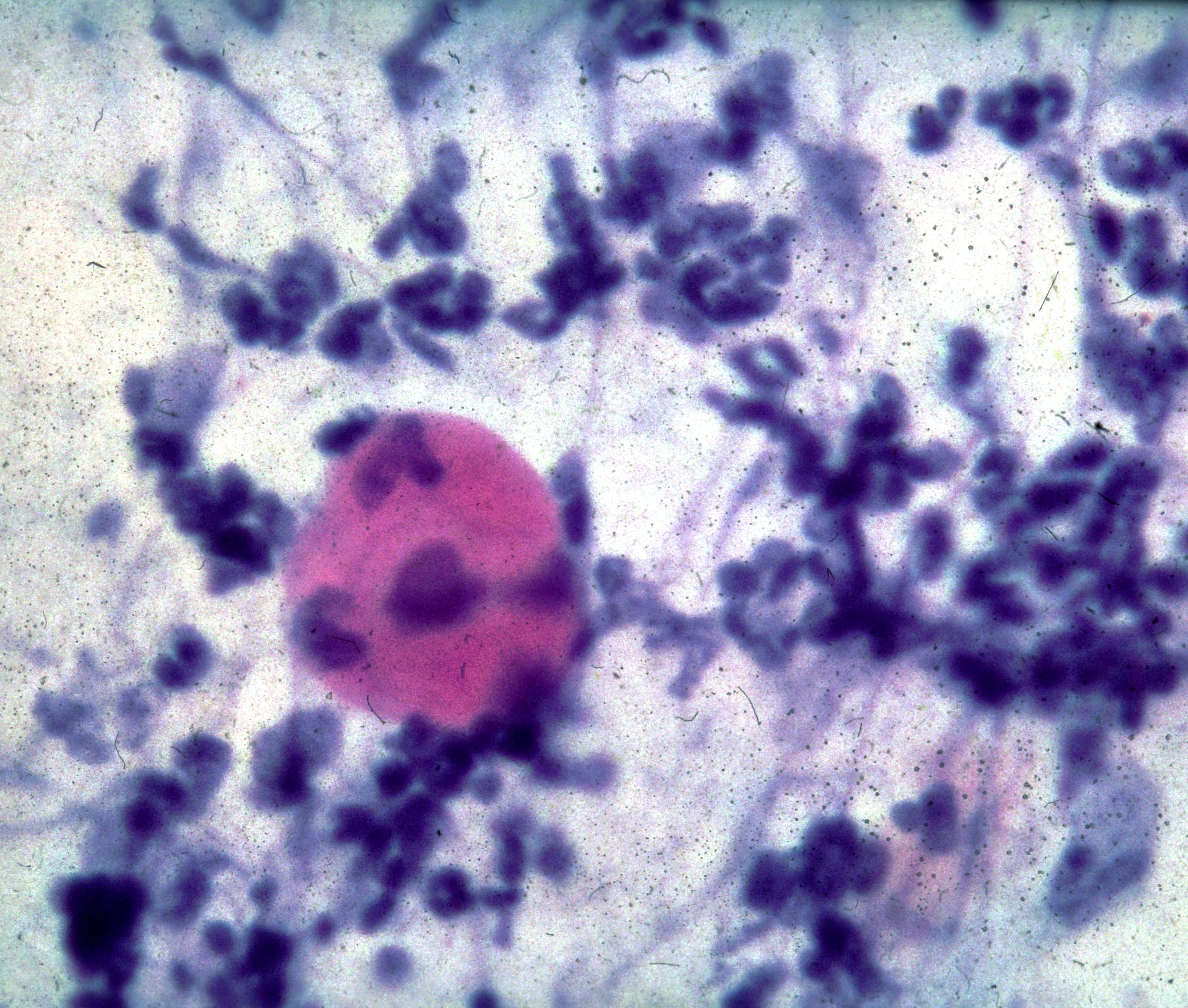 Round cell metaplasia, numerous granulocytes…
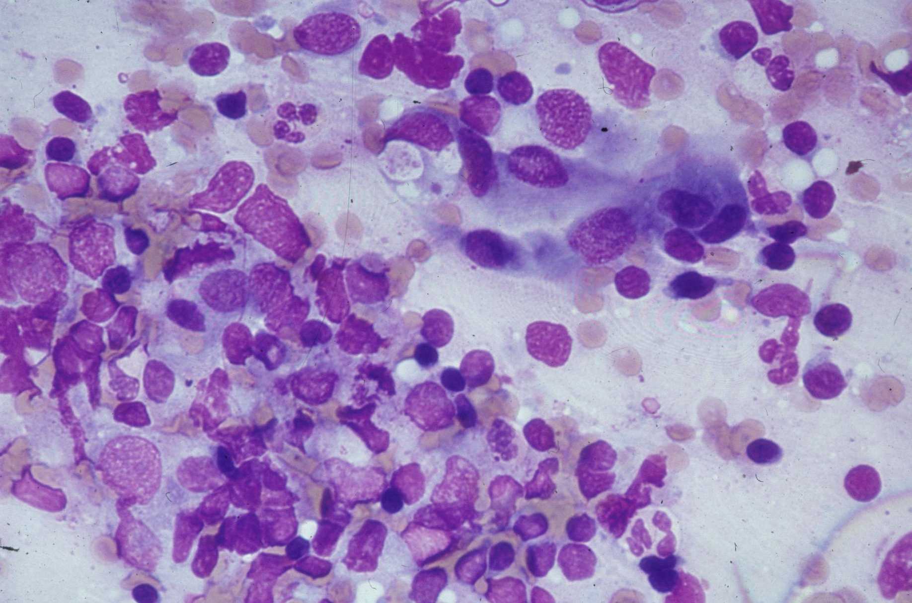 Lung, brush smear, numerous lymphocytes…
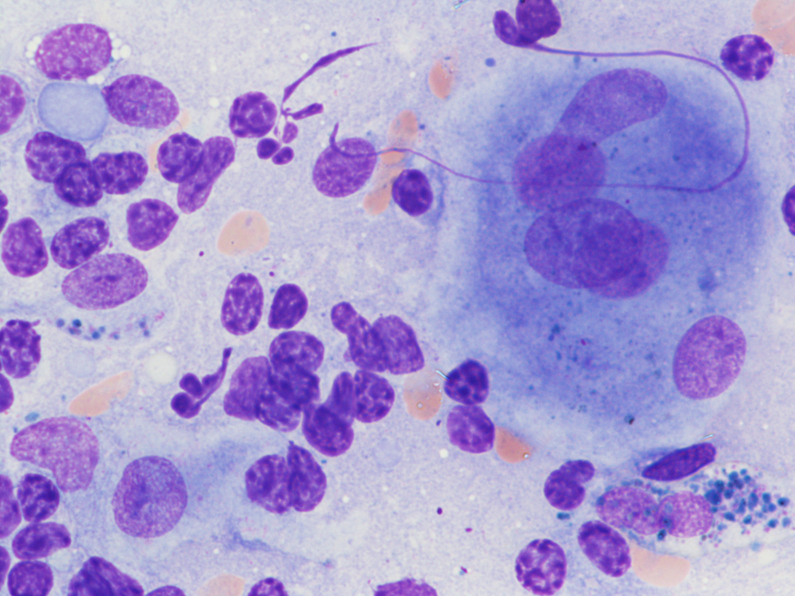 Numerous lymphocytes, a small epithelioid…
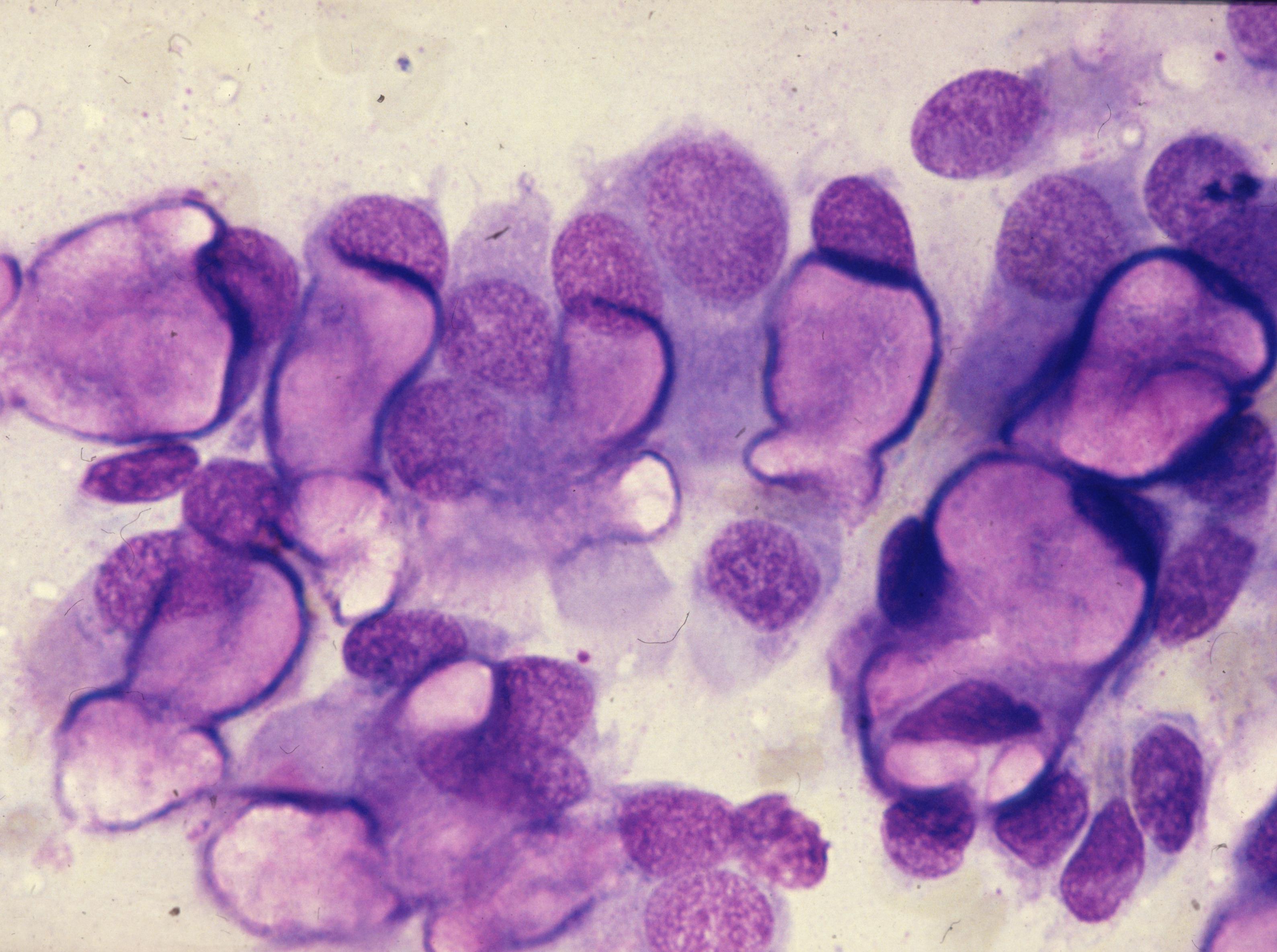 Non-specific reaction with numerous goblet…
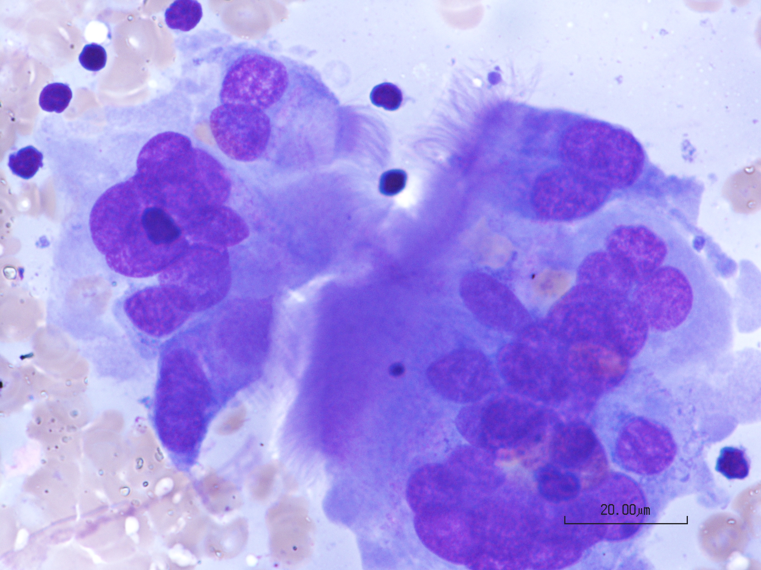 Non-specific reaction with multinucleated…
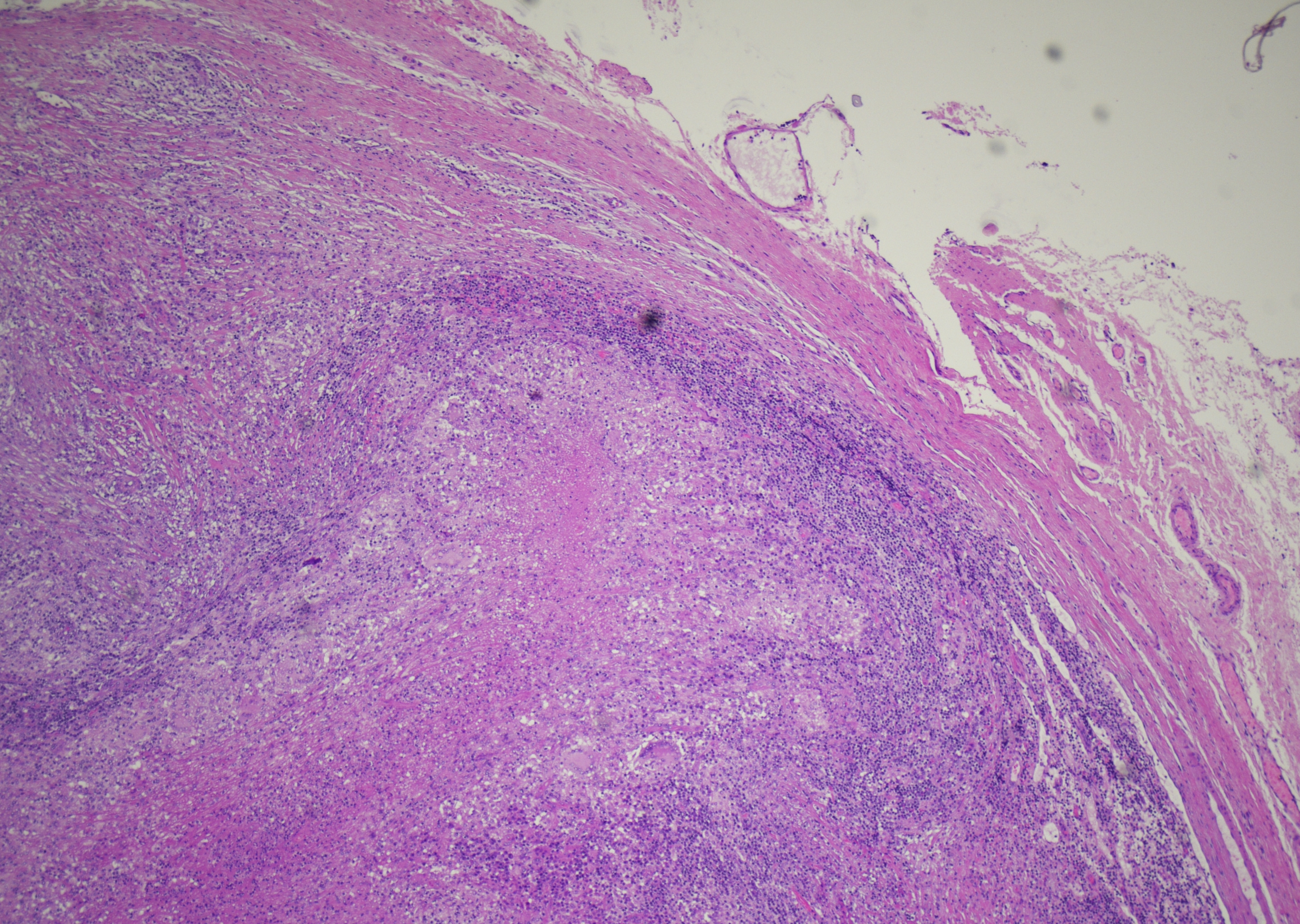 Lymph node with epitheloid granuloma and…
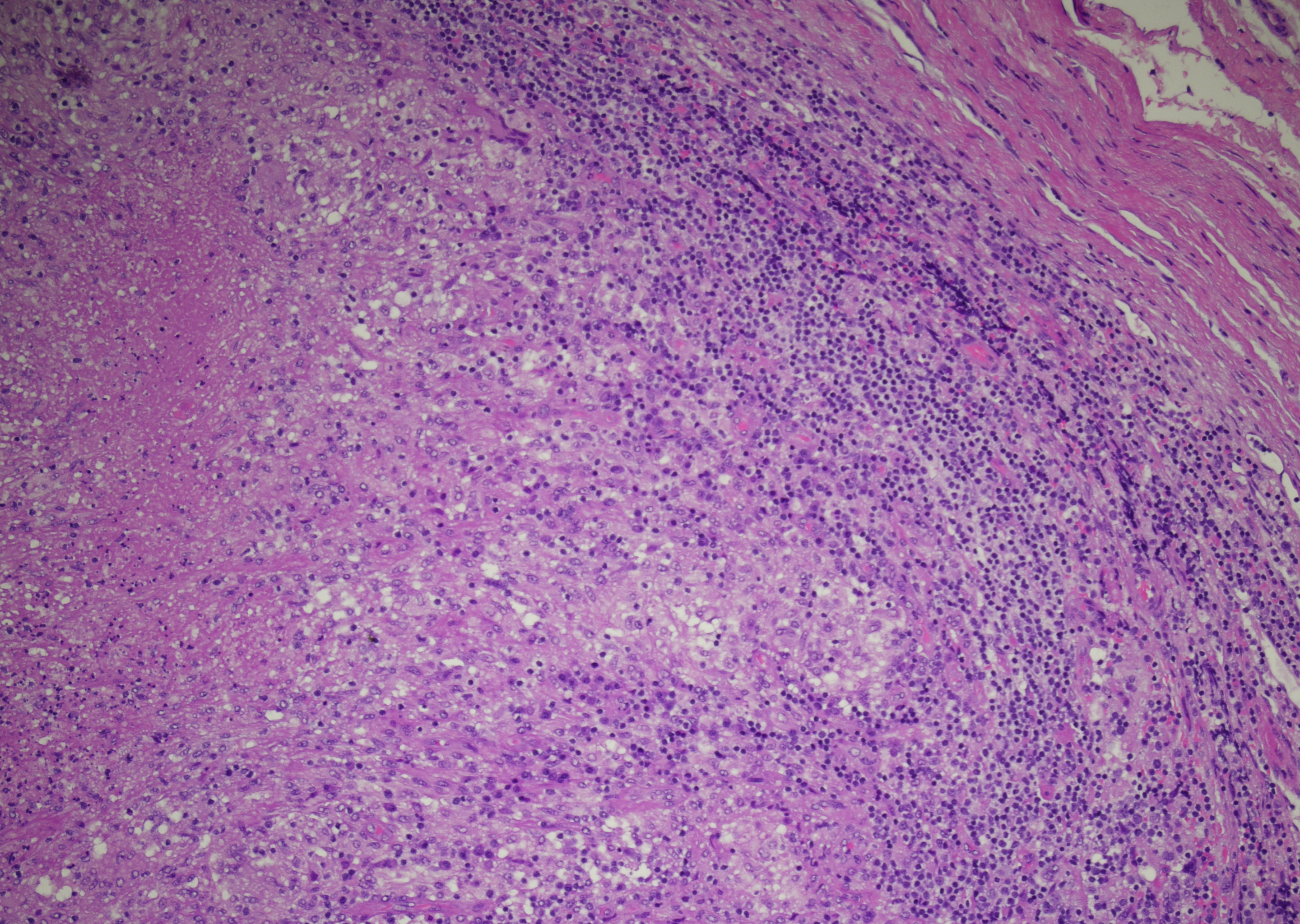 Lymph node with epitheloid granuloma and…
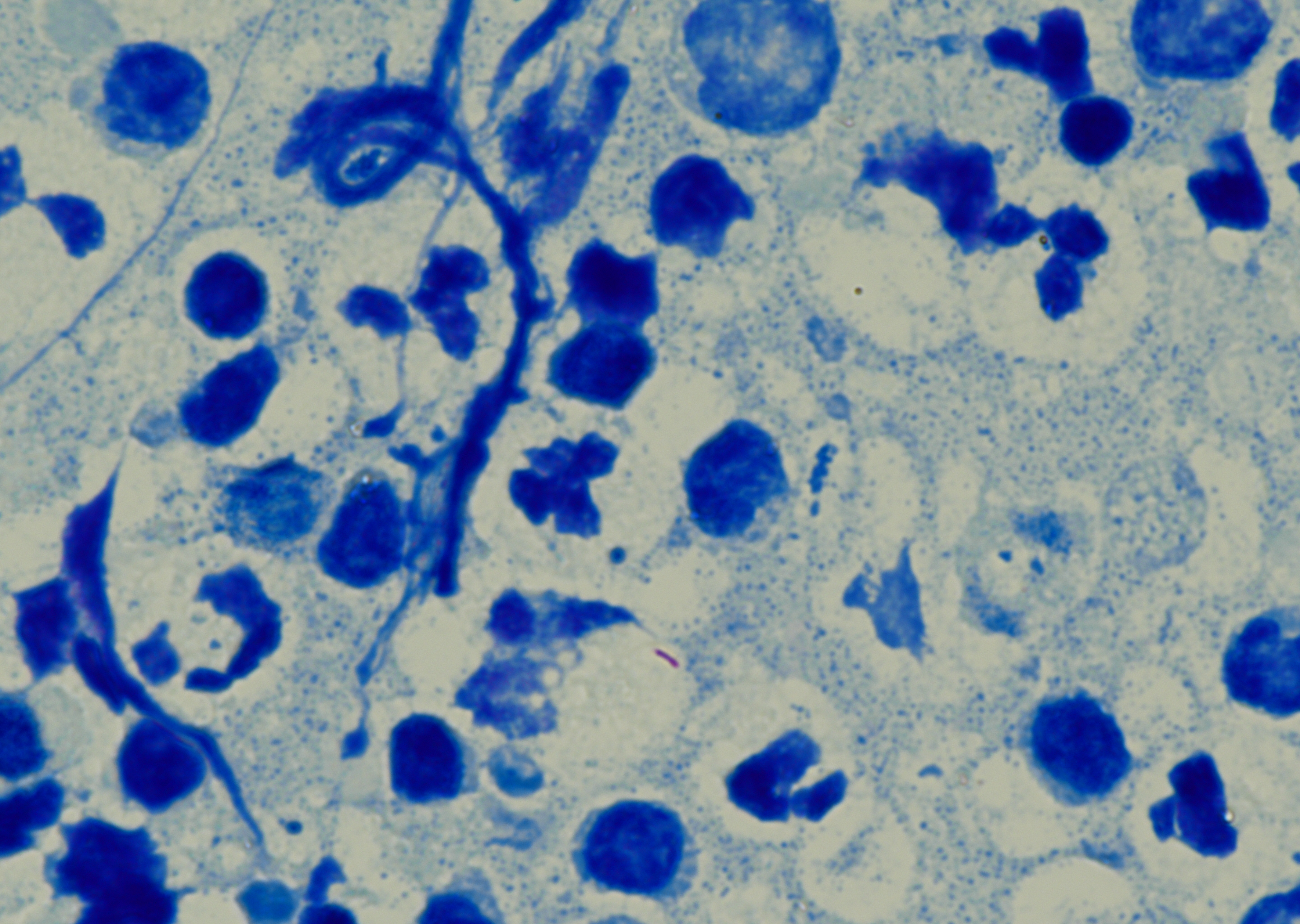 Lymph node with one red-stained acid-fast…
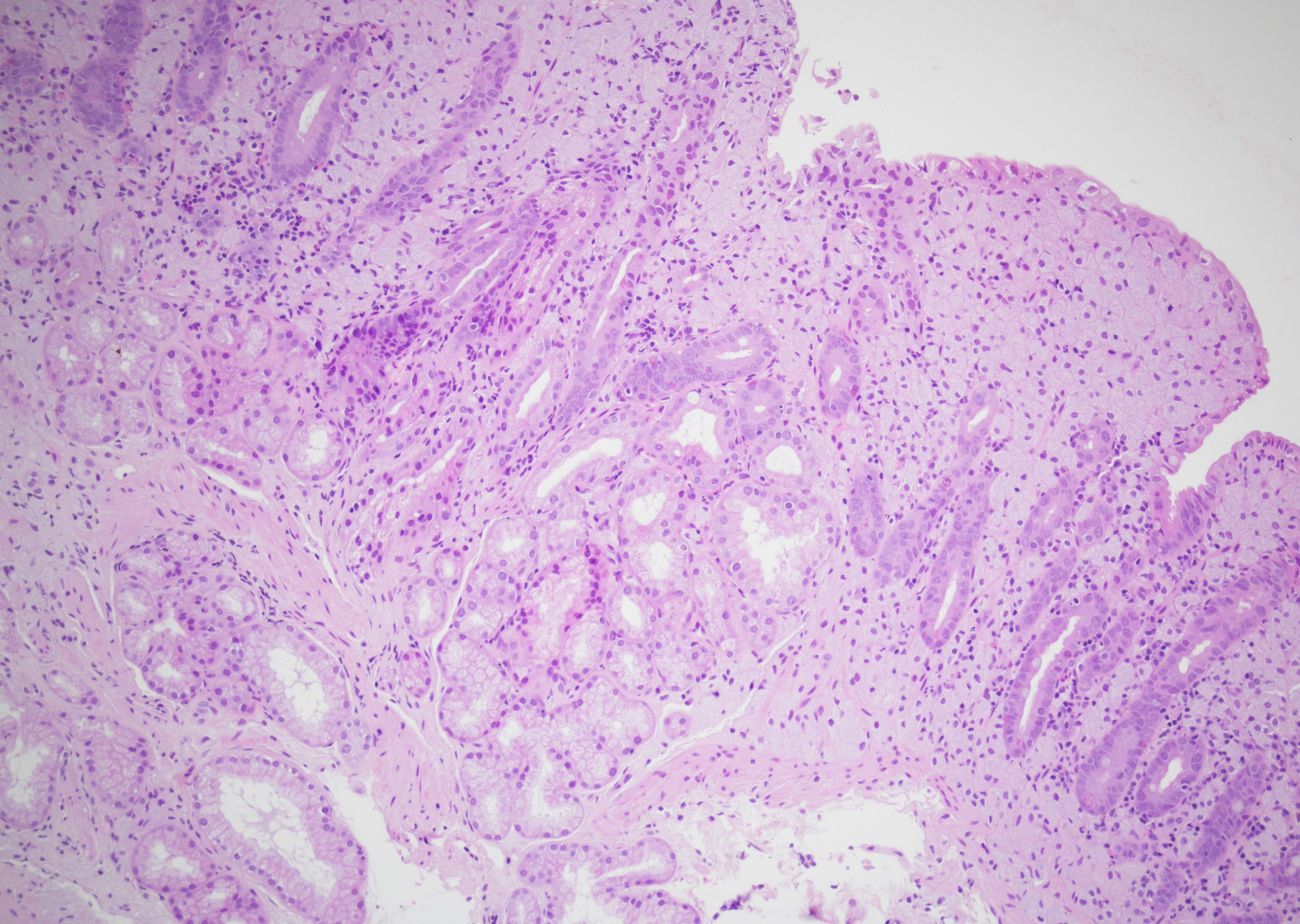 Duodenal biopsy of a HIV patient, the lamina…
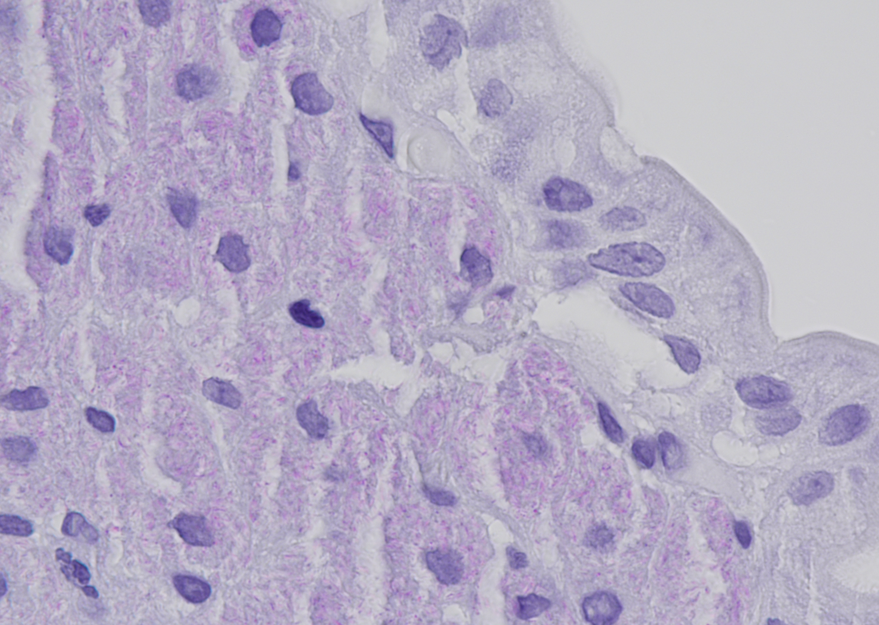 Duodenal biopsy of a HIV patient, the…
Morphology
Cytology:
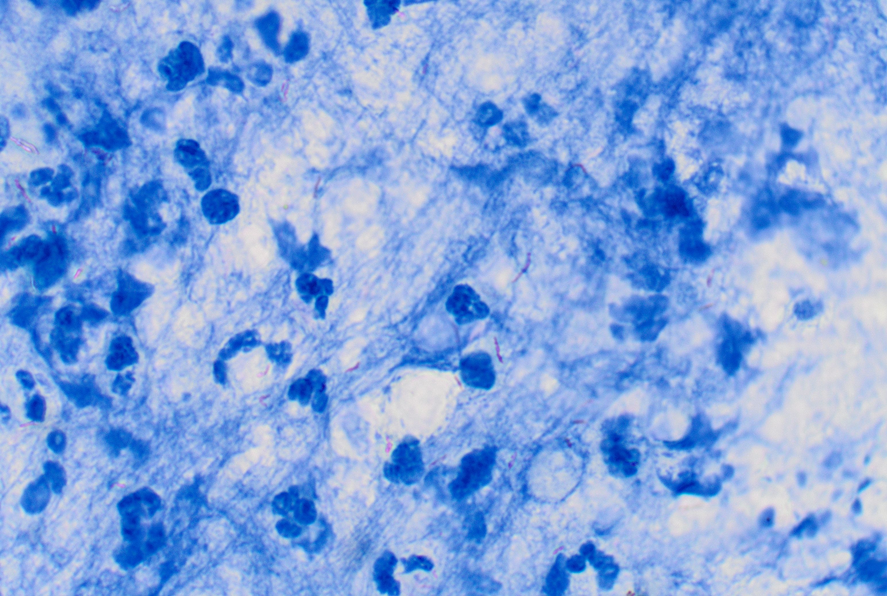
- Fine, slender acid-fast rods
- Seen singly or in small clusters
- Either intracellularly within histiocytes or free in the background detritus
- Reliable detection requires Ziehl-Neelsen or fluorescence staining
- Bacillary load is variable and often low in non-tuberculous mycobacterial infections
- Usually epitheloid histiocytes are seen on a necrotic greasy background
- Multinucleated giant cells are seen more infrequently
- In immunosuppressed patients, the typical cytomorphology of caseating granulomatous inflammation may not be seen but suppurative necrotic inflammation, mycobacterial spindle pseudotumour or a specimen comprised entirely of necrosis may be seen instead (1)
Histology:
- Usually not visible or only barely detectable on H&E staining
- Ziehl-Neelsen or auramine stains demonstrate red or fluorescent bacilli located intracellularly within macrophages or extracellularly in necrotic debris
- Typically associated with granulomatous inflammation, epithelioid cell granulomas and Langhans-type giant cells, sometimes with central necrosis
- Positive staining for PAS and/or Grocott indicate non-tuberculous mycobacteria
Surrounding Reaction:
- Smudgy detritus with degenerating granulocytes, which may be absent in non-tuberculous mycobacterial infections
- Numerous granulocytes
- Groups of epithelioid cells and Langhans-type giant cells
- Surrounding reaction with alveolar cells showing elongated nuclei; variable numbers of lymphoid cells, partly reactive, suggestive of lymph node involvement
- Reactive changes of the respiratory epithelium with goblet cell hyperplasia and round cell as well as squamous metaplasia
Clinical notes
Primary tuberculosis caused by Mycobacterium tuberculosis is usually self-limiting and predominantly affects the middle or lower lobes of the lungs. Radiologically, it presents as a pulmonary infiltrate, often accompanied by hilar lymphadenopathy. In individuals with impaired immune defense, progressive primary tuberculosis with hematogenous generalization may develop.
BCG-adenitis needs to be distinguished from Mycobacterium tuberculosis and other virulent mycobacterial infections. In BCG-adenitis, the infant is well and has attained appropriate milestones, unlike tuberculosis usually only one lymph node is involved (usually axilla ipsilateral to the arm where the vaccine was administered but has been described in ipsilateral neck lymph nodes as well) and acid fast stains are positive in both conditions. Culture and PCR can differentiate vaccine-derived Mycobacterium bovis from Mycobacterium tuberculosis.
Post-primary tuberculosis arises either from endogenous reactivation of persistent mycobacteria or from exogenous reinfection and may manifest months to years after the initial infection. Approximately 90% of post-primary cases result from hematogenous dissemination and typically involve the apical segments of the lungs, although extrapulmonary sites may also be affected. Clinical presentation varies with disease stage, ranging from asymptomatic infection to low-grade fever, weight loss, and night sweats. Chest radiographs commonly show ill-defined infiltrates, hilar lymphadenopathy, and, in some cases, cavitation. Calcification, fibrosis, and cavitary lesions represent post-specific residual changes.
Miliary tuberculosis is a rare form characterized by high fever and, if the meninges are involved, meningeal signs. Radiologically, it presents as a disseminated, fine nodular pattern. A particular form of disease progression is the development of a tuberculoma, which is considered either a residual lesion of primary tuberculosis or the result of appositional growth of disseminated foci. Imaging typically reveals a well-circumscribed, rounded lesion.
Infection with non-tuberculous mycobacteria (MOTT) may also cause significant disease, including pulmonary infections, lymphadenitis, and infections of the skin, soft tissues, and joints.
FNA lymph node or other sites provides a cost-effective and reliable diagnosis of mycobacterial infection especially with the addition of ancillary tests like acid fast staining, culture and NAAT.
Ancillary Testing
Culture (prolonged incubation), PCR including NAAT, line probe assays, MALDI-TOF / 16S rRNA, drug susceptibility testing, X-ray / CTAdditional Information
The genus Mycobacterium (M.) includes the Mycobacterium tuberculosis complex, which consists of M. tuberculosis, M. bovis, M. caprae, M. africanum, M. microti, M. canettii, and M. pinnipedii, as well as M. leprae. Mycobacteria spp. also include those referred to as non-tuberculous mycobacteria (NTM, MOTT), such as M. abscessus, M. kansasii, M. xenopi, and the M. avium complex, which can also cause serious diseases. Predominantly, patients with immunodeficiency (e.g., AIDS patients) and those with chronic lung diseases are affected.
Bacille Calmette-Guérin (BCG) remains the sole vaccine currently approved for preventing tuberculosis. Each year, it is administered to more than 100 million newborns. Evidence from meta-analyses suggests that BCG provides around 50% protection against pulmonary TB in adults, while its effectiveness against severe forms such as tuberculous meningitis and disseminated (miliary) disease in infants is closer to 80%. This higher level of protection explains its continued widespread use in regions where TB is endemic.
Although generally considered safe, BCG is a live-attenuated vaccine, which means there is a small but real risk of adverse effects. These can range from mild, localized reactions to rare but serious cases of disseminated infection.
Management is controversial. Most BCG-adenitis will resolve within a few months without the need for antimicrobial medication. However, some infants are treated with a single anti-tuberculous agent while others are treated with multiple anti-tuberculous agents.
Differential Diagnosis
References
- Michelow P, Omar T, Field A, Wright C. The cytopathology of mycobacterial infection. Diagn Cytopathol. 2016 Mar;44(3):255-62.