
- EN
- DE

Virus
Human Papillomavirus (HPV)


- Organ System
- Genitourinary Tract, Skin and Mucosa, Respiratory Tract
- Diagnostic Method
- Cytology, Histology

- Geographical Region
- Dry Areas,
- Islands,
- Tropical and Subtropical Region,
- Polar and Subpolar Zone,
- Temperate Zone
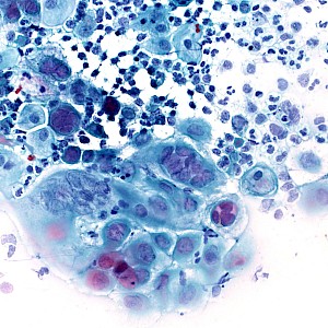
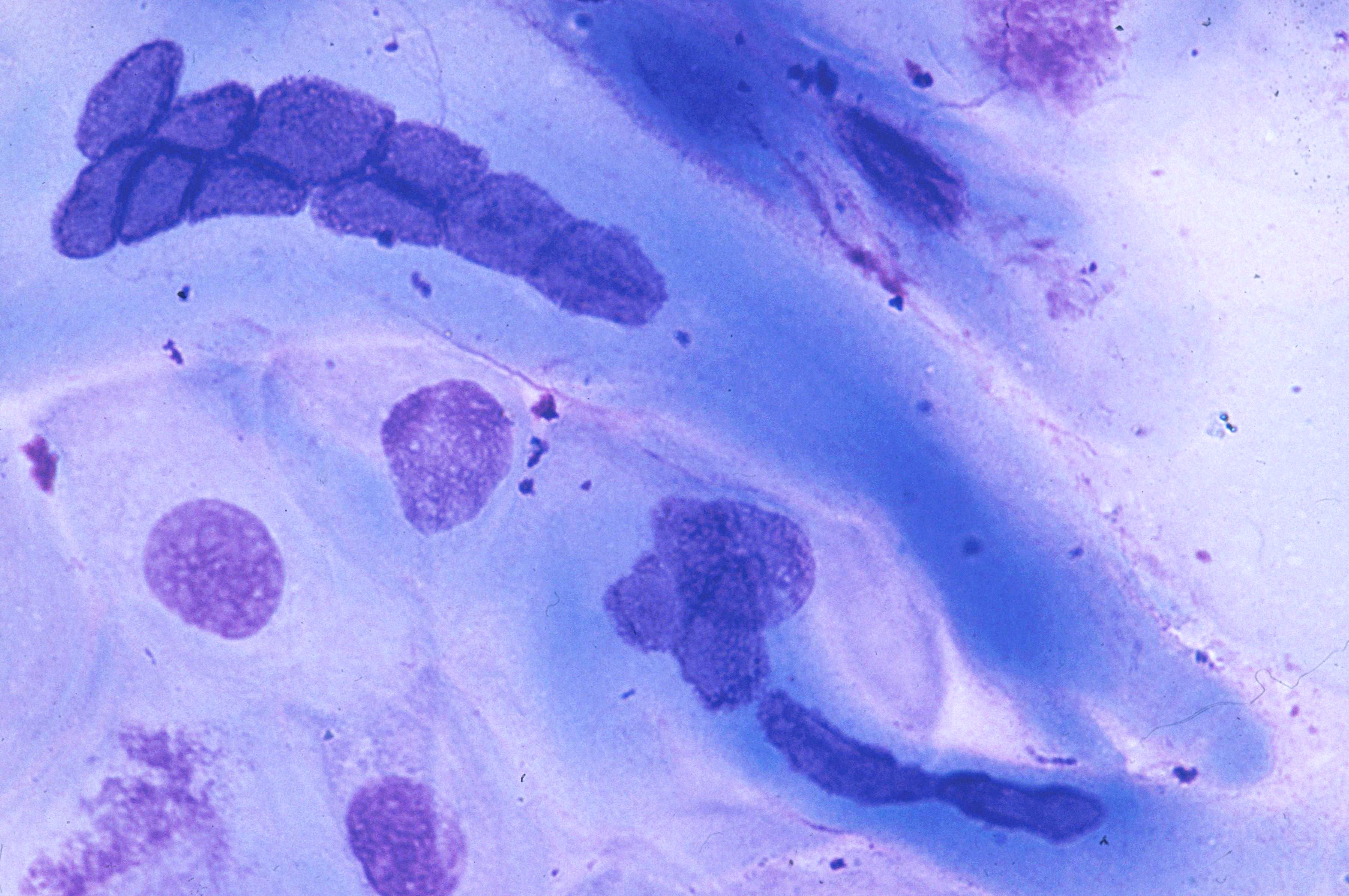
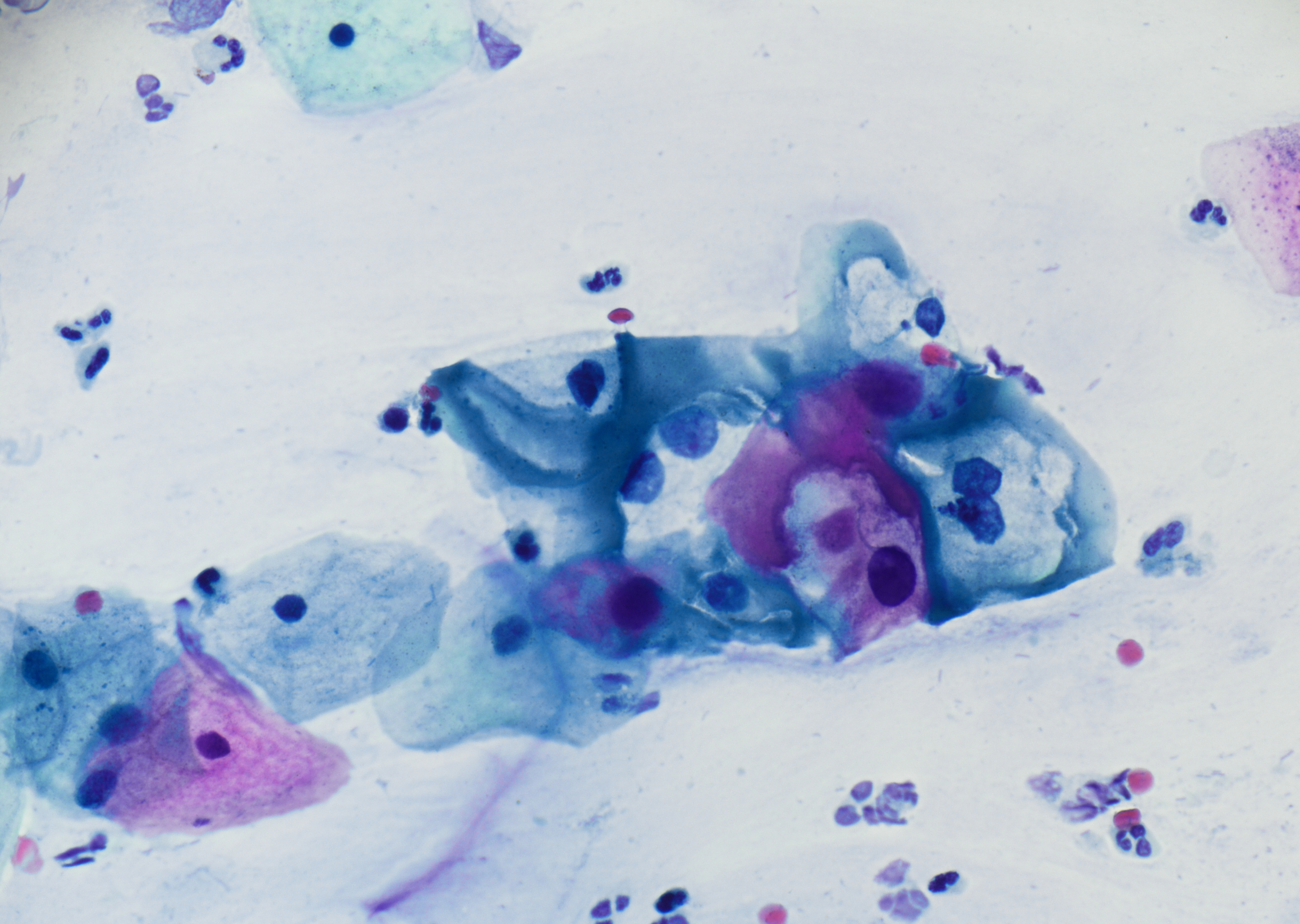 Cervical smear, koilocytic cell group with…
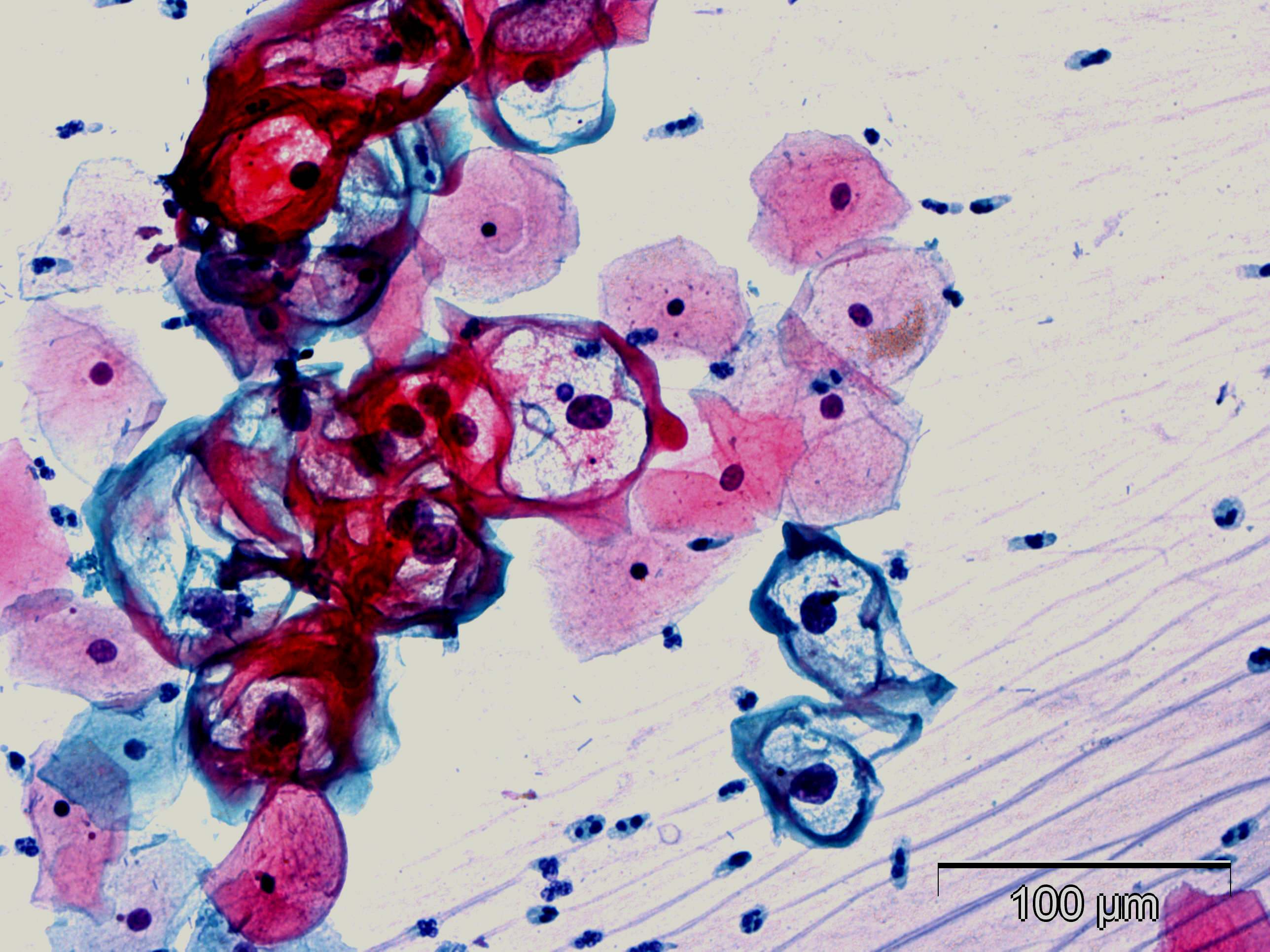 Cervical smear, numerous koilocytes with…
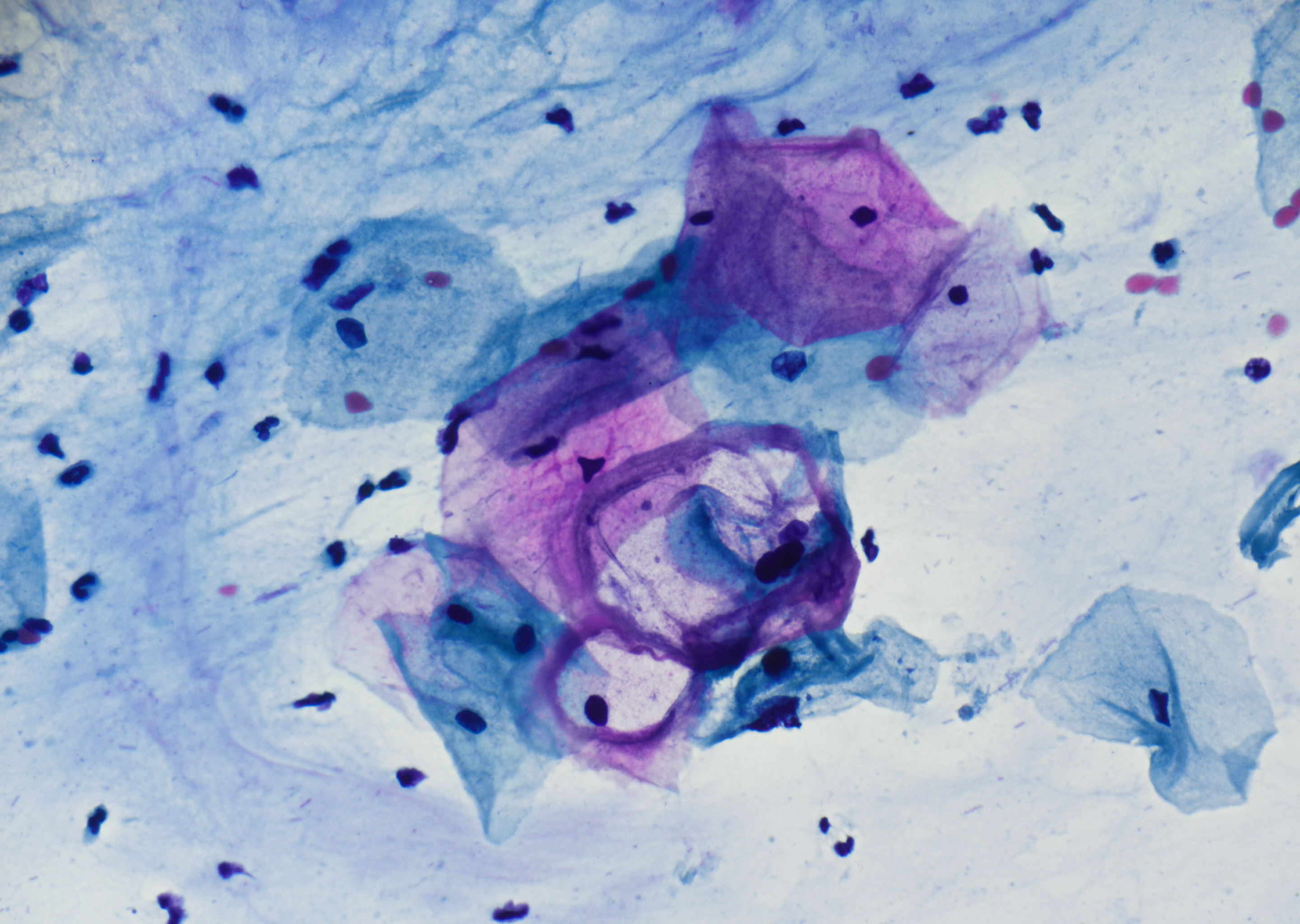 Cervical smear, koilocytes showing a sharply…
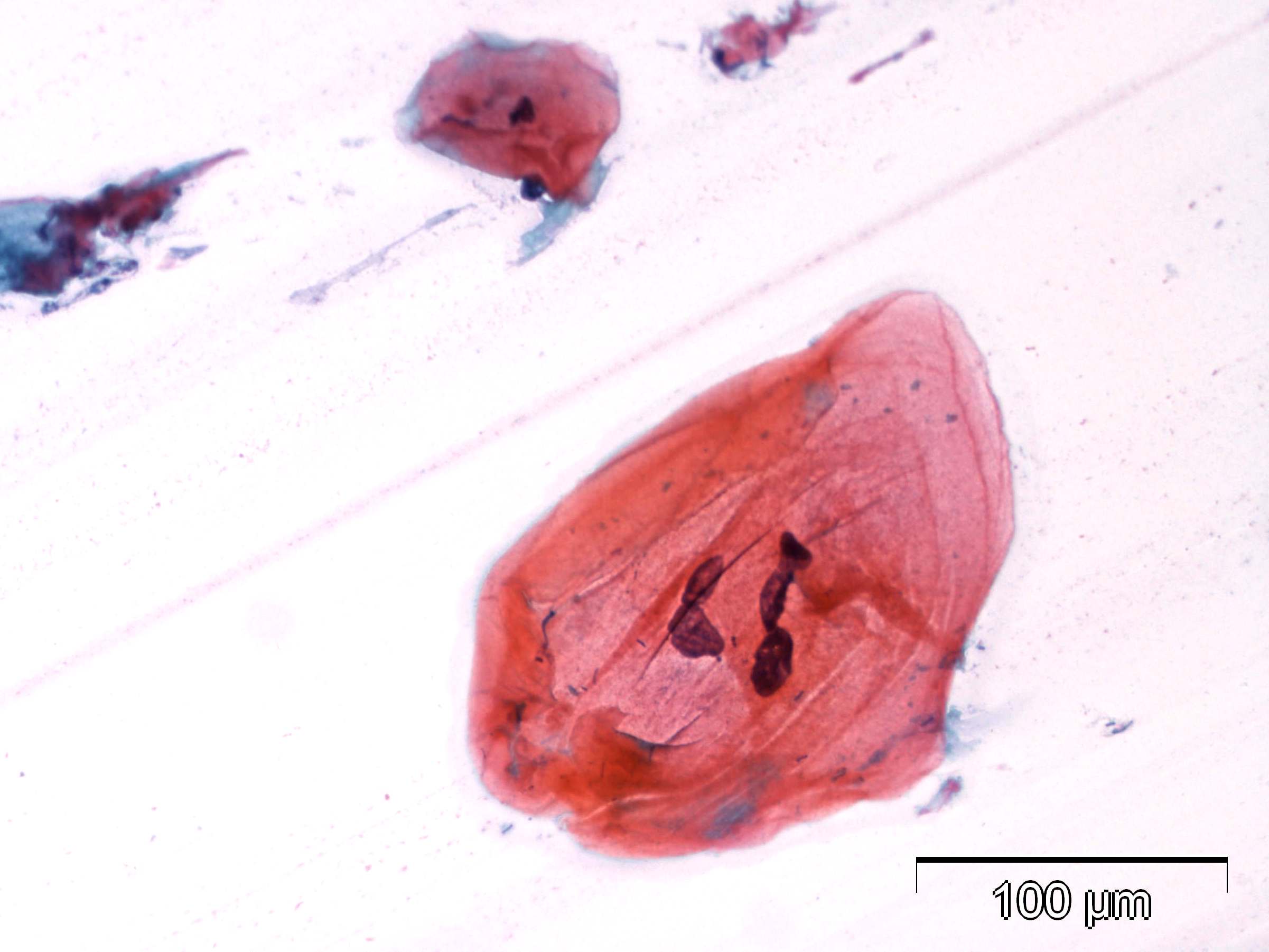 Cervical smear, markedly enlarged…
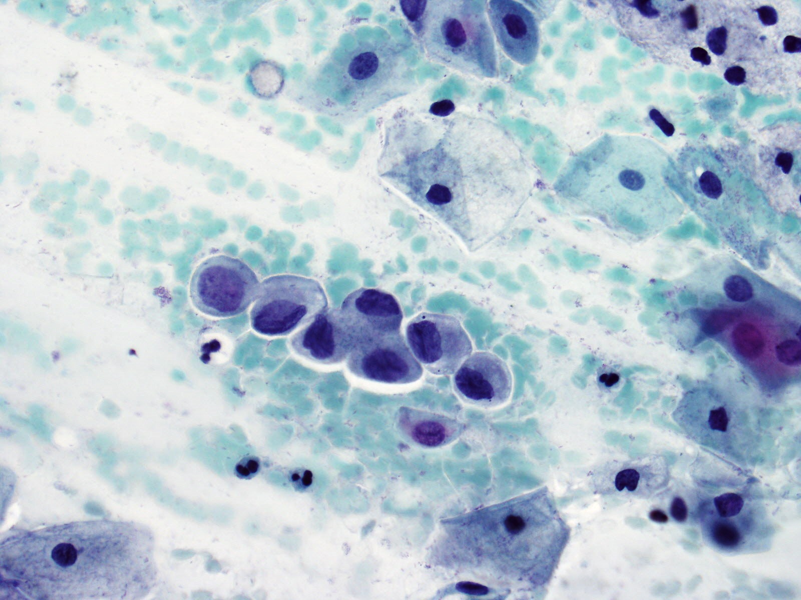 Cervical smear, high-grade dysplastic cells…
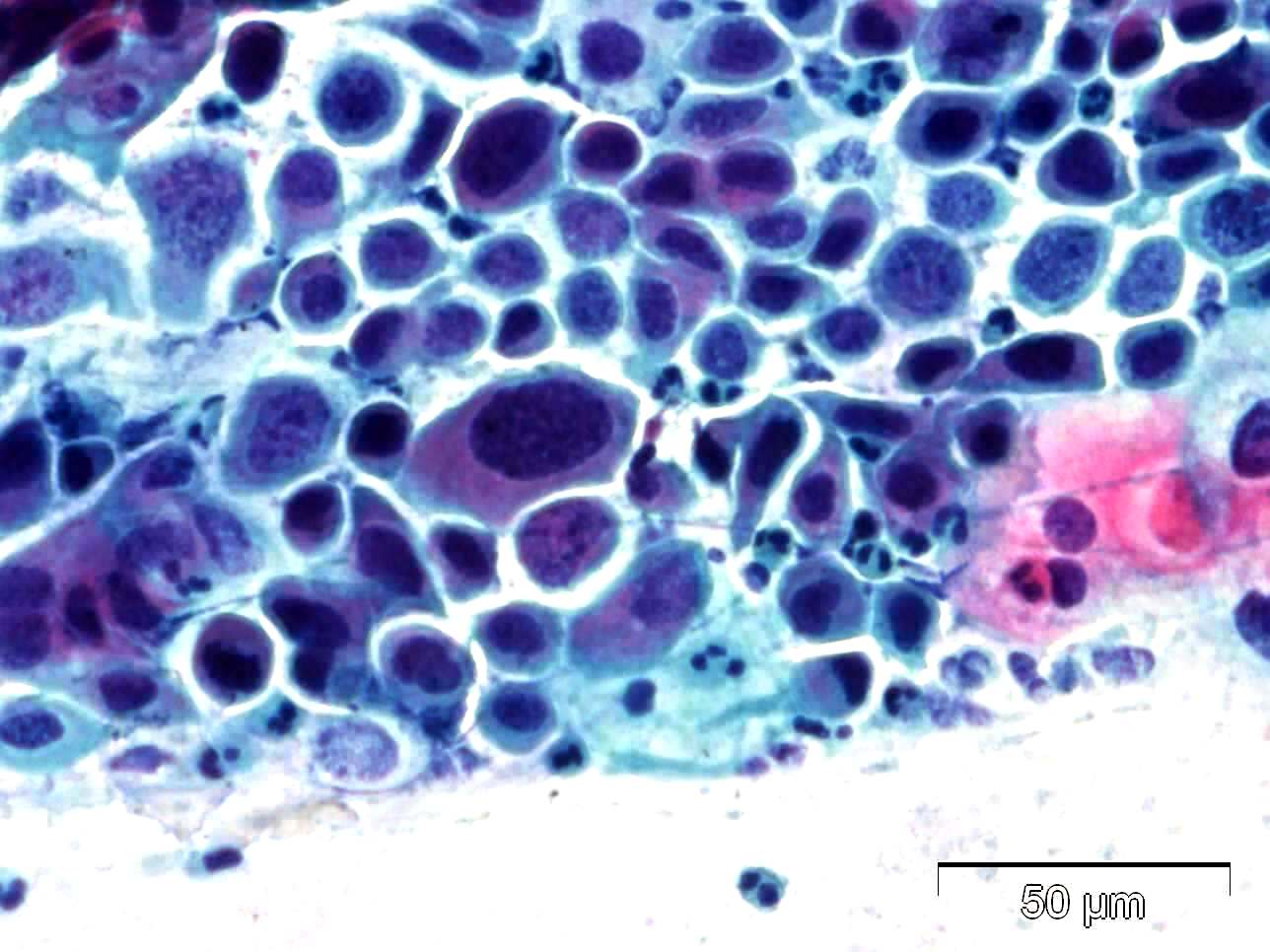 Cervical smear, high-grade dysplastic cells…
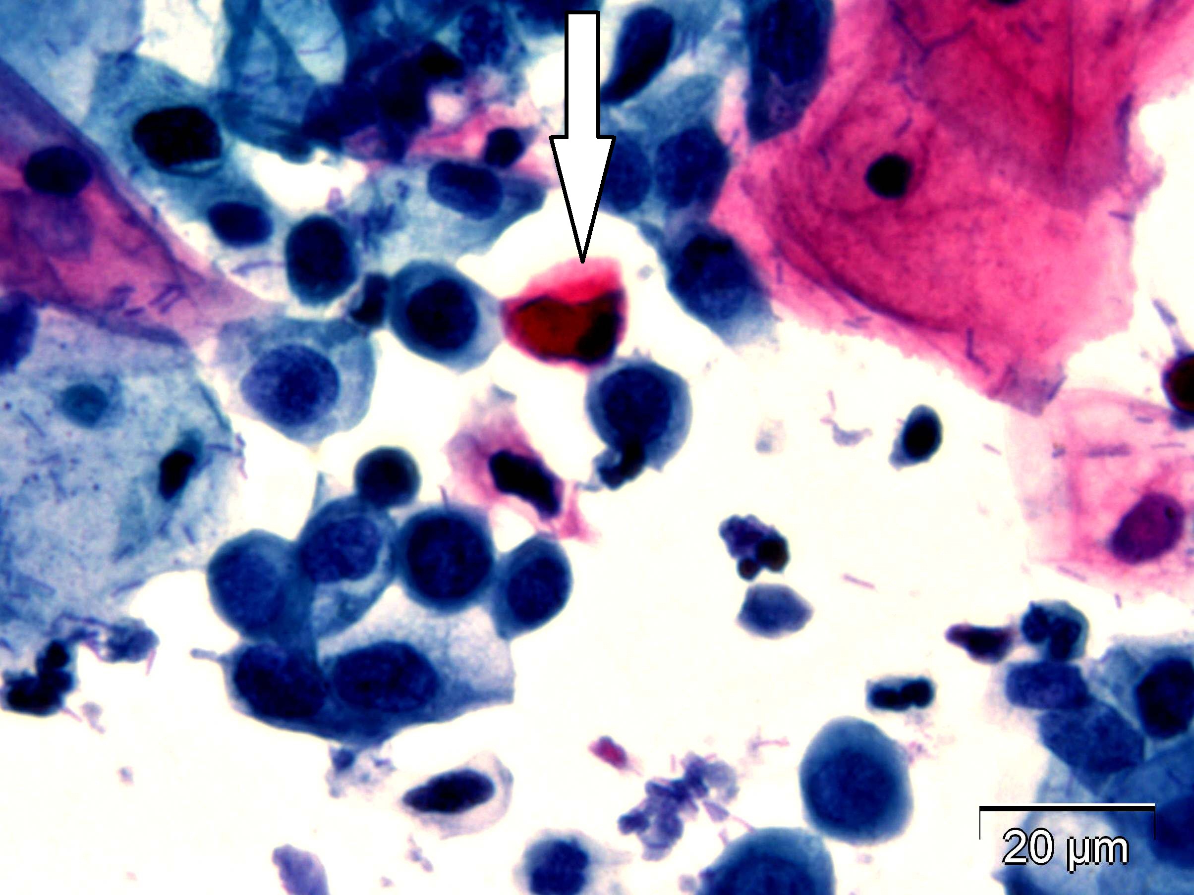 Cervical smear, high-grade dysplastic cells…
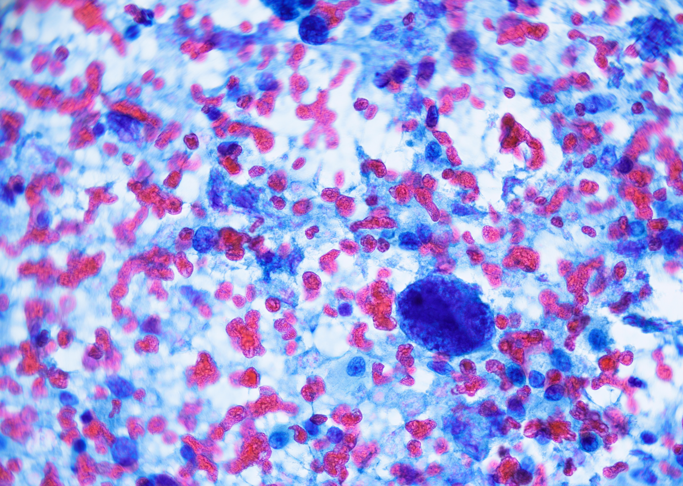 Cervical smear, SCC with tumor diathesis and…
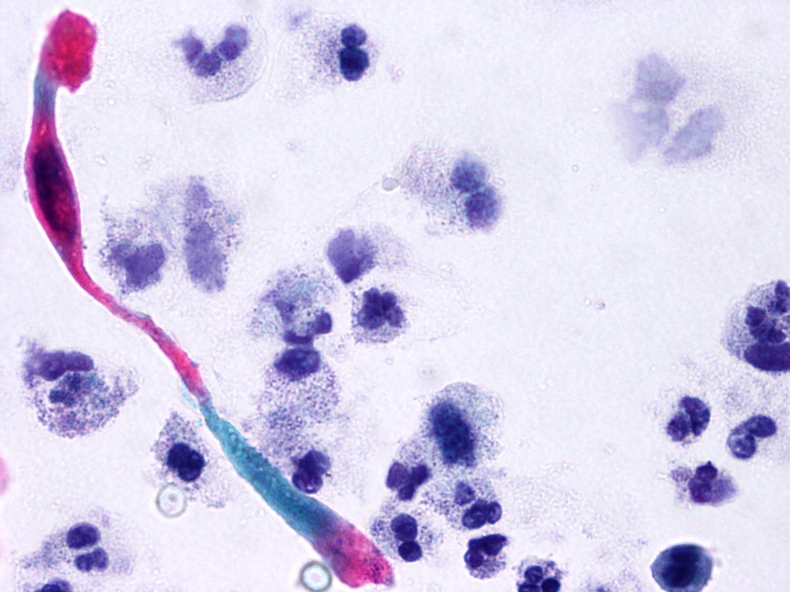 Cervical smear, a spindle-shaped cell with…
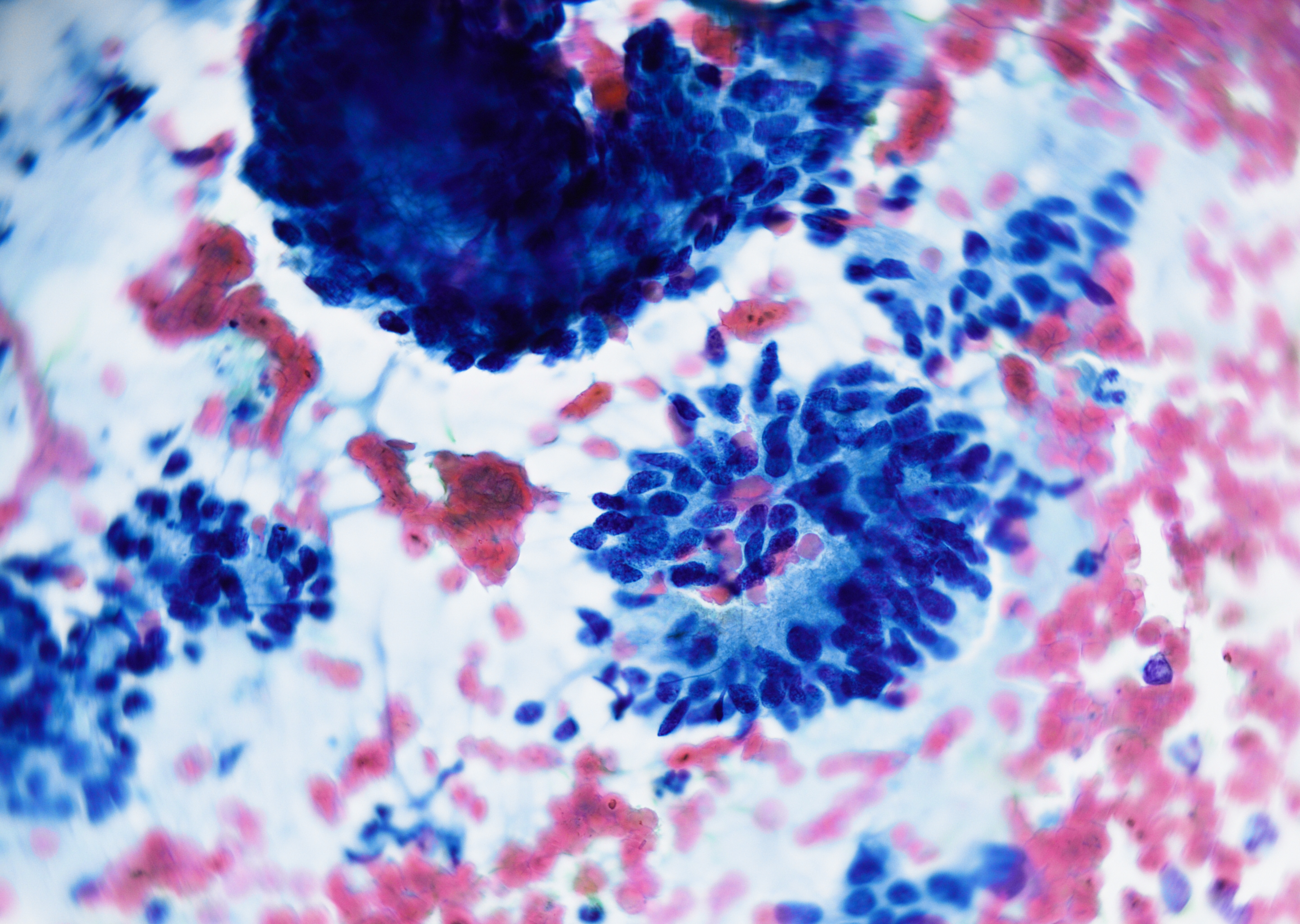 Cervical smear, atypical glandular cells…
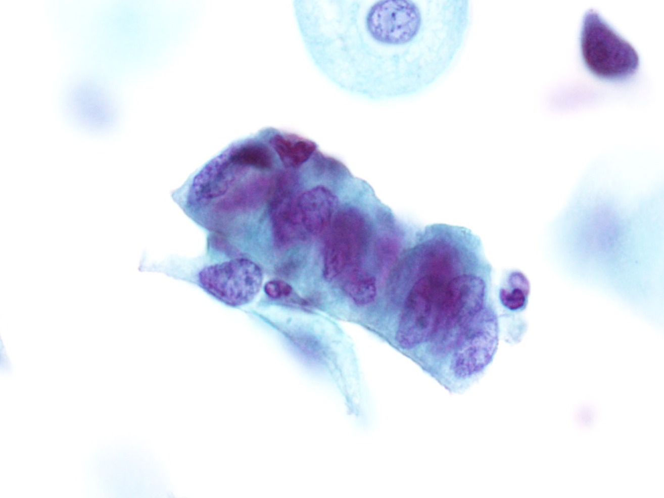 Cervix LBC, epithelial strip of columnar…
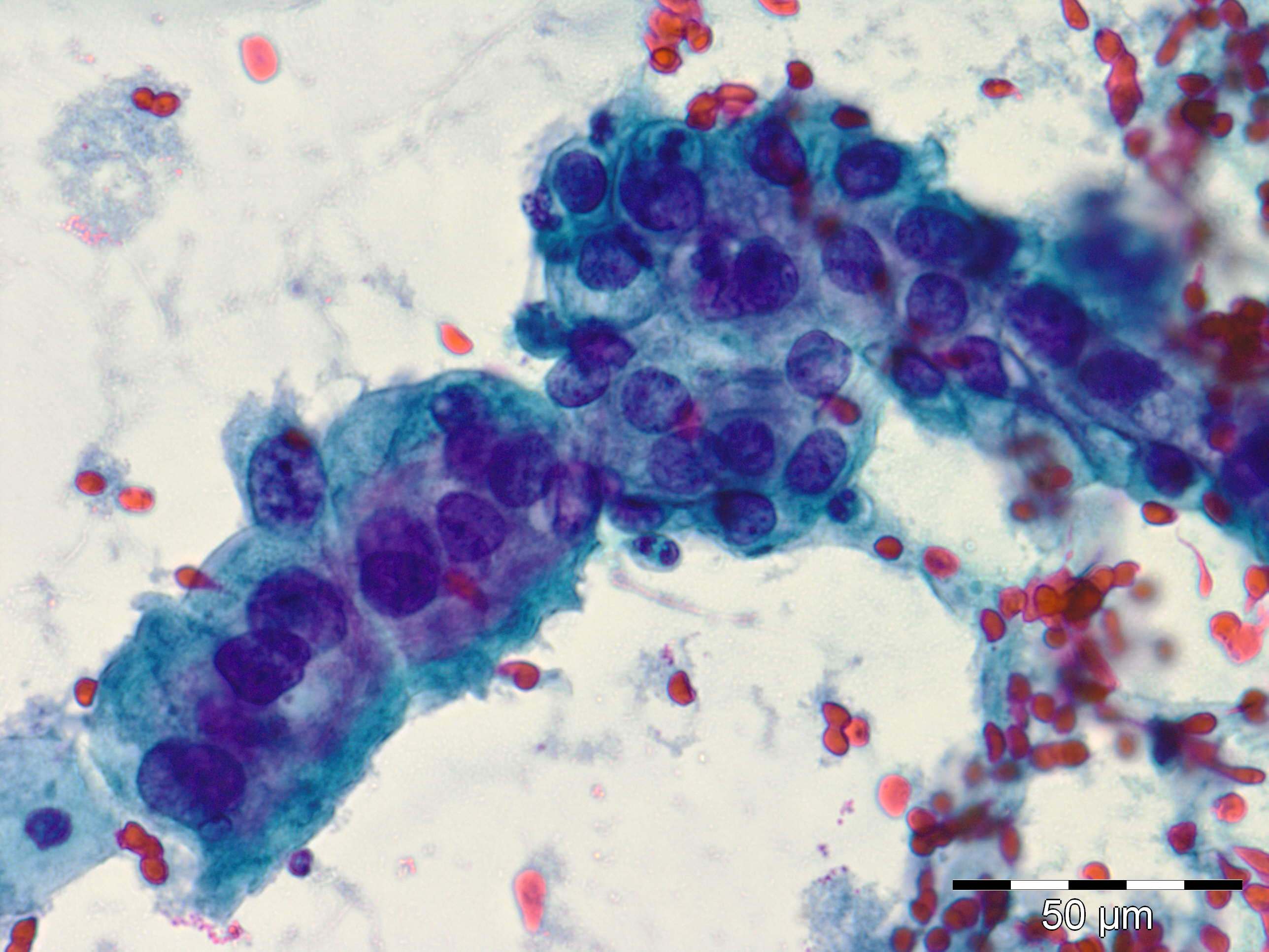 Cervical smear, atypical glandular cell…
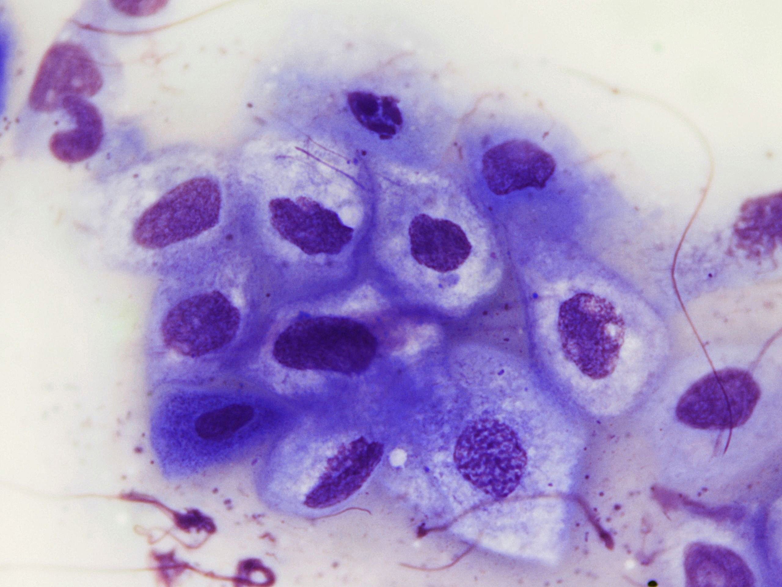 Lung, brush cytology, group of koilocytes…
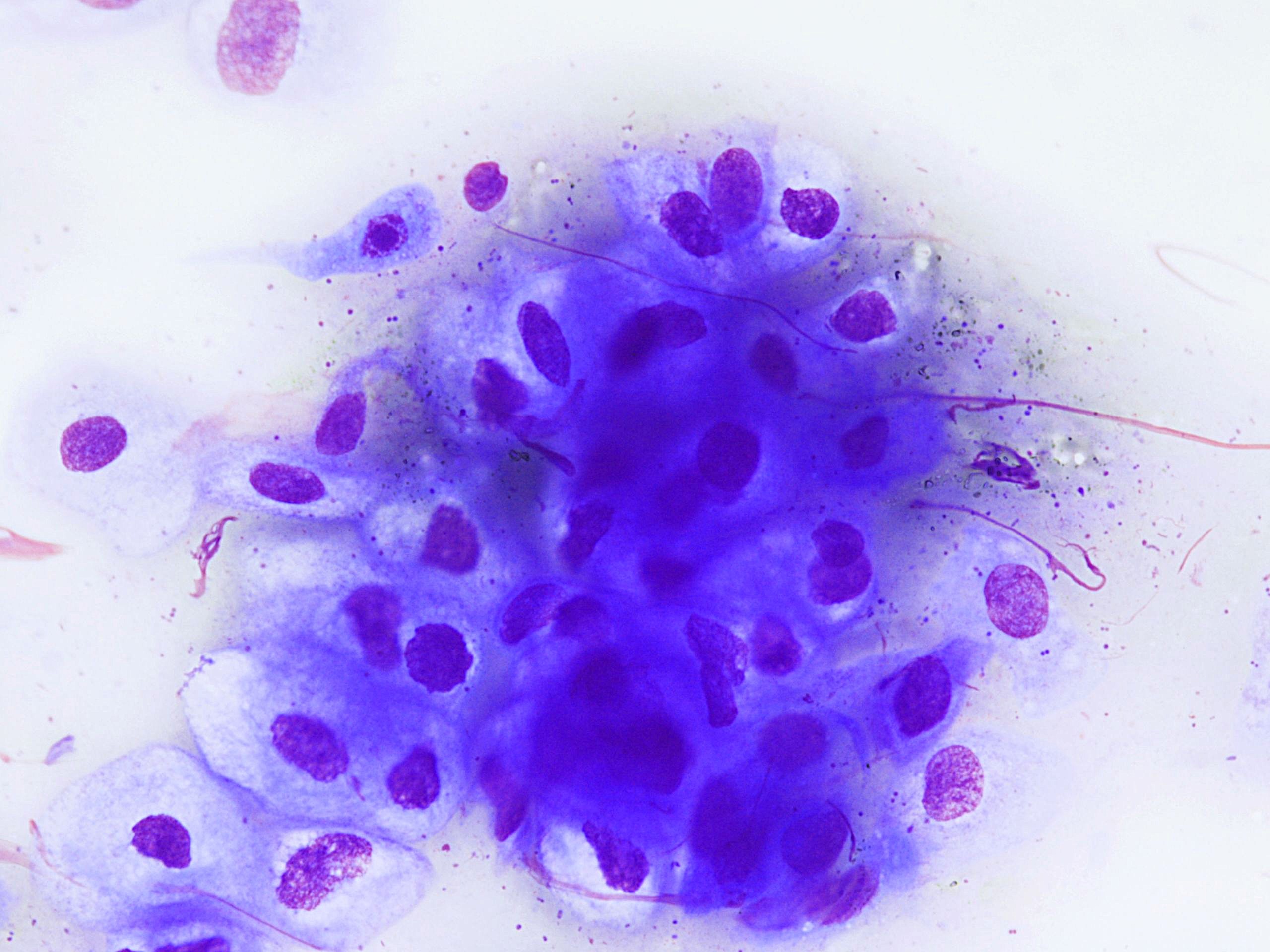 Lung, brush cytology, group of koilocytes…
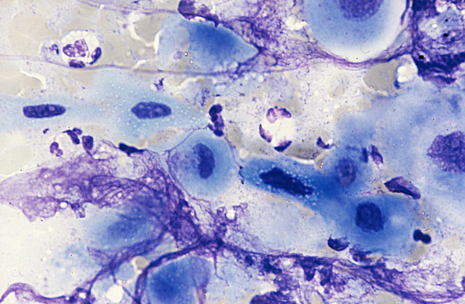
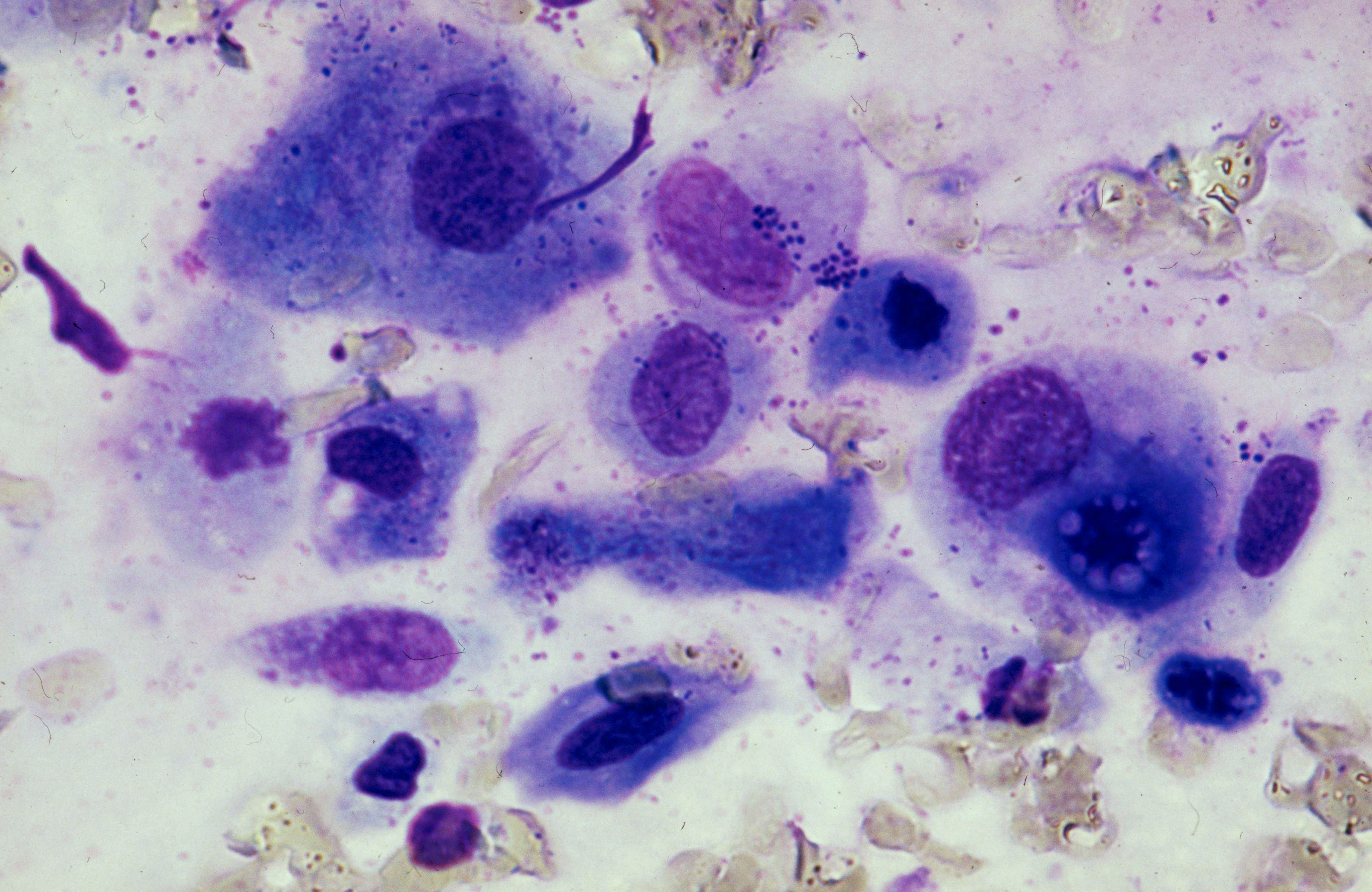 Lung, brush cytology, metaplastic cells…
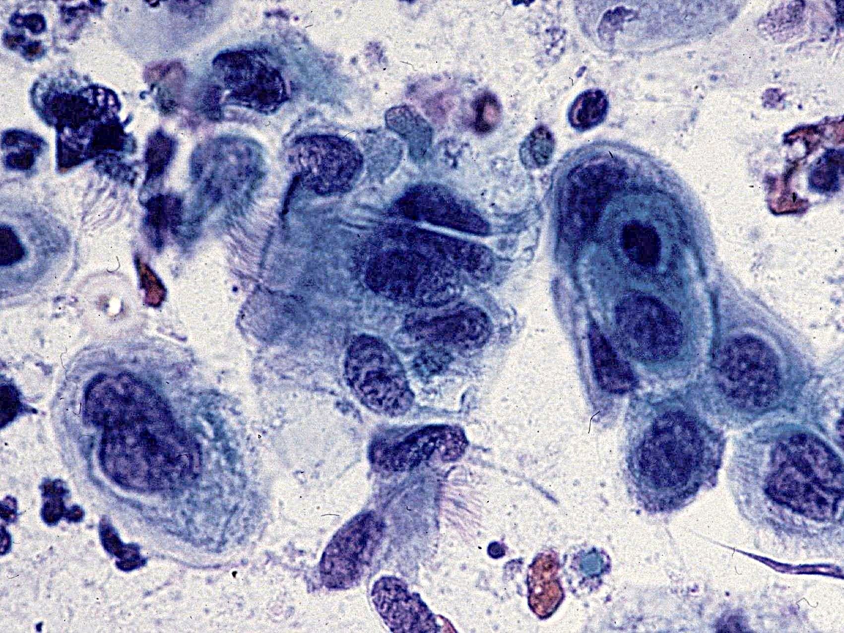 Lung, brush cytology, squamous metaplastic…
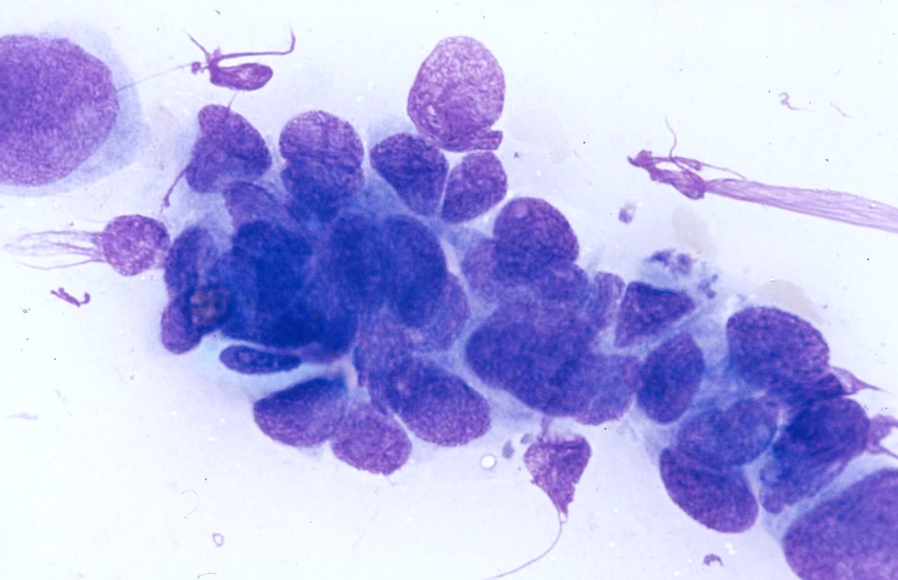
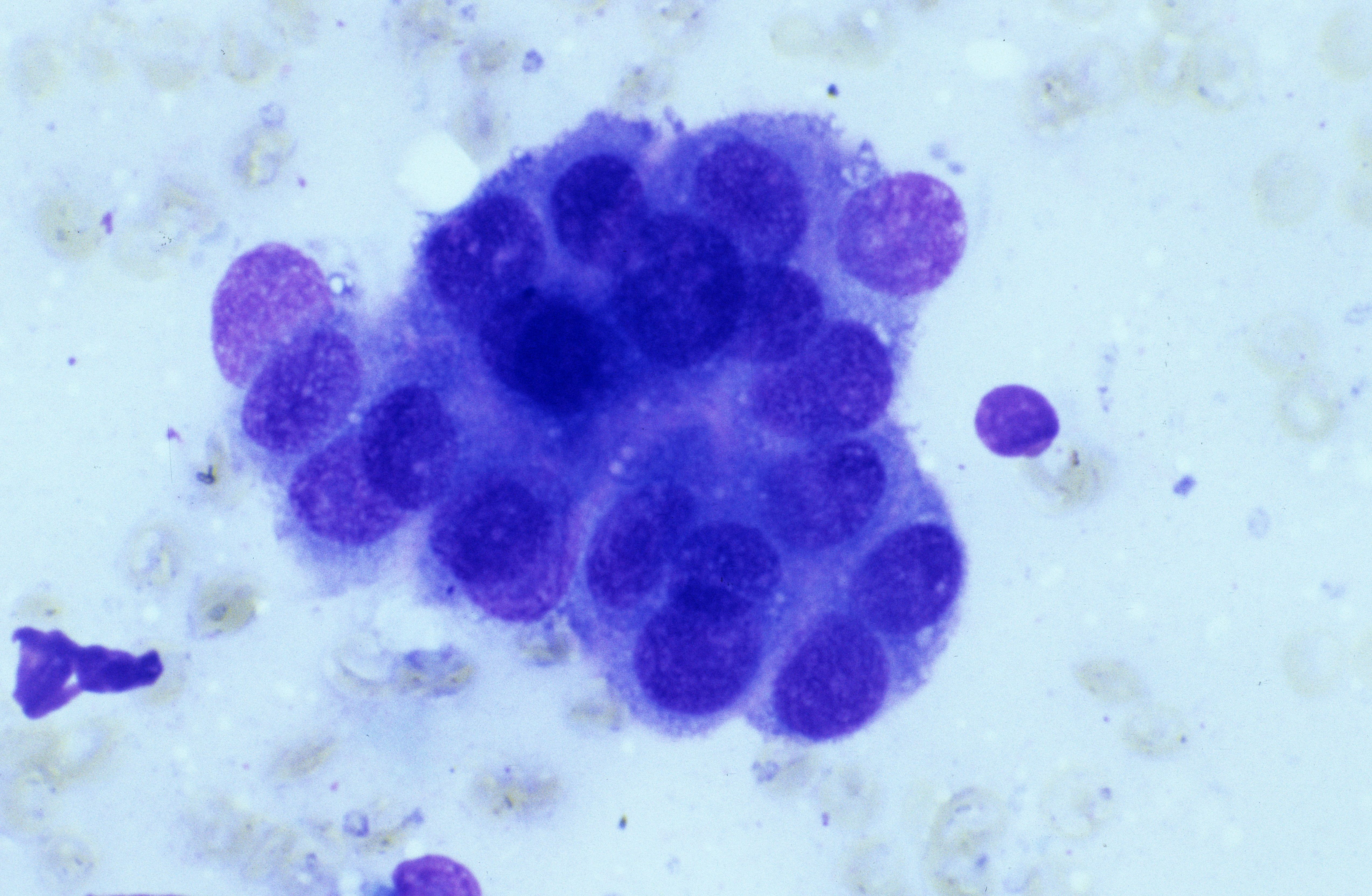
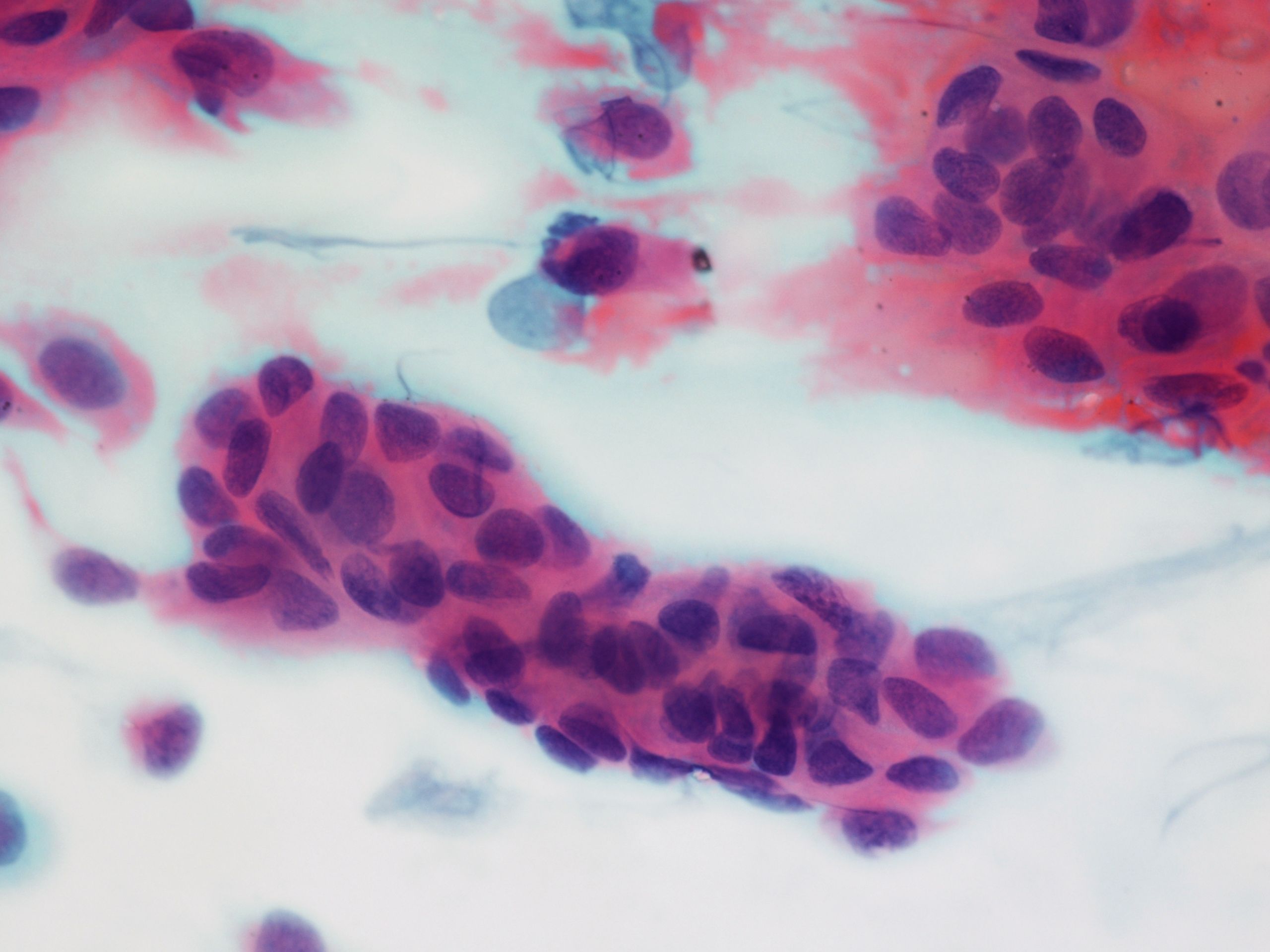 Lung, brush smear, cohesive, partly…
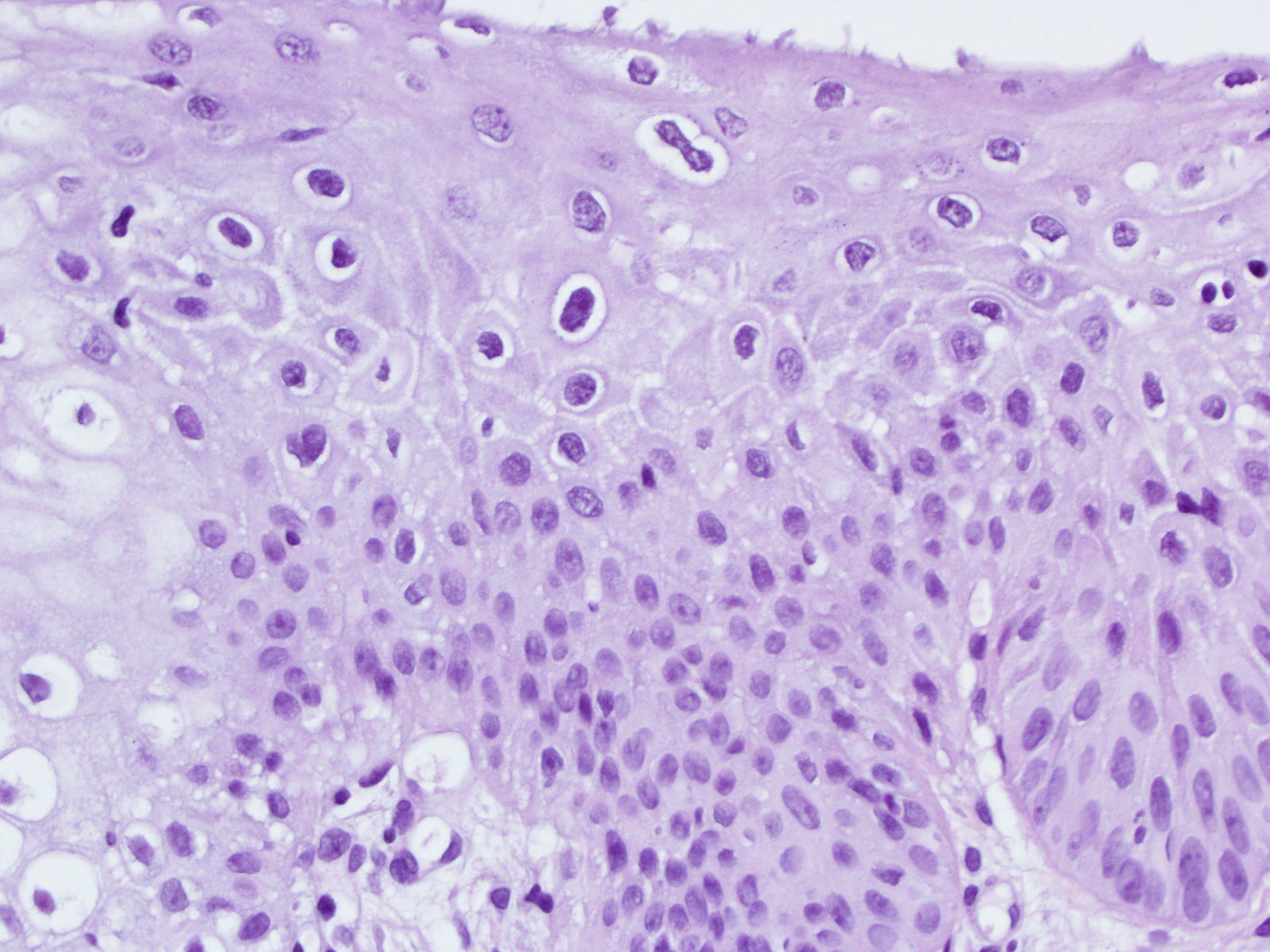 Cervix, LSIL with loss of nuclear polarity…
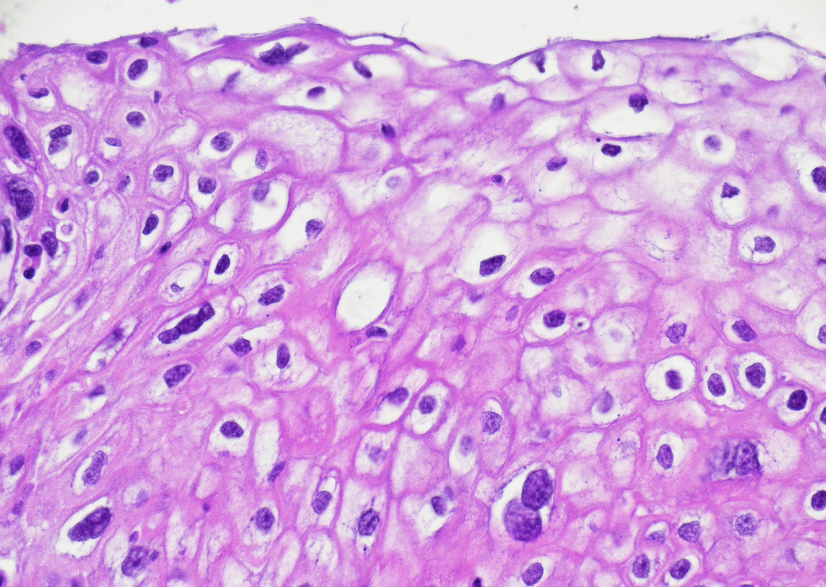 Cervix, LSIL with multinucleated nuclei…
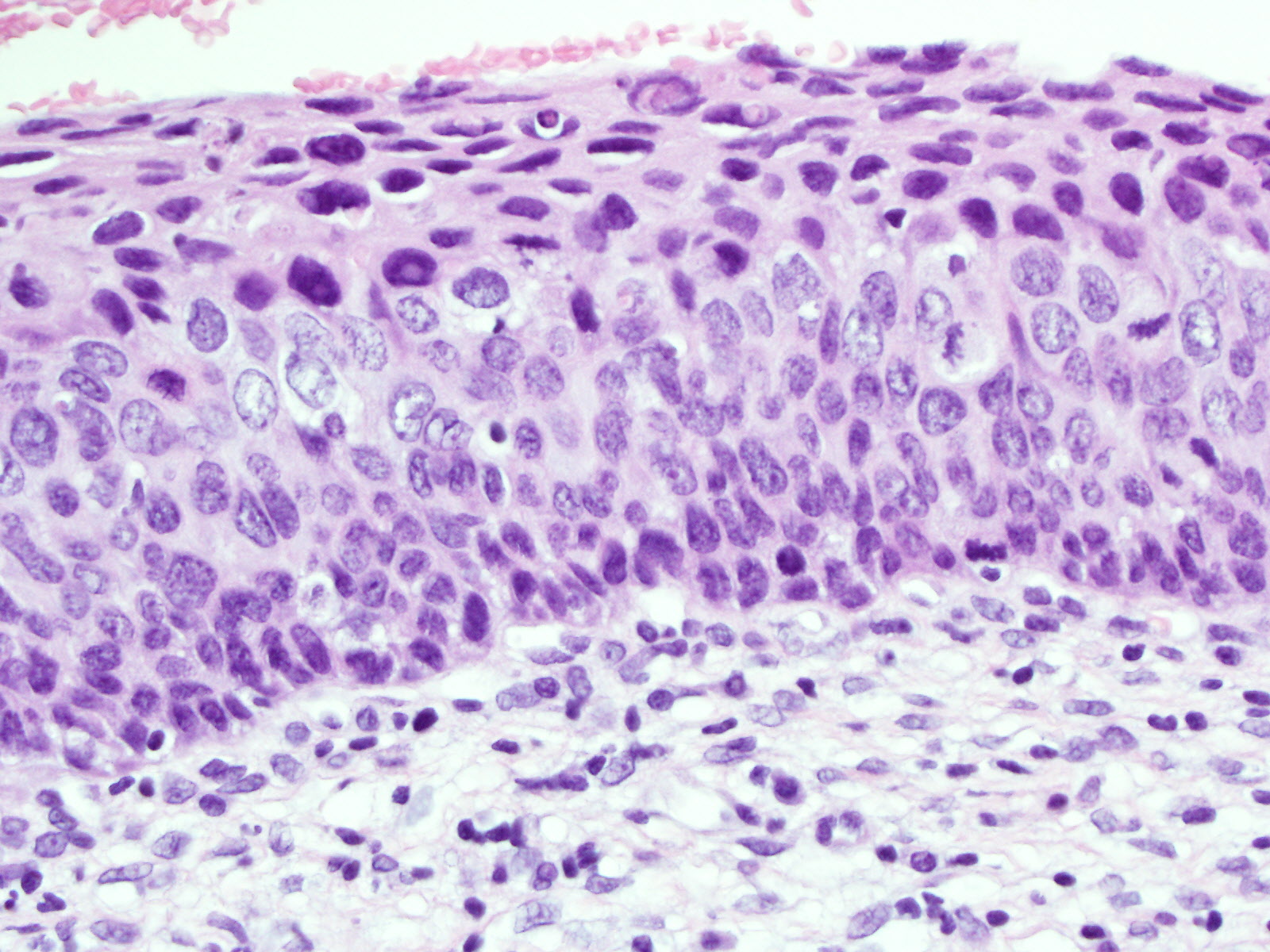 Cervix, HSIL with loss of nuclear polarity…
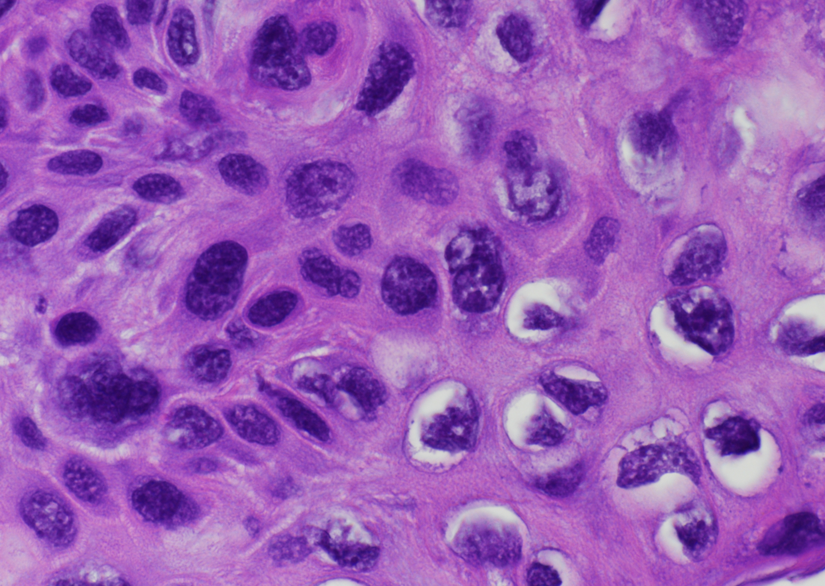 Cervix, HSIL, high-grade dysplasia with…
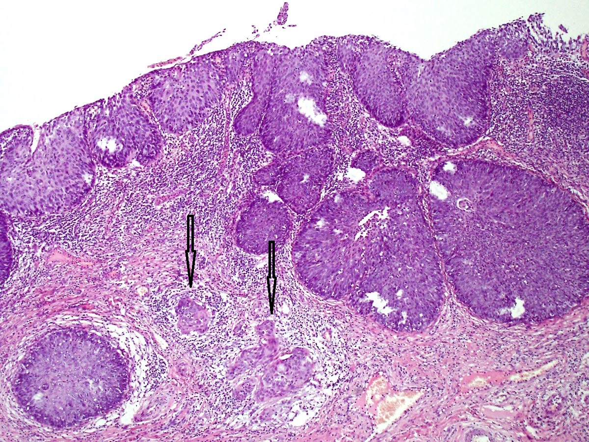 Cervix, HSIL with expansive crypt…
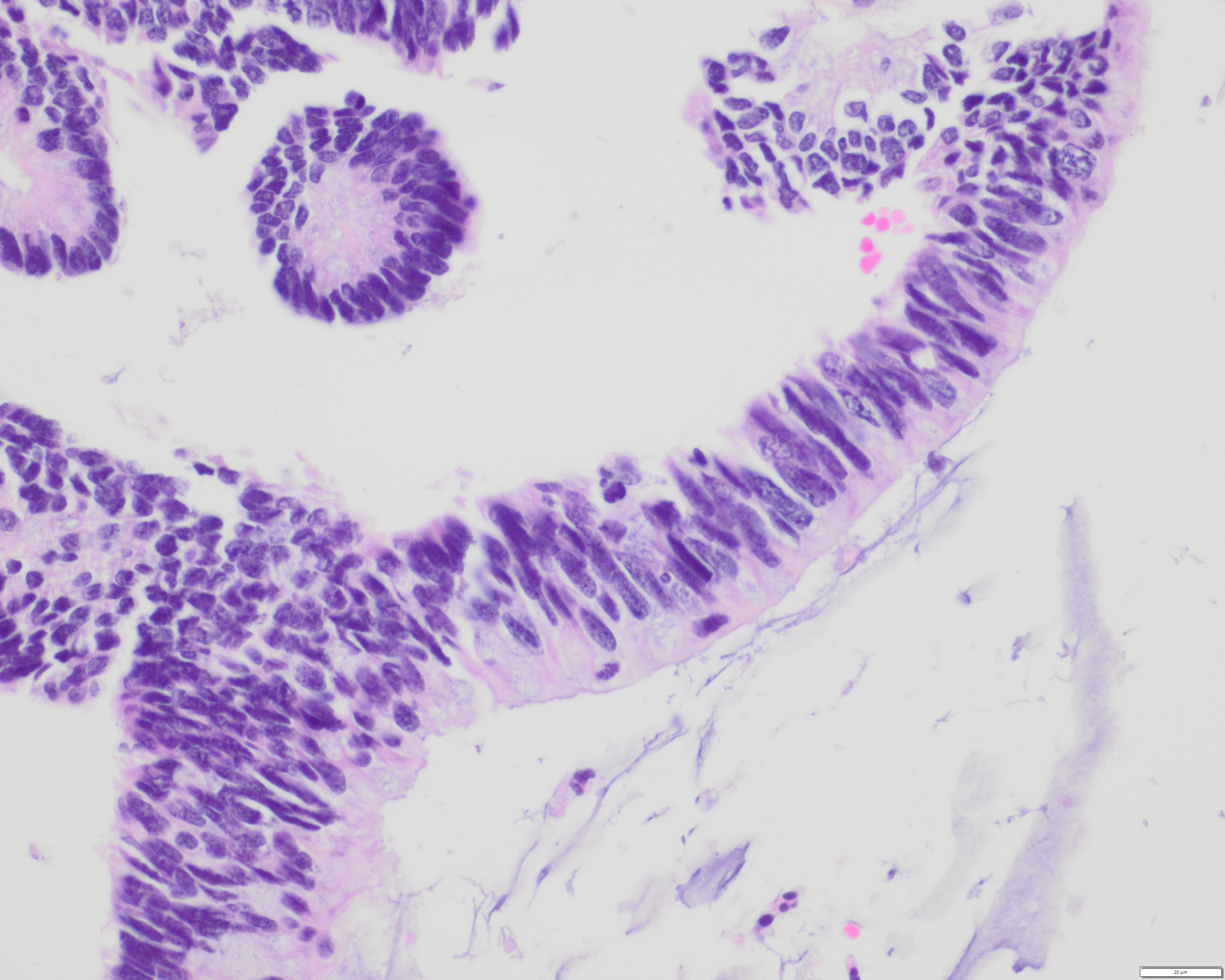 Cervix, AIS with enlarged, hyperchromatic…
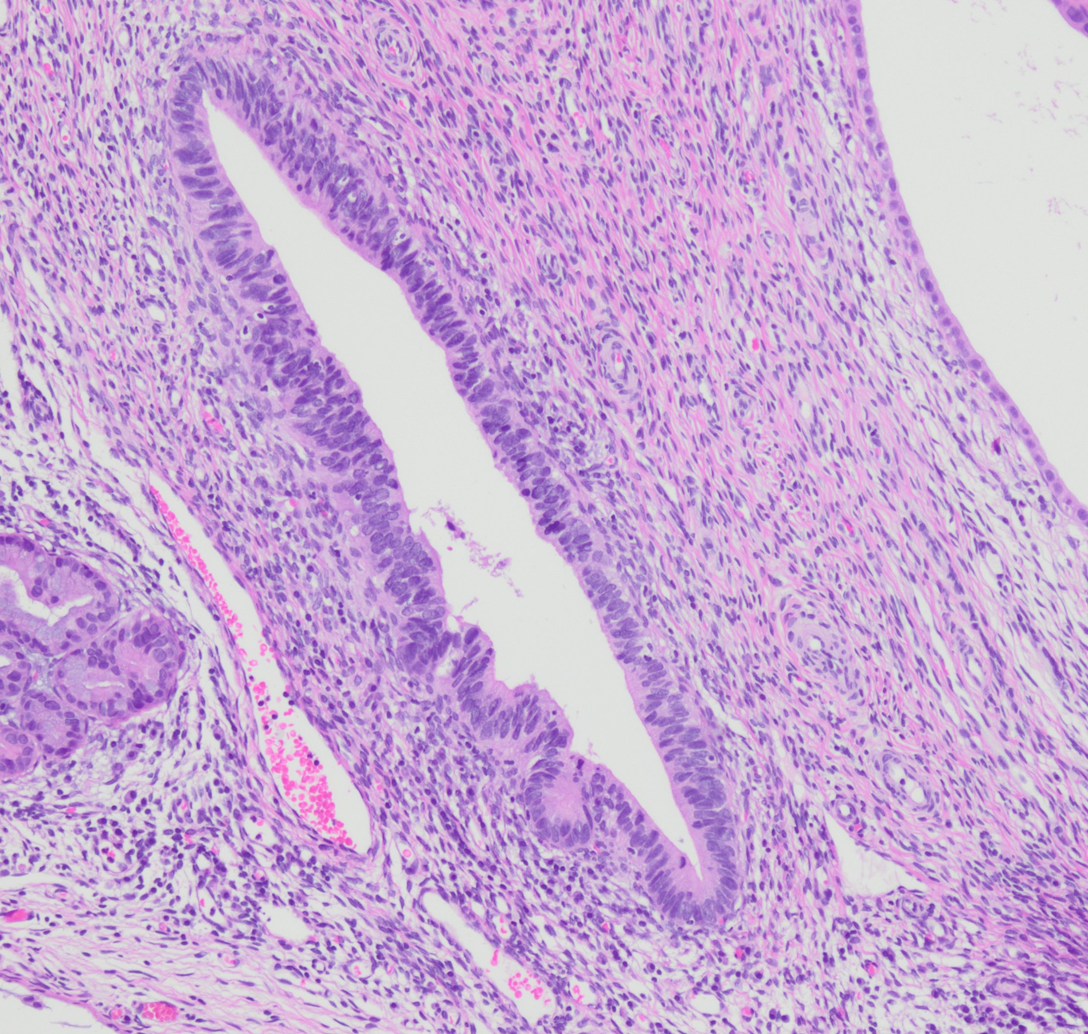 Cervix, AIS in an individual cervical crypt…
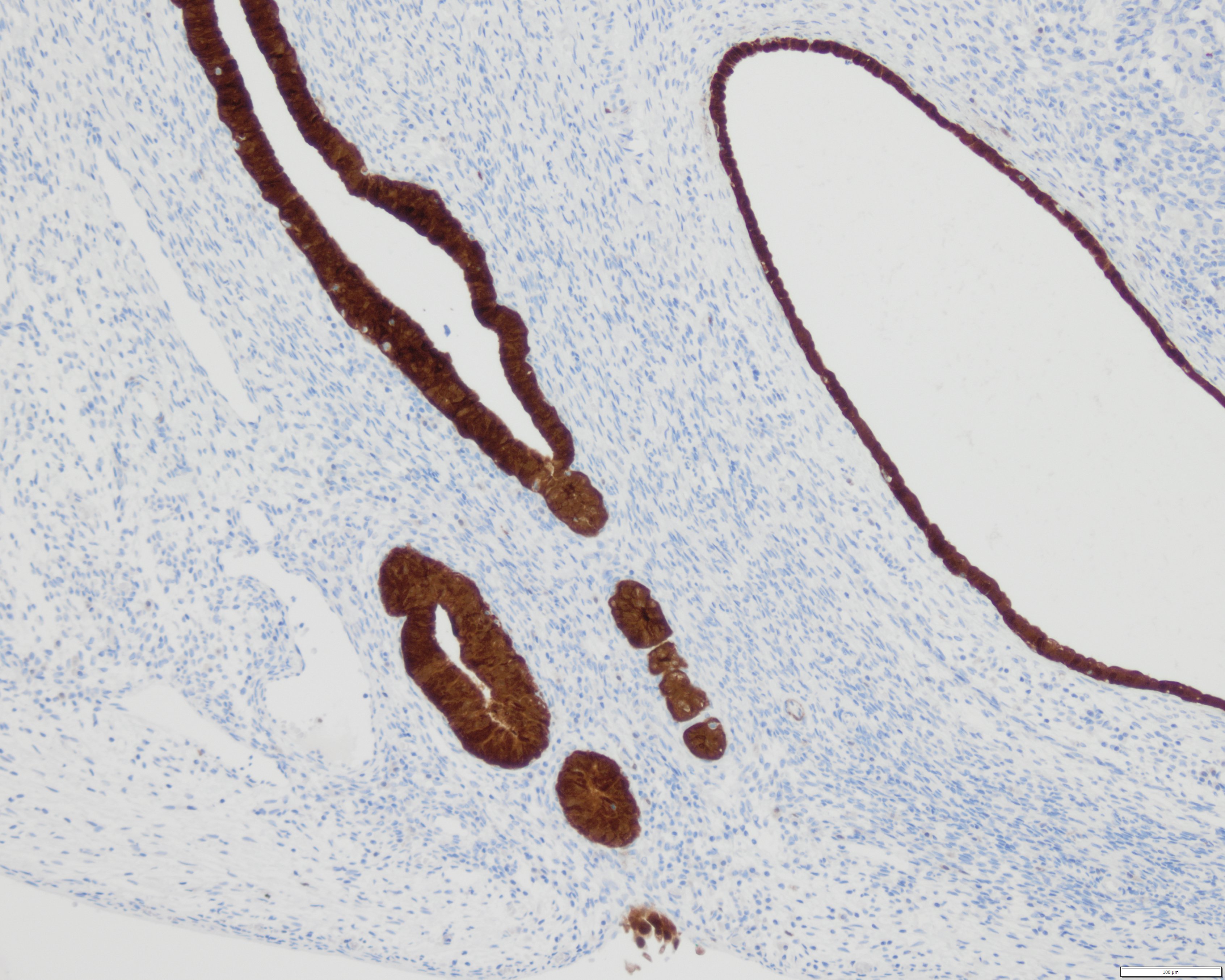 Cervix, AIS with strongly diffuse p16…
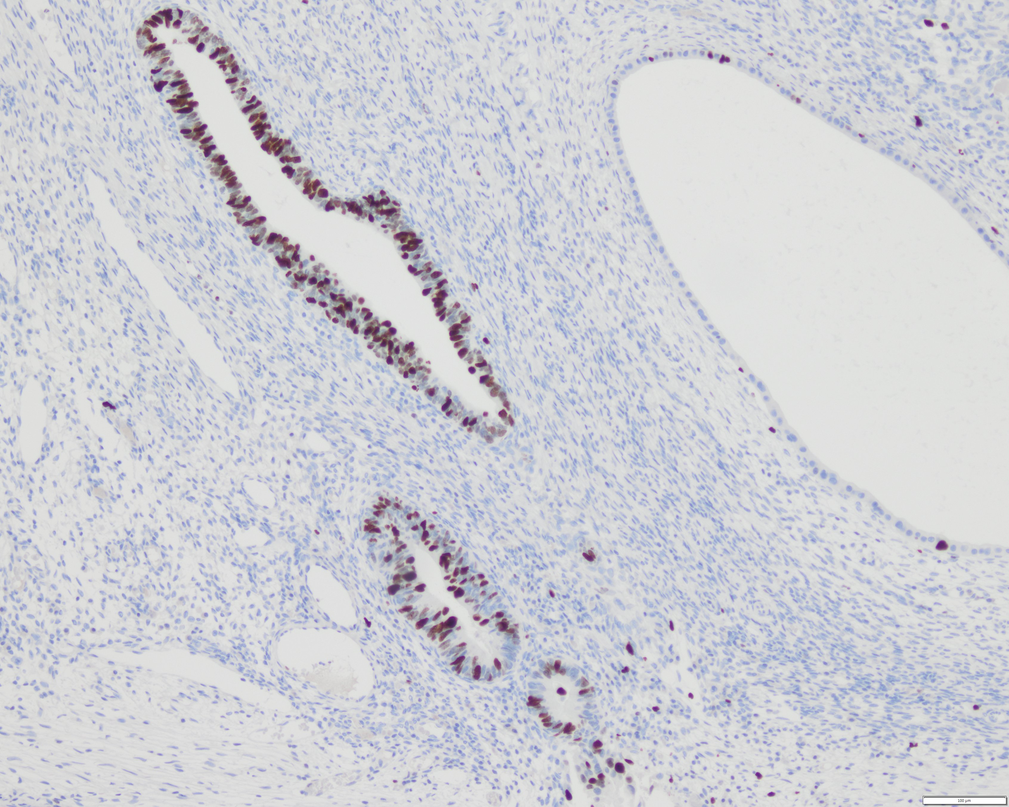 Cervix, AIS, high Ki-67 proliferation index…
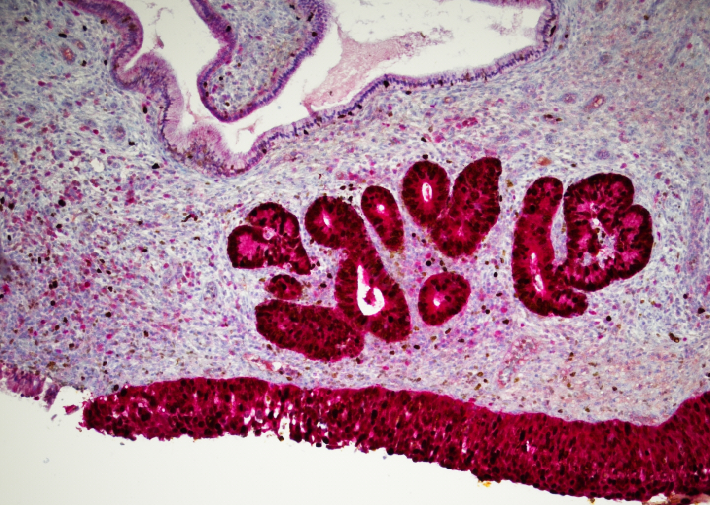 Cervix, p16 (red) and Ki-67 (brown)…
Morphology
Cytological features of HPV associated lesions (1)
Variable HPV-associated changes depending on site, infection phase, and lesion grade. Koilocytic changes are mainly seen in productive infection; high-grade dysplasia often dominated by nuclear atypia, with classical koilocytes possibly absent. Cytological features are mainly described for the cervix; anal and vulvar samples may show somewhat different morphology.
Low-grade squamous intraepithelial lesion (LSIL):
- Superficial and intermediate cells with nuclear enlargement (up to 1/3 of cell size and > 2,5 up to 3 times the size of an intermediate cell nucleus) and discrete, irregular nuclear form and contour
- Koilocytic changes - mature squamous cells with sharply demarcated perinuclear halos, a dense peripheral cytoplasmic rim, and enlarged hyperchromatic nuclei with partly irregular nuclear membranes, coarse to irregularly distributed chromatin, and occasional bi- or multinucleation
High-grade squamous intraepithelial lesion (HSIL):
- Atypical basal and parabasal cells, partly arranged in 3D clusters and syncytial aggregates
- High N:C ratio with barely visible cytoplasm
- Hyperchromatic nuclei with irregular form, contour, and grooves
- Thickened nuclear contour with coarse chromatin
Squamous cell carcinoma (SCC):
- Keratinizing vs. non-keratinizing SCC
- Keratinizing SCC with severely atypical keratinizing cells, nuclear pleomorphism, hyperchromatic nuclei, and some pyknotic nuclei; variable cell forms (e.g., spindly, tadpole-like); malignant horn pearls can be seen
- Non-keratinizing SCC usually with tumor diathesis, relatively monomorphic round cells with high N:C ratio; enlarged nuclei with coarsely clumped chromatin, moderate pleomorphism, and thickened, irregular nuclear contours; often "naked" nuclei visible; usually single cells or in syncytial aggregates
Adenocarcinoma in situ (cervix):
- Atypical glandular cells in groups/sheets, occasionally single cells
- Enlarged, hyperchromatic, stratified, and palisading nuclei ("chrysanthemum-like", "feathering")
- High N:C ratio
Adenocarcinoma (cervix):
- Usually tumor diathesis
- Syncytial or papillary aggregates, rosettes, and 3D clusters
- Usually round to oval, enlarged hyperchromatic nuclei with high N:C ratio, coarse chromatin, macronucleoli
- Vacuolated cytoplasm can be seen
- In the background of AIS and/or HSIL features
Histological features of HPV associated lesions (2)
LSIL:
- Koilocytic changes in superficial layers, consisting of nuclear enlargement and perinuclear clearing
- Loss of polarisation in the lower half of the epithelium, preserved cell maturation in the upper half of the epithelium
- Condyloma (skin): exophytic lesion with marked acanthosis of the squamous epithelium and papillary or verrucous architecture, fibrovascular cores, parakeratosis, hyperkeratosis; may show koilocytes
- Often p16 negative
HSIL (moderate to severe dysplasia):
- Loss of cell maturation affecting at least two-thirds (moderate) to full thickness (severe) of the epithelium
- High N:C ratio and nuclear abnormalities consisting of irregular nuclear contours and hyperchromasia
- Frequent mitoses, even in the upper half of the epithelium, and atypical mitoses can be present
- Strong block-type staining with p16
HPV-associated SCC:
- Several patterns: keratinizing, non-keratinizing, basaloid, warty (condylomatous), papillary, and lymphoepithelioma-like SCC
- Infiltrative, irregularly sized and shaped nests, anastomosing cords or solid sheets
- Nuclear pleomorphism, frequent mitoses, and individual cell keratinization
- Early stromal invasion/microinvasion associated with paradoxical maturation, showing increased epithelial cell eosinophilia, nests with ragged contours, and stromal loosening
- Strong block-type staining with p16
Adenocarcinoma in situ (cervix):
- Abrupt change from normal to atypical endocervical epithelium of the surface and individual crypts
- Enlarged, hyperchromatic, pseudostratified nuclei with frequent apical mitotic figures
- Variable loss of mucin
- Often occurs alongside HSIL
- Strongly p16 positive with a high proliferation index (Ki67)
HPV-associated Adenocarcinoma (cervix):
- Can have non-destructive (AIS-like, Silva pattern A) or destructive (Silva pattern B and C) growth patterns
- Histological subtypes: usual type (papillary, villoglandular, micropapillary) and mucinous type (mucinous NOS, intestinal, signet-ring cell, stratified mucin-producing)
- Enlarged, hyperchromatic, elongated nuclei with frequent apical mitotic figures and karyorrhexis
- Strongly p16 positive with a high proliferation index (Ki67) and absent or focally weak ER/PR expression, wildtype p53 (CAVE: rare p16-negative HPV-associated Adenocarcinoma)
HPV and papillomatosis of the respiratory tract
- Koilocytes
- Metaplasia without but also with severe atypia
- Keratinizing cells
- Spindle-shaped cells, some arranged in dense clusters
- Papillary cell clusters
- In case of transition to squamous cell carcinoma (SCC), its typical morphology will be seen, malignant transformation is rare and occurs in patients with known pulmonary recurrent respiratory papillomatosis (3)
Clinical notes
Transmission via direct skin-to-skin contact (genital area) and via vaginal, anal, or oral sex. Co-infection with multiple HPV types is possible. HPV-associated malignancies include cervix, vagina, vulva, anus, penis, and oropharynx.
HPV-associated squamous epithelial papilloma and papillomatosis are rare, mainly pediatric conditions with frequent recurrences. Laryngeal papillomatosis is most commonly associated with HPV types 6 and 11. Juvenile papillomatosis carries an increased risk of squamous cell carcinoma, which may develop on average about 15 years after the initial diagnosis.
Ancillary Testing
IHC (p16), ISH, HPV-DNA-testingAdditional Information
Clinically, the key distinction is between high-risk and low-risk HPV types.
High-risk: 16, 18, 31, 33, 35, 39, 45, 51, 52, 56, 58, 59.
Low-risk: 6, 11, 40, 43, 54, 69, 70.
The main driver of progression to invasive carcinoma is persistent infection with high-risk HPV. SCC and adenocarcinoma of the cervix uteri are HPV-associated in 90-95% and 99%, respectively (2).
HPV is seen in some SCC of the lung, but the role of the virus is not fully understood (4).
HPV vaccination reduces the risk of persistent HPV vaccine-type attributable infection and precancerous lesions and carcinomas, with the greatest benefit before sexual exposure. A prior infection does not confer reliable immunity (5).
Differential Diagnosis
References
- Nayar R, Wilbur DC (eds): The Bethesda System for Reporting Cervical Cytology, ed 3. Definitions, Criteria, and Explanatory Notes. New York, Springer, 2015.
- WHO Classification of Tumours Editorial Board. Female genital tumours. Lyon (France): International Agency for Research on Cancer; 2020. (WHO classification of tumours series, 5th ed.; vol. 4). https://publications.iarc.fr/592.
- Pai SI, Wasserman I, Ji YD, Gilman M, Hung YP, Faquin WC, Mino-Kenudson M, Muniappan A. Pulmonary manifestations of chronic HPV infection in patients with recurrent respiratory papillomatosis. Lancet Respir Med. 2022 Oct;10(10):997-1008. doi: 10.1016/S2213-2600(22)00008-X. Epub 2022 Jul 18. PMID: 35863360; PMCID: PMC10634185.
- Osorio JC, Andrade-Madrigal C, Gheit T, Corvalán AH, Aguayo F. SLC7A11 Expression Is Up-Regulated in HPV- and Tobacco-Associated Lung Cancer. Int J Mol Sci. 2024 Dec 10;25(24):13248. doi:
- Markowitz LE, Unger ER. Human papillomavirus vaccination. N Engl J Med. 2023;388(19):1790-1798. doi:10.1056/NEJMcp2108502