
- EN
- DE

Parasites
Giardia lamblia (syn. G. duodenalis, G. intestinalis)

- Organ System
- Digestive Tract
- Diagnostic Method
- Cytology, Histology

- Geographical Region
- Dry Areas,
- Islands,
- Tropical and Subtropical Region,
- Polar and Subpolar Zone,
- Temperate Zone
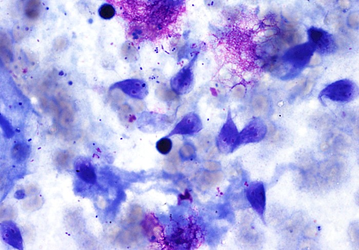
 Bile duct brushing with hemorrhagic…
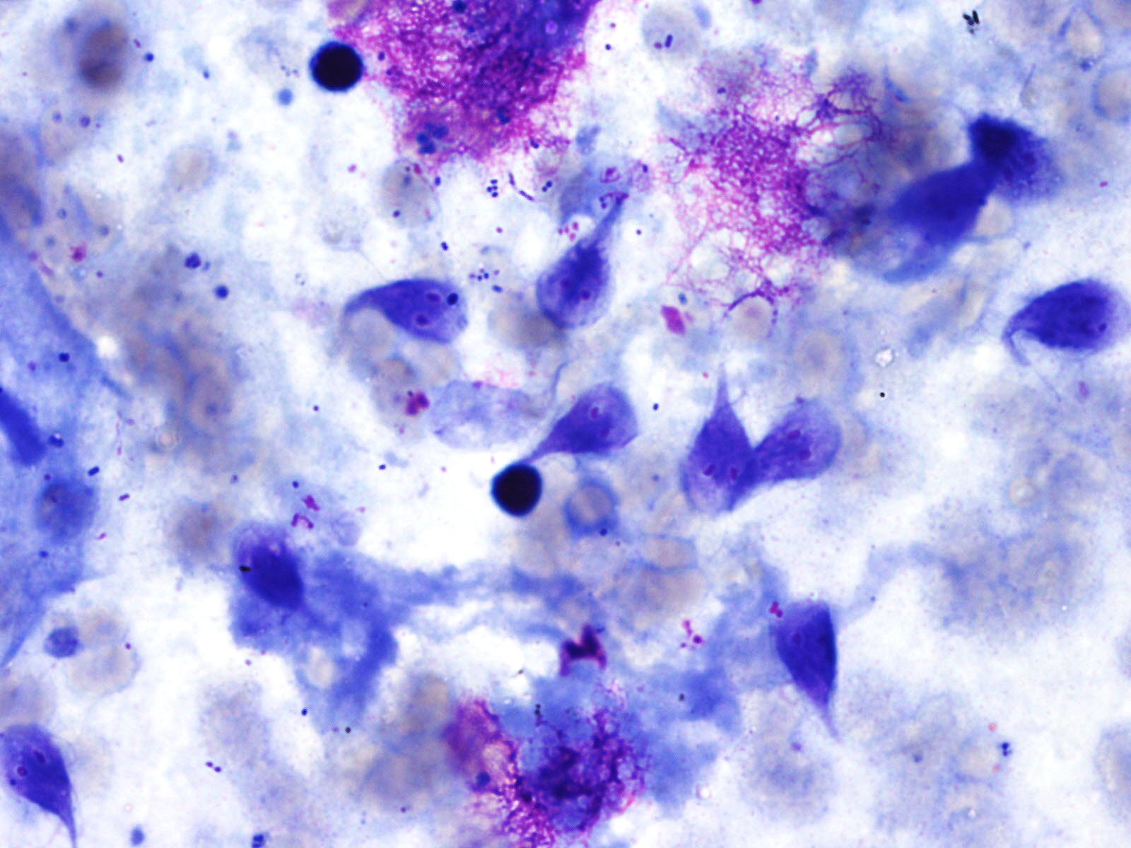
 Bile duct brushing with hemorrhagic…
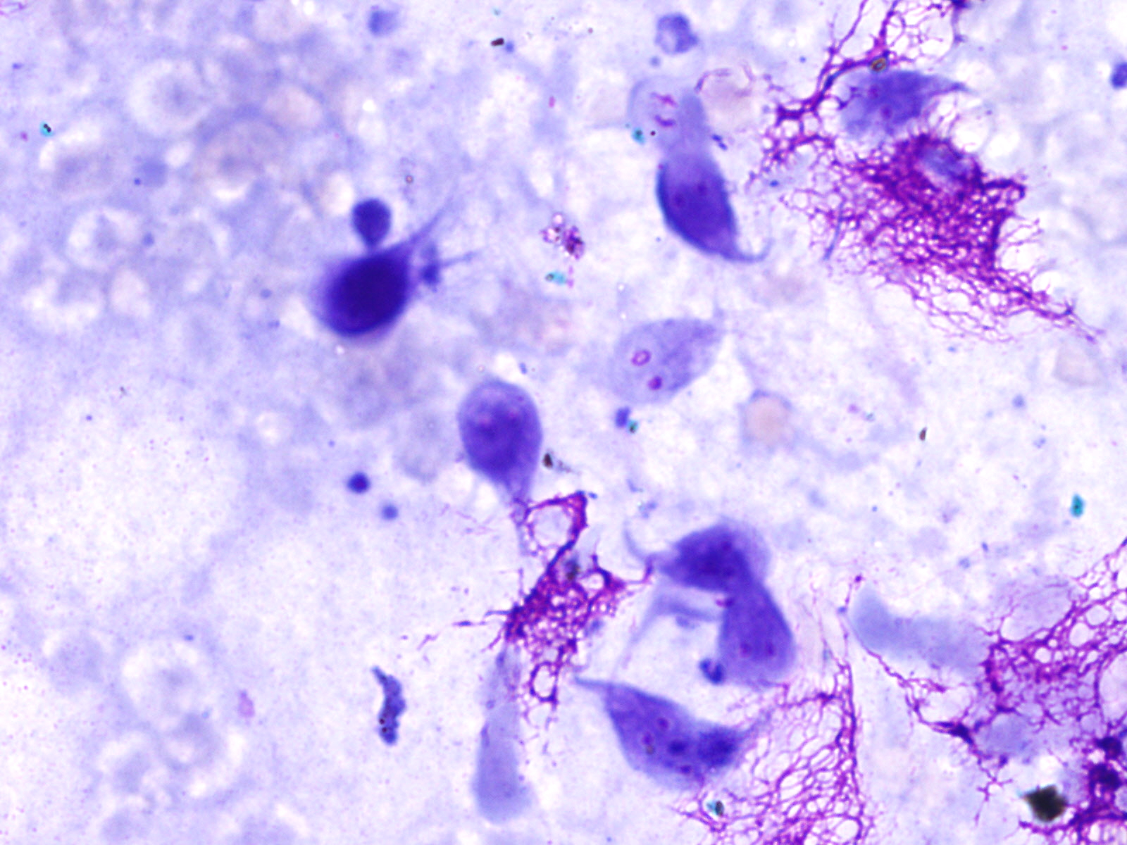
 Bile duct brushing with hemorrhagic…
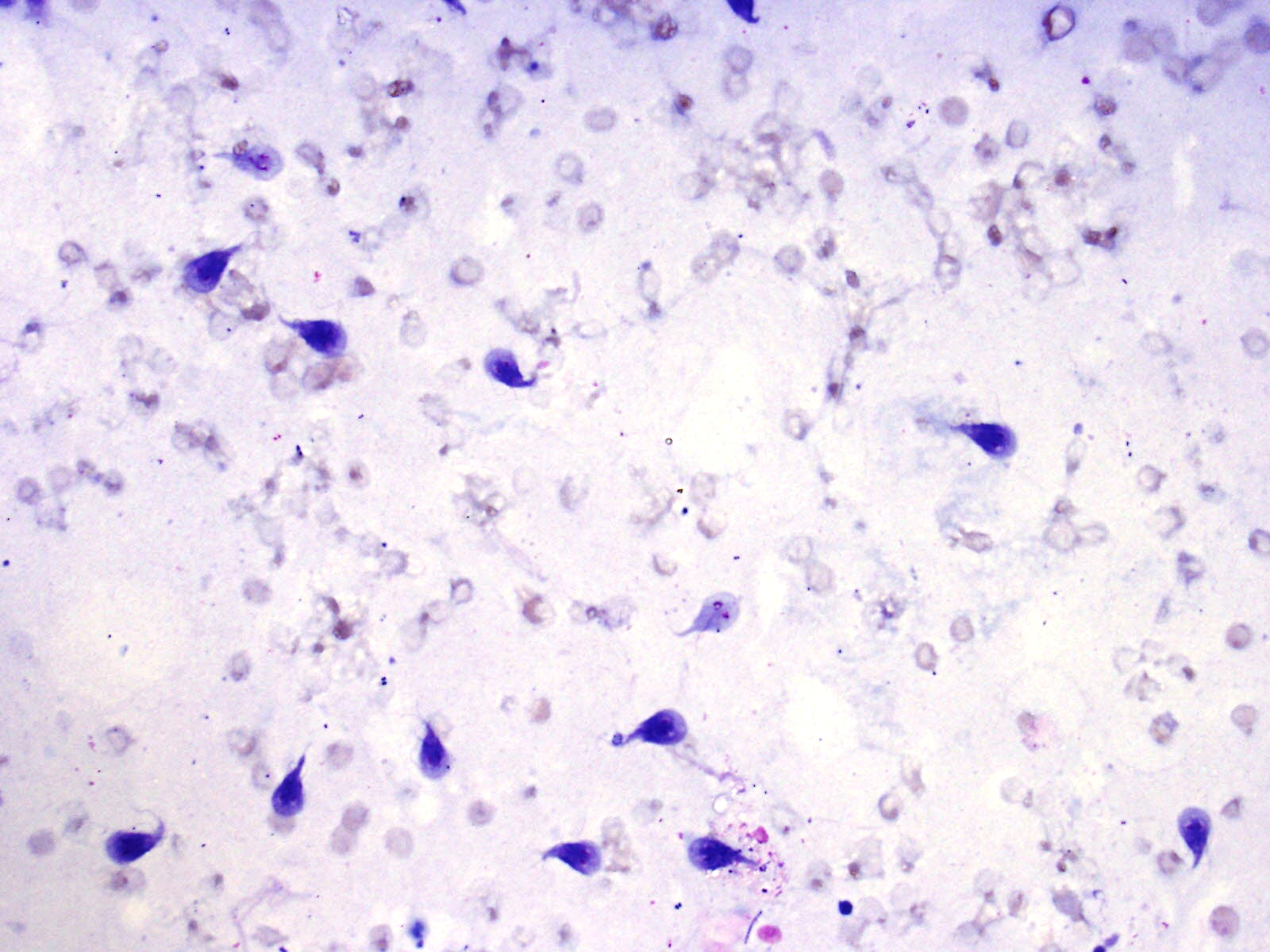
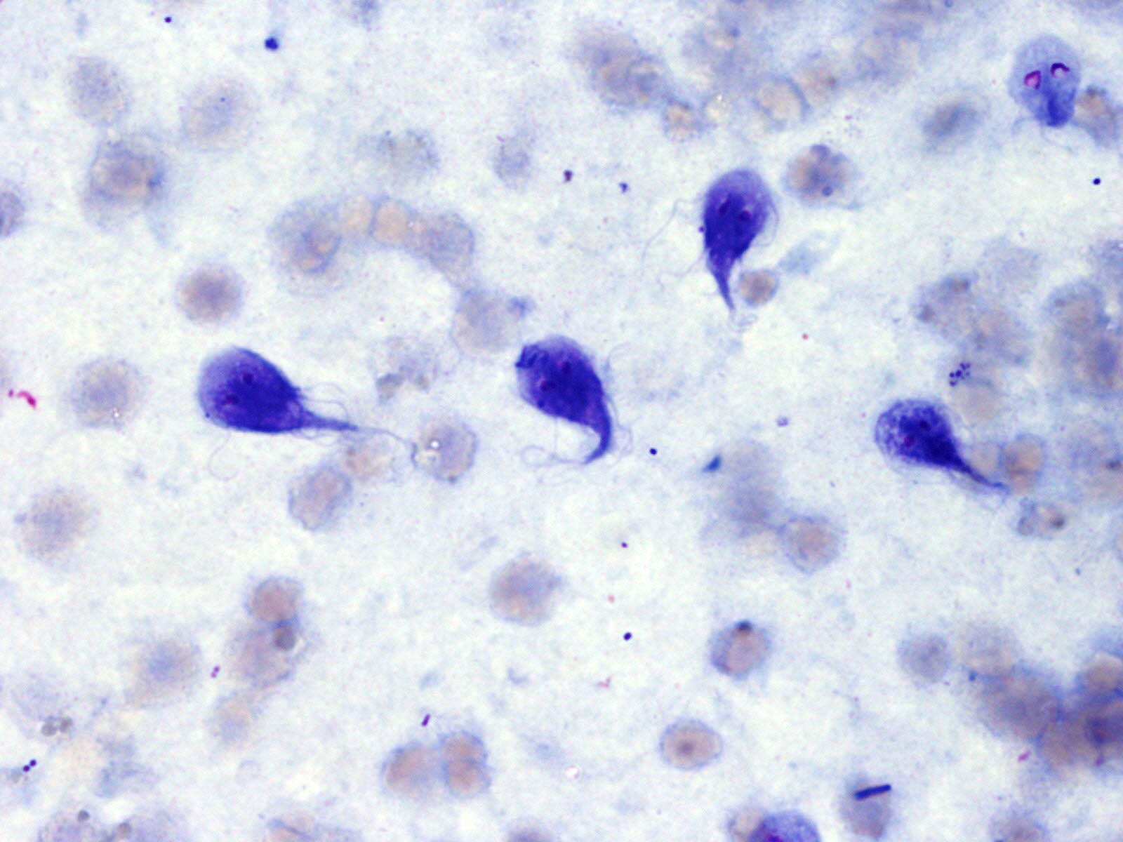 Bile duct brushing with hemorrhagic…
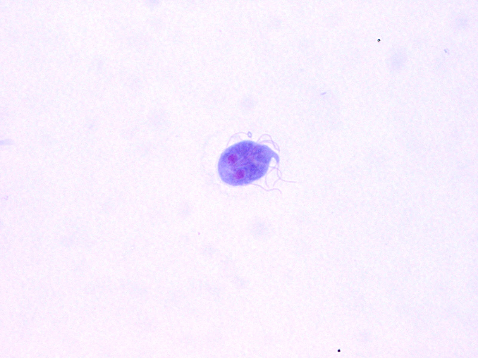 Bile duct brushing, pear- or kite-shaped…
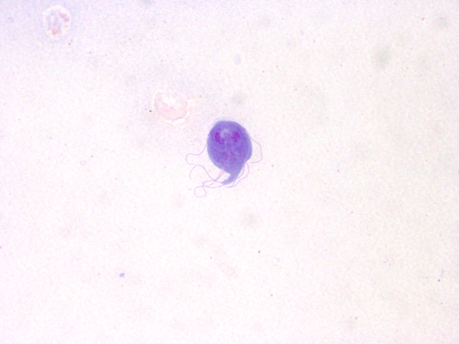 Bile duct brushing, pear- ore kite-shaped…
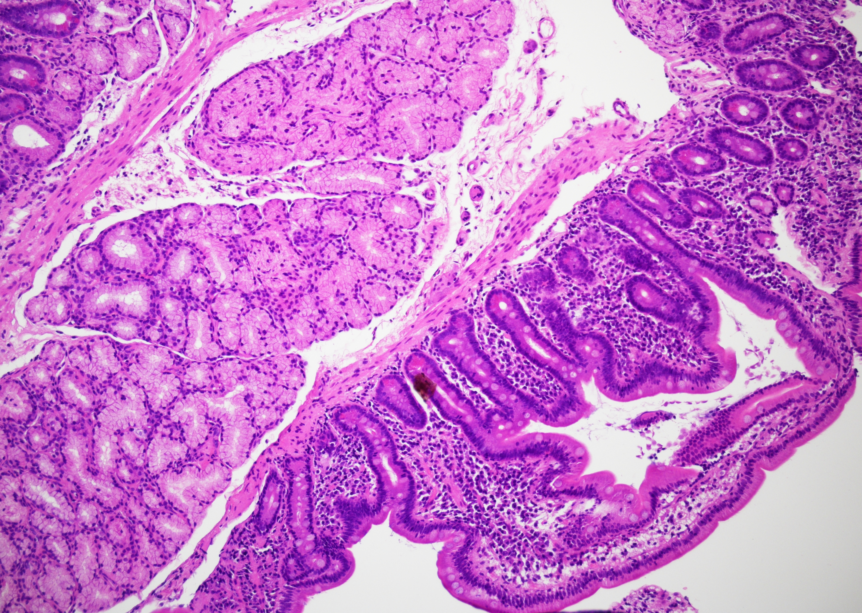 Duodenal biopsy with small pathogens near…
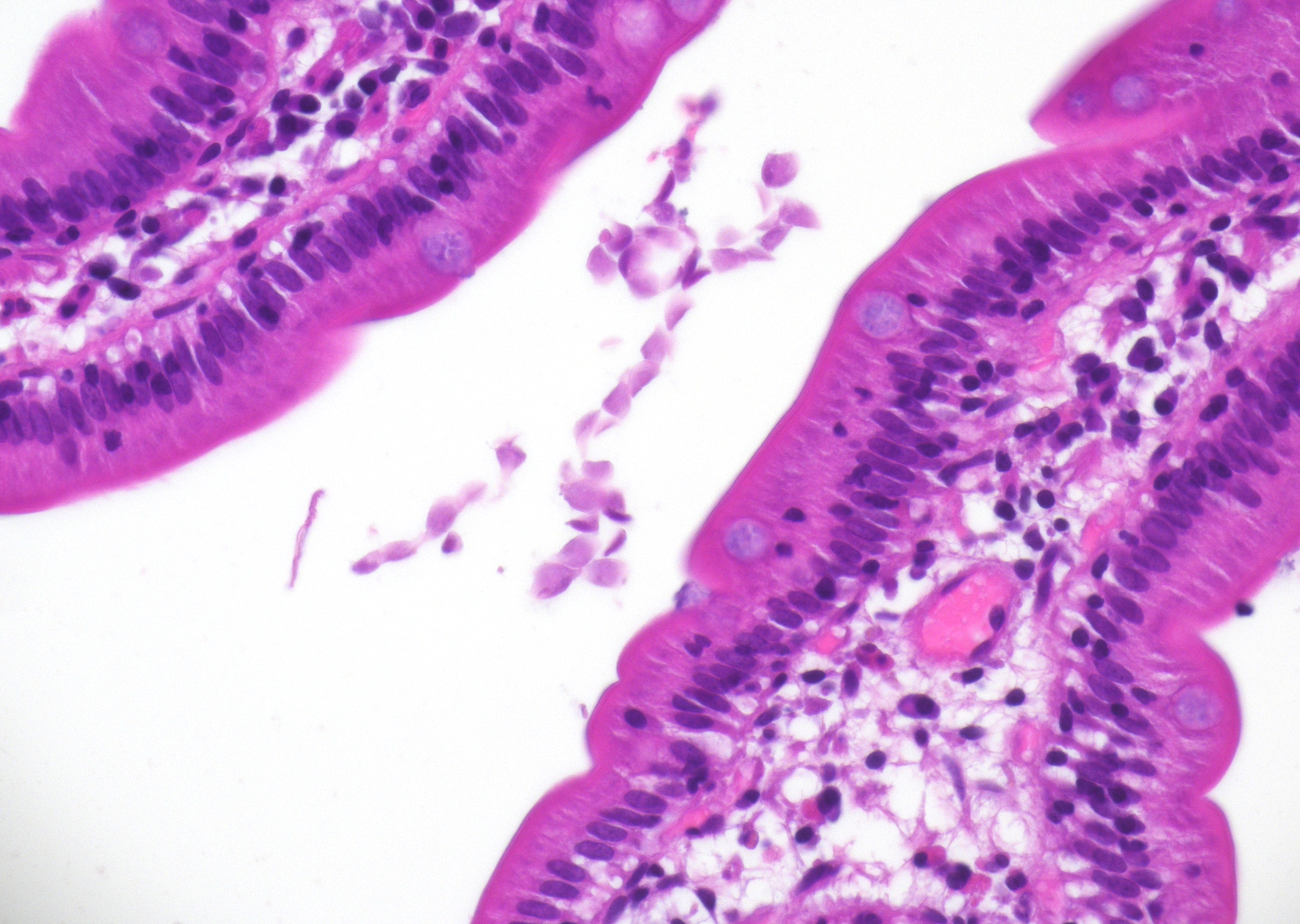 Duodenal biopsy with leaf-like pathogens…
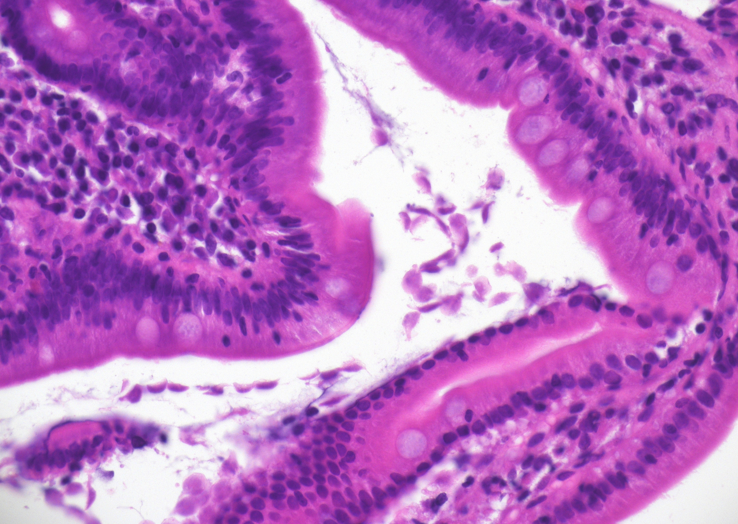 Duodenal biopsy with leaf-like pathogens…
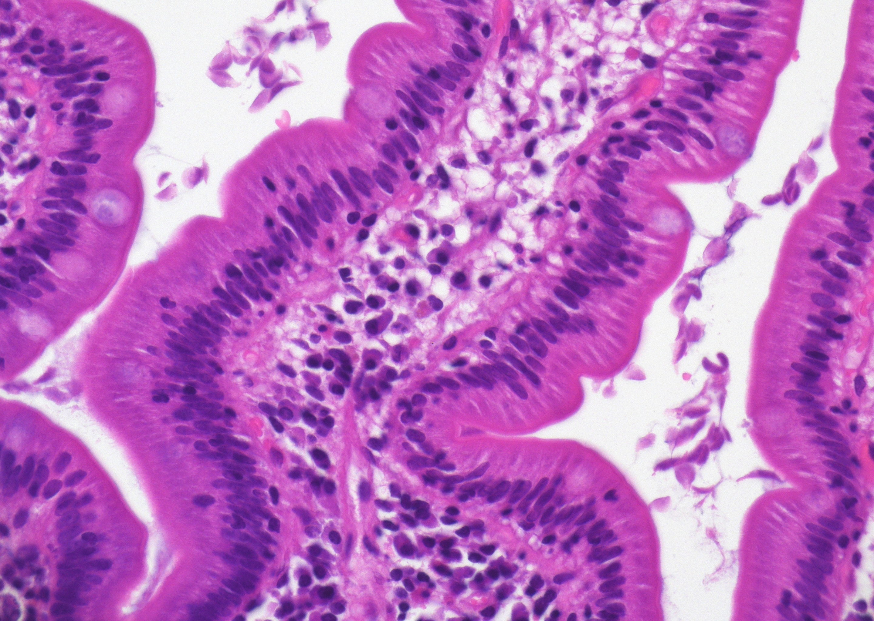 Duodenal biopsy with leaf-like pathogens…
Morphology
Cytology:
- Trophozoites 10-20 µm, pear- or kite-shaped
- Bilaterally symmetric - two nuclei with central karyosomes giving a characteristic "face-like" / "eyeball" appearance
- The anterior portion is broad and rounded, whereas the posterior extremity gradually tapers to a pointed termination
- Ventral adhesive disk, paired axonemes and median bodies, eight flagella
- Characteristic falling-leaf motility in fresh wet mounts
- Cysts 8-12 µm (up to 19 µm), oval to ellipsoid
- mature forms with 4 nuclei and visible fibrils
- thin wall, refractile
- best visualized in fresh wet prep, iodine wet mount, trichrome, or iron hematoxylin stains
Histology:
- Trophozoites adhere to the mucosal surface and between villi in the duodenum and proximal jejunum, without tissue invasion
- Duodenal histology is often normal or shows minimal changes; when present, findings may include mild to moderate villous blunting, increased intraepithelial lymphocytes, crypt hyperplasia, and eosinophilic infiltration of lamina propria
- Severe cases can mimic celiac disease
Differential Diagnosis:
- Balantidium coli
- Amebiasis
- Cryptosporidiosis
- Celiac disease
Clinical notes
Acute or chronic watery or greasy, foul-smelling diarrhea, bloating, flatulence, weight loss, malabsorption, steatorrhea; post-infectious lactose intolerance common; asymptomatic carriage occurs.
Rarely, Giardia can spread to the biliary and pancreatic ducts, leading to gallbladder, liver, and pancreatic complications (1).
Ancillary Testing
Stool antigen tests (EIA or DFA), multiplex PCR panels, stool ova and parasites (O&P) test on 2–3 specimens on different days, trichrome or iron hematoxylin stains, immunocyto-/-histochemistry: CD117 positiveAdditional Information
Transmission is fecal-oral, primarily through contaminated water; direct person-to-person and foodborne spread also occur.
Cysts are infectious immediately upon excretion and environmentally hardy, remaining viable for at least 3 months in cold water. As few as 10–100 cysts can cause infection. Cysts are moderately chlorine-tolerant; boiling (≥1 minute) and appropriate filtration (≤3–5 µm pore size) are more reliable for inactivation than routine chlorination alone (2, 3).
References
Ross, A. G. P., Olds, G. R., Cripps, A. W., Farrar, J. J., & McManus, D. P. (2013). Enteropathogens and chronic illness in returning travelers. New England Journal of Medicine, 368(19), 1817–1825. https://doi.org/10.1056/NEJMra1207777
- Pyzocha N, Cuda A. Common Intestinal Parasites. Am Fam Physician. 2023 Nov;108(5):487-493. PMID: 37983700.
- Backer, H. D., & Hill, V. (2026). Water disinfection for travelers. In CDC yellow book: Health information for international travel (2026 ed.). Centers for Disease Control and Prevention. https://wwwnc.cdc.gov/travel/yellowbook
- NIH. (2025). Guidelines for the prevention and treatment of opportunistic infections in children with and exposed to HIV. https://clinicalinfo.hiv.gov/en/guidelines/pediatric-opportunistic-infection