- EN
- DE

Parasites
Enterobius vermicularis (Pinworm)

- Organ System
- Digestive Tract, Genitourinary Tract
- Diagnostic Method
- Cytology, Histology

- Geographical Region
- Dry Areas,
- Islands,
- Tropical and Subtropical Region,
- Polar and Subpolar Zone,
- Temperate Zone
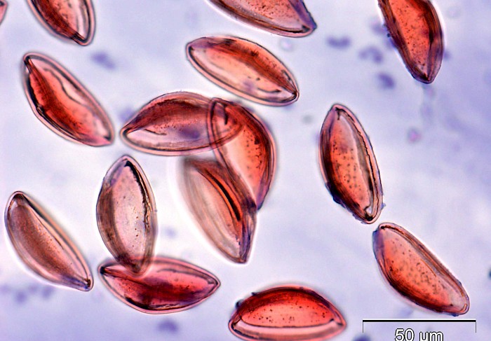
 Perianal tape test, transparent oval ova…
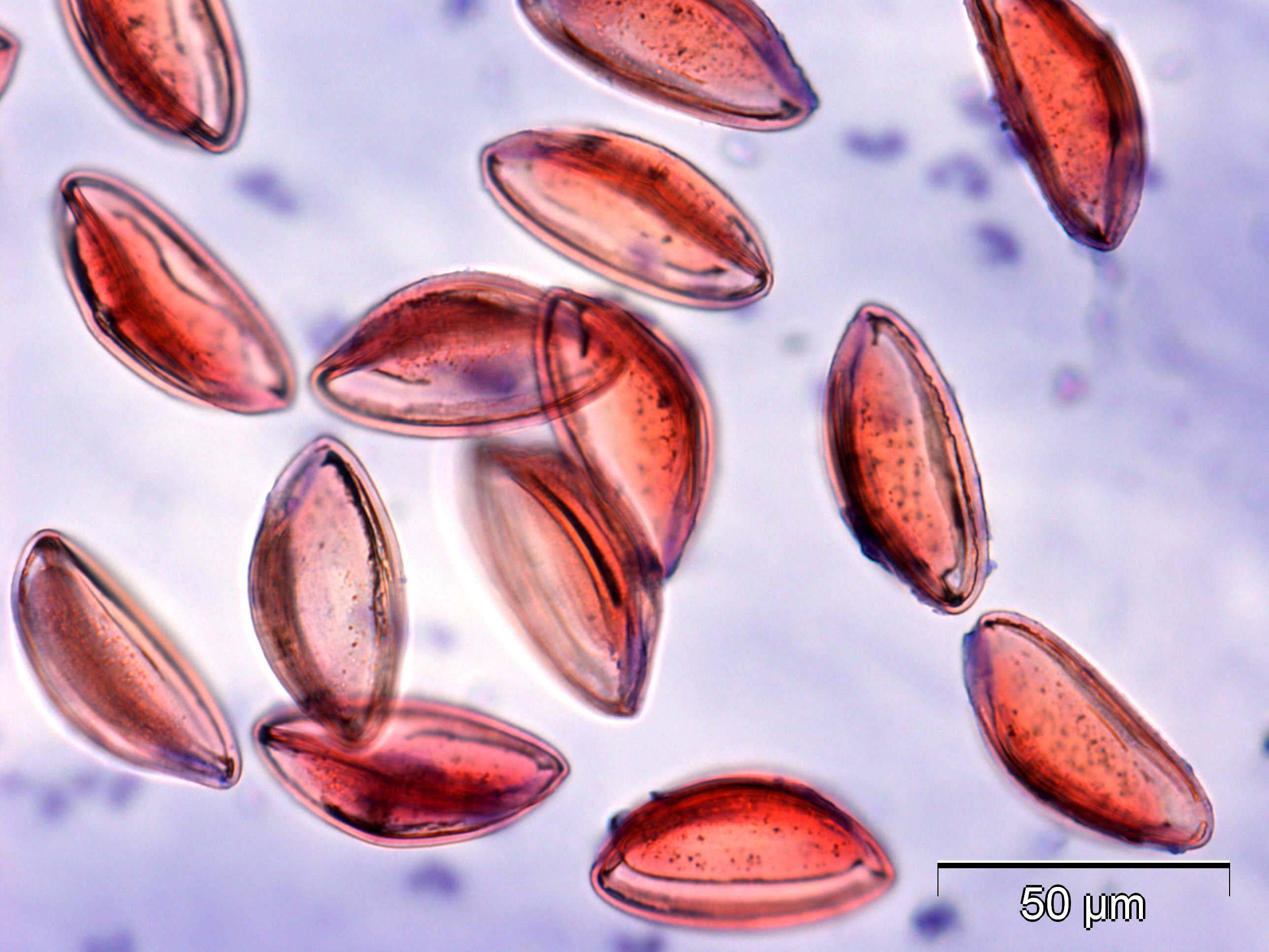
 Perianal tape test, transparent oval ova…
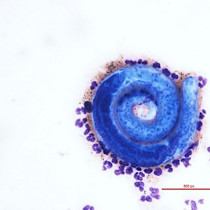
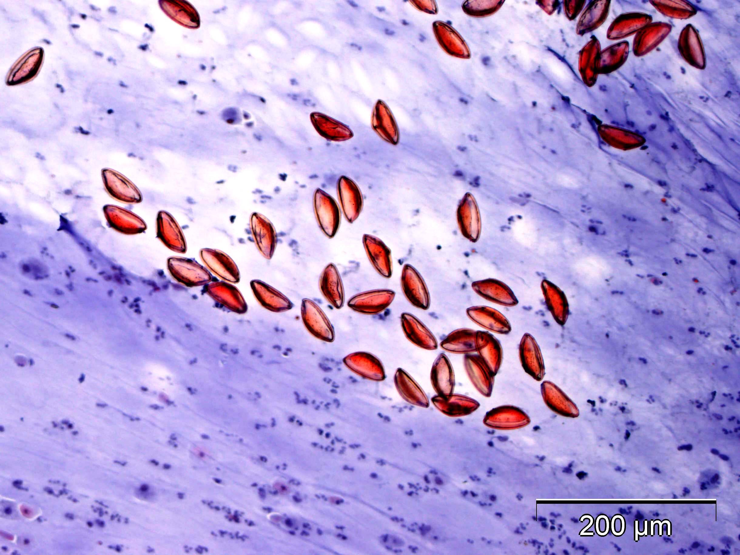
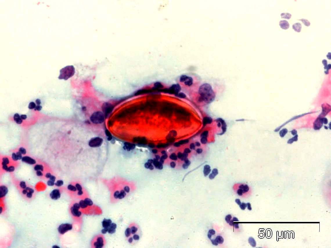 Cervical Pap smear, ovum surrounded by…
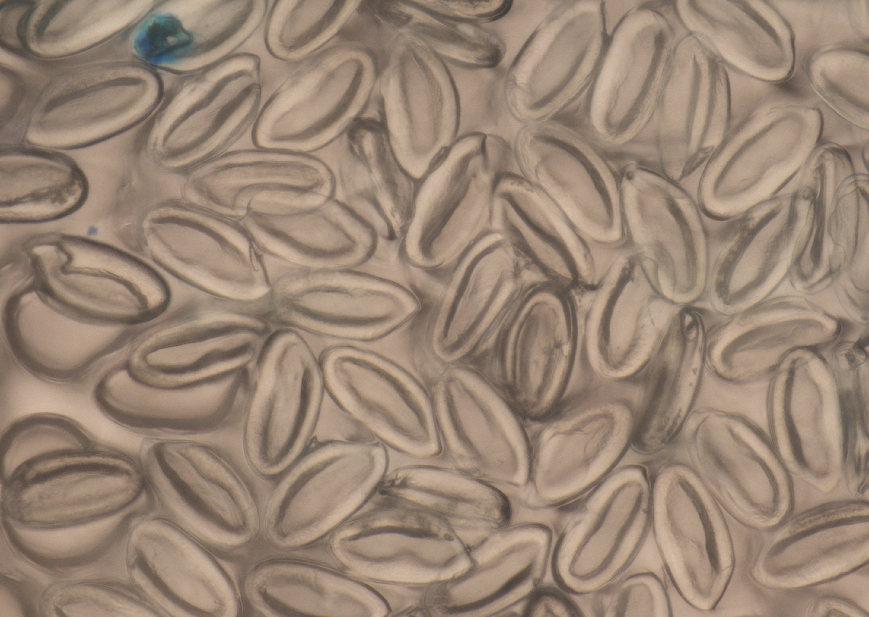 Perianal tape test, ova in the uterus of a…
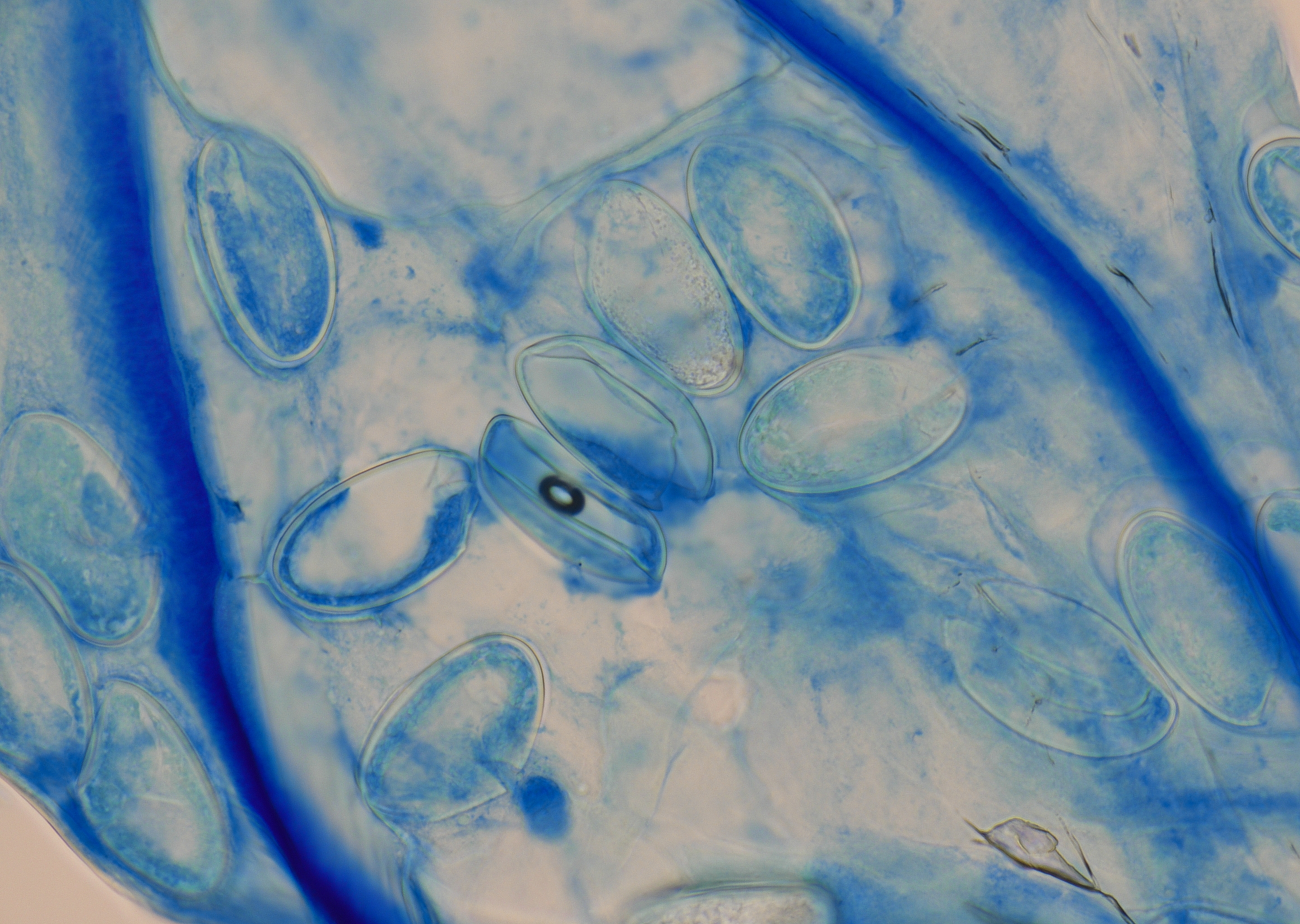 Perianal tape test, ova in gravid uterus of…
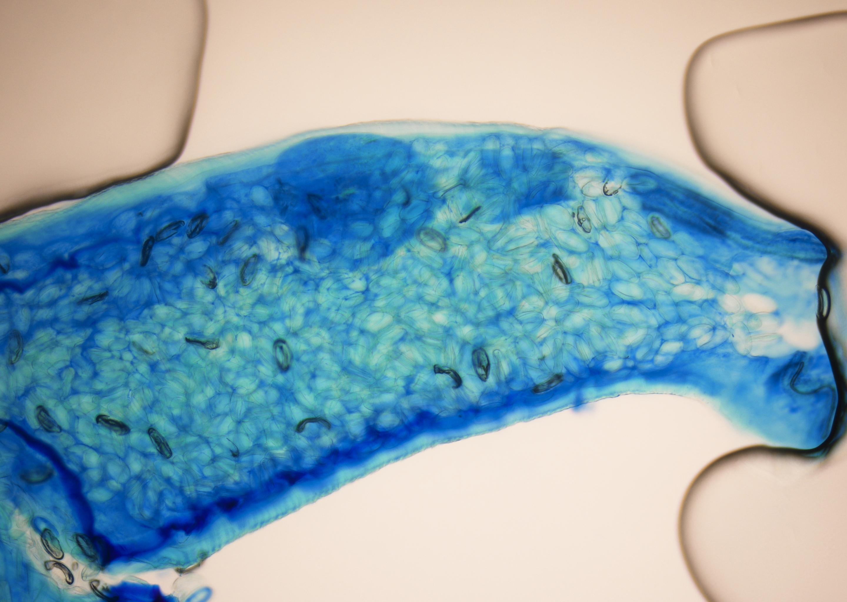 Perianal tape test, ova in gravid uterus of…
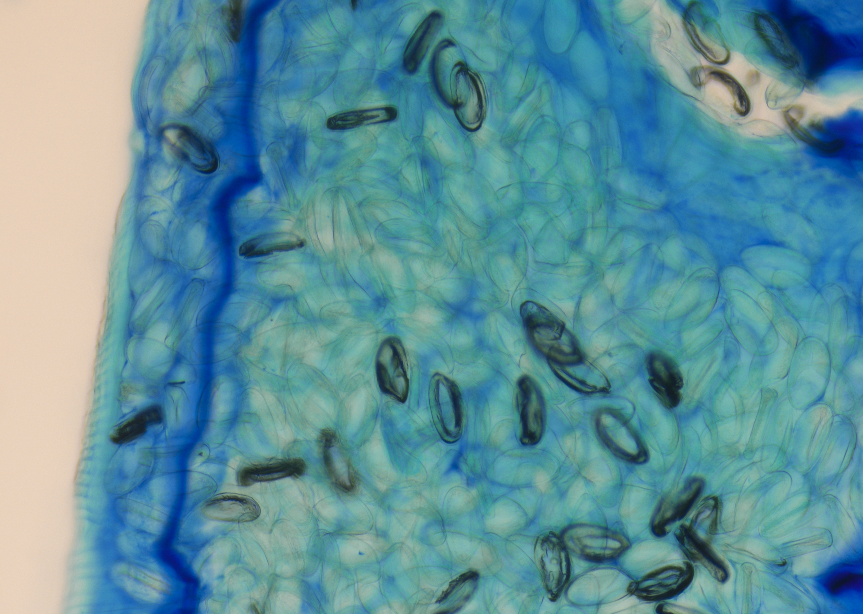 Perianal tape test, ova in gravid uterus of…
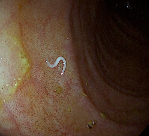 Visible adult worm during colonoscopy
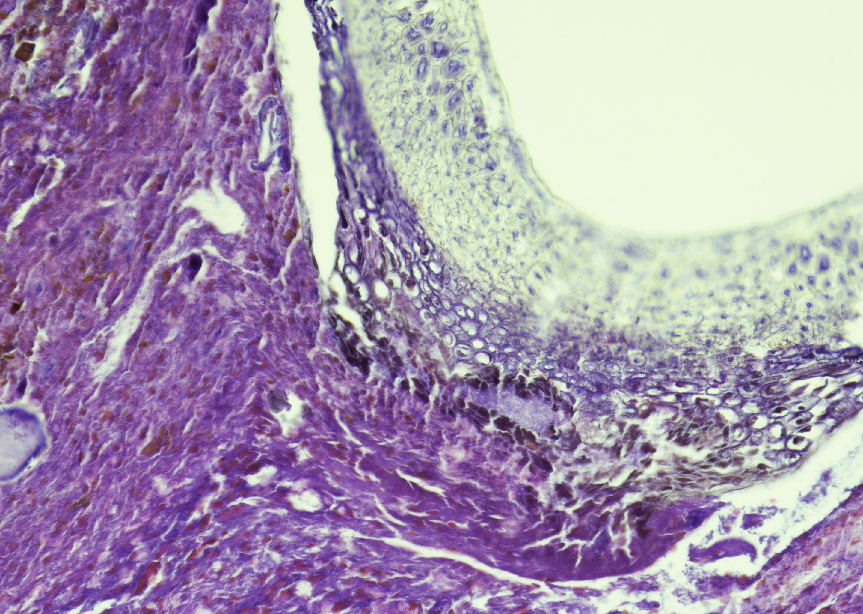
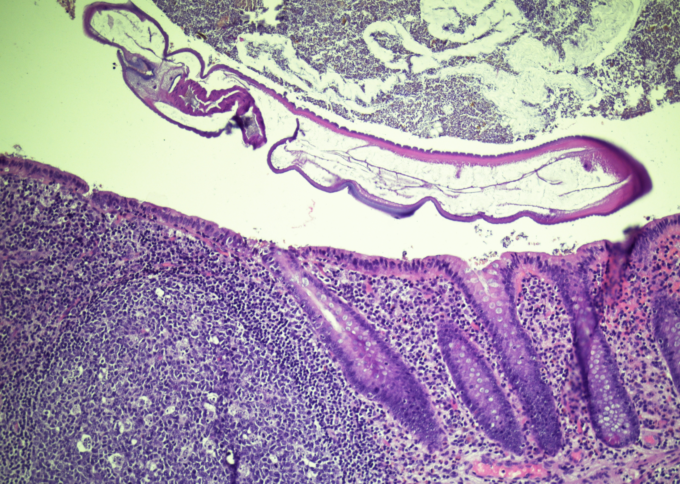 Appendectomy, part of a longitudinal section…
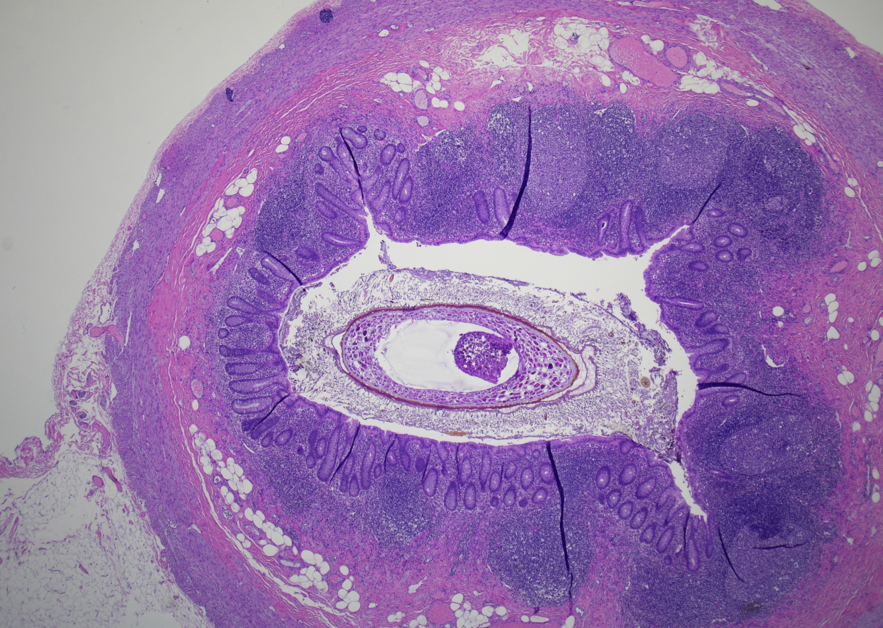
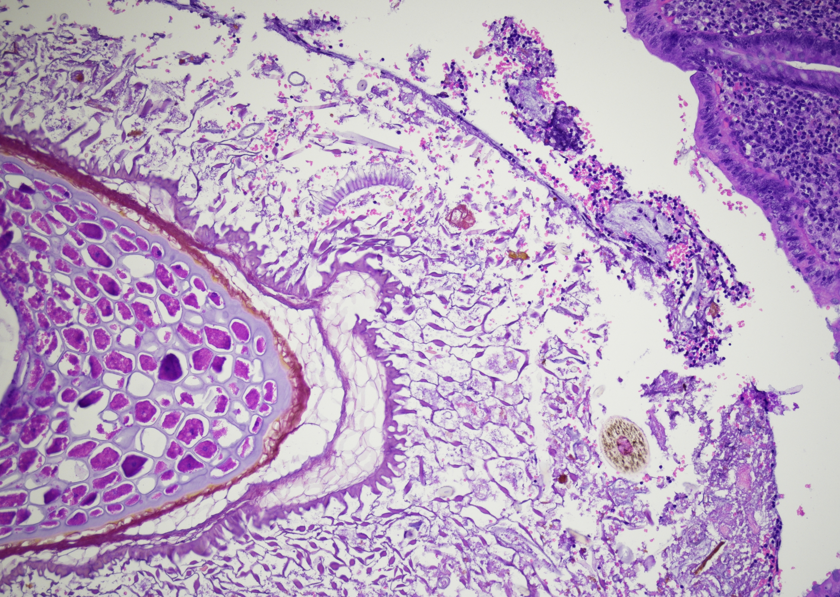
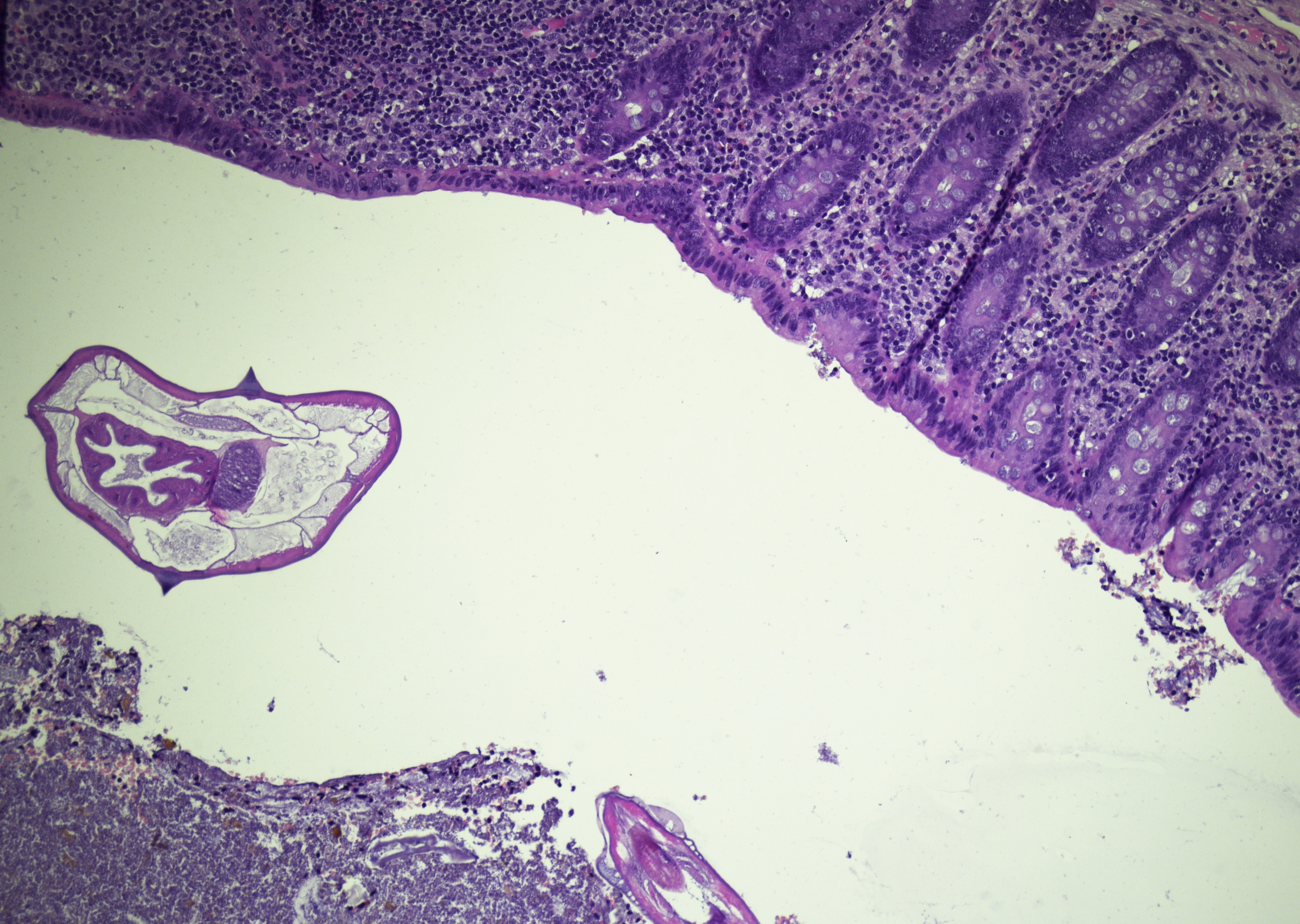 Appendectomy, transverse section of E.
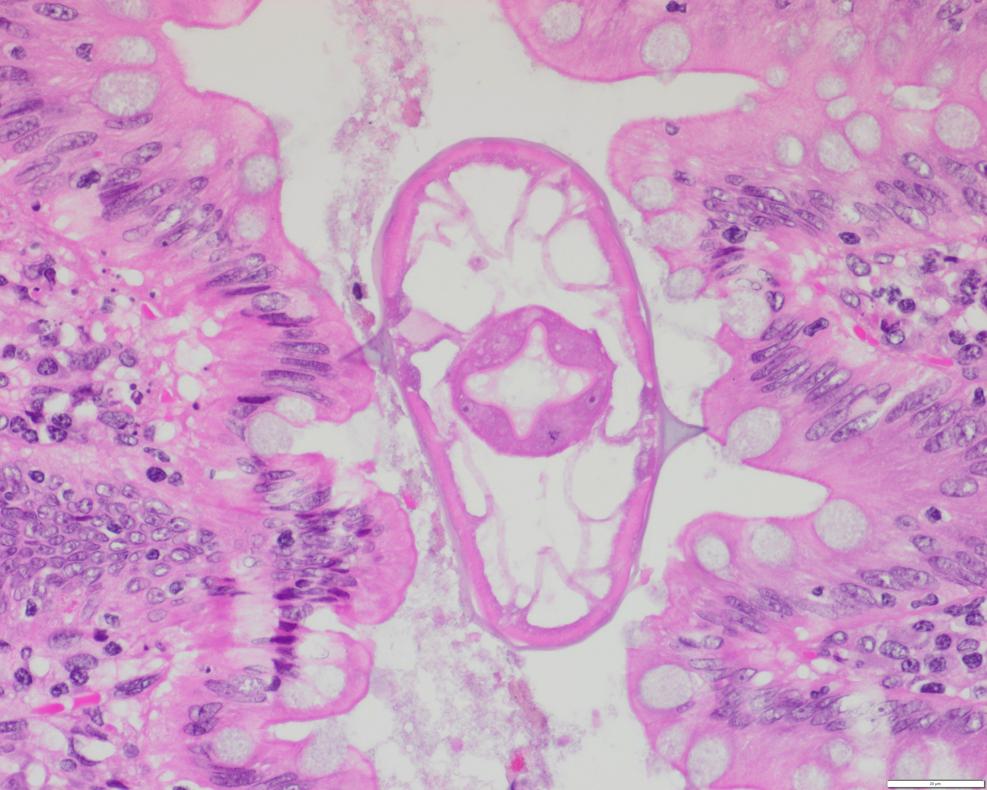 Appendectomy, transverse section of E.
Morphology
Cytology:
- Perianal adhesive tape test / cellulose tape preparation:
- Eggs of Enterobius vermicularis
- Transparent
- Oval to plano-convex, slightly flattened on one side, with a double-contoured refractile shell
- Approximately 50-60 × 20-30 µm
- Containing a coarsely granular embryo or curved larva
- Eggs of Enterobius vermicularis
- Rare finding in cervicovaginal Pap smears:
- Eggs, embryos, or larvae
- Often in an acute inflammatory background
- Contamination from the perineal region possible, especially in a clean background
Histology:
- Usually incidental finding, most commonly in the appendix or large bowel:
- Adult worms white to ivory-white
- Female worms 8-13 mm long, with pointed tail
- Male worms smaller, with blunt posterior end
- Histological features:
- Cuticle with prominent lateral alae
- Platymyarian musculature
- Gastrointestinal tract
- Reproductive structures
- Ovaries, gravid uterus with eggs
- Male reproductive tubules with spermatozoa
Differential Diagnosis:
- Strongyloidiasis - usually embedded within epithelium, small nematode
- Ancylostomiasis/Necatoriasis - attaches to small intestine, eggs are thin-shelled and colorless
- Trichuriasis - adult worm longer than E. vermicularis, embedded into mucosa of colon, eggs are barrel-shaped and possess polar "plugs" at each end
- Ascariasis - large adult worms reside in small intestine, eggs are ovoid
Clinical notes
Enterobius vermicularis is a common human parasite found worldwide, but most often in temperate regions. Infections are most common among school- and preschool-aged children and in crowded settings.
Infections caused by E. vermicularis are usually asymptomatic, but some patients may experience non-specific symptoms that can resemble appendicitis, such as abdominal pain, loss of appetite, and irritability. In rare instances, it can lead to appendicitis due to obstruction.
The most common associated symptom is nocturnal perianal pruritus.
Infection occurs after ingesting the eggs. Transmission can happen directly through self-inoculation of eggs via anus-to-mouth transfer with contaminated hands, or indirectly through contaminated surfaces or exposure to eggs in the environment.
After ingestion, the larvae hatch from the eggs in the small intestine, where they develop into adults (three to four weeks) and reside in the colon, primarily in the cecum. Mostly at night, the gravid female worms migrate to the anal area and deposit their eggs on the perianal folds.
Extraintestinal sites of disease are rare.
Gynecologic disease can occur, most likely due to ectopic migration of gravid female worms.
Ancillary Testing
Perianal tape test (eggs)Additional Information
Humans are considered the only natural host for E. vermicularis.