
- EN
- DE

Parasites
Echinococcus Species

- Organ System
- Digestive Tract, Respiratory Tract, Serous Cavities
- Diagnostic Method
- Cytology, Histology

- Geographical Region
- Dry Areas,
- Islands,
- Tropical and Subtropical Region,
- Polar and Subpolar Zone,
- Temperate Zone
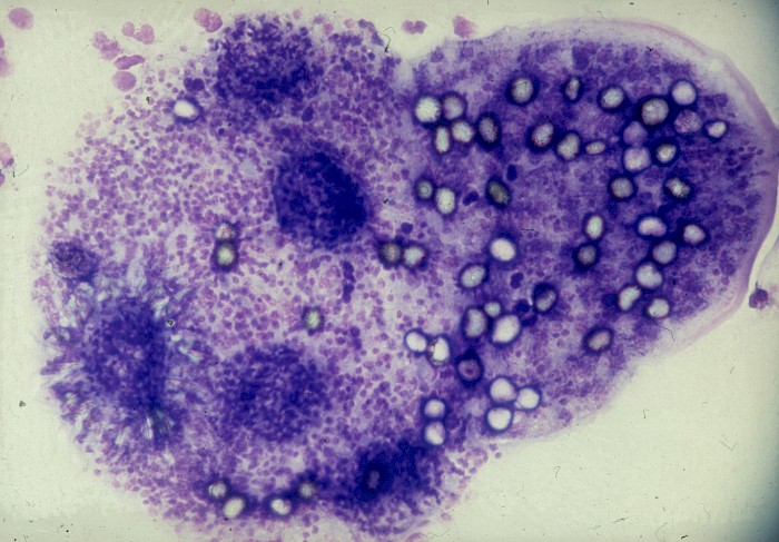
 Lung, biopsy, protoscolex with rostral hooks…
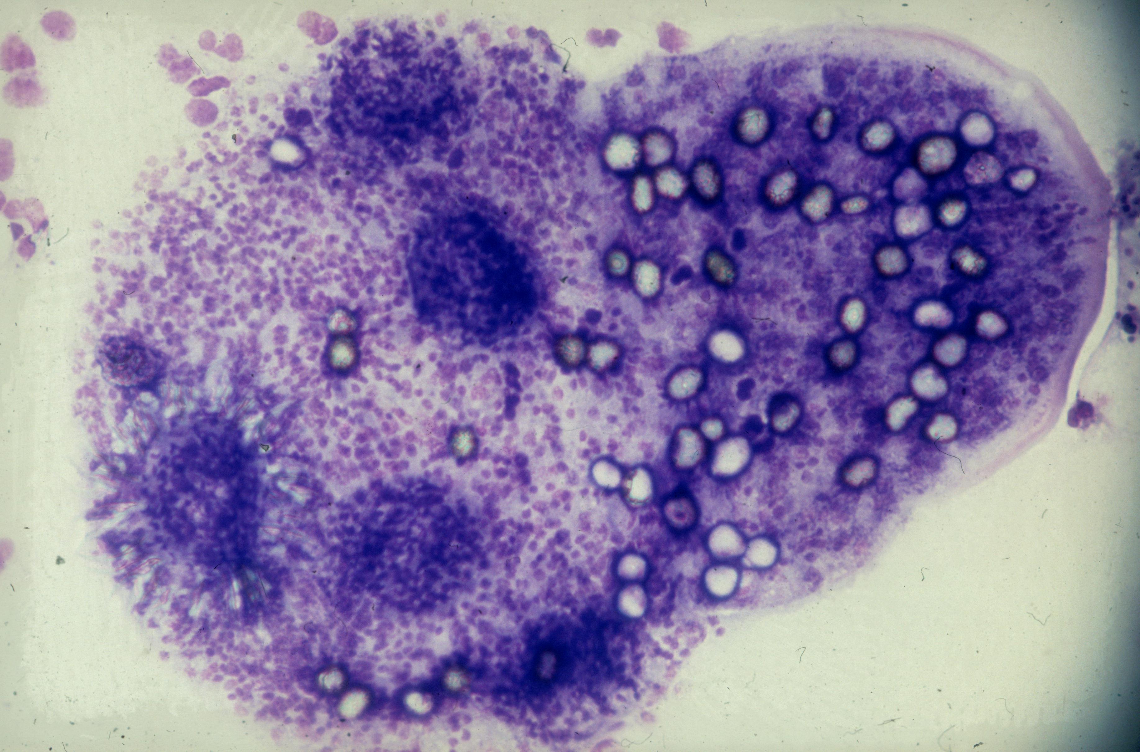
 Lung, biopsy, protoscolex with rostral hooks…
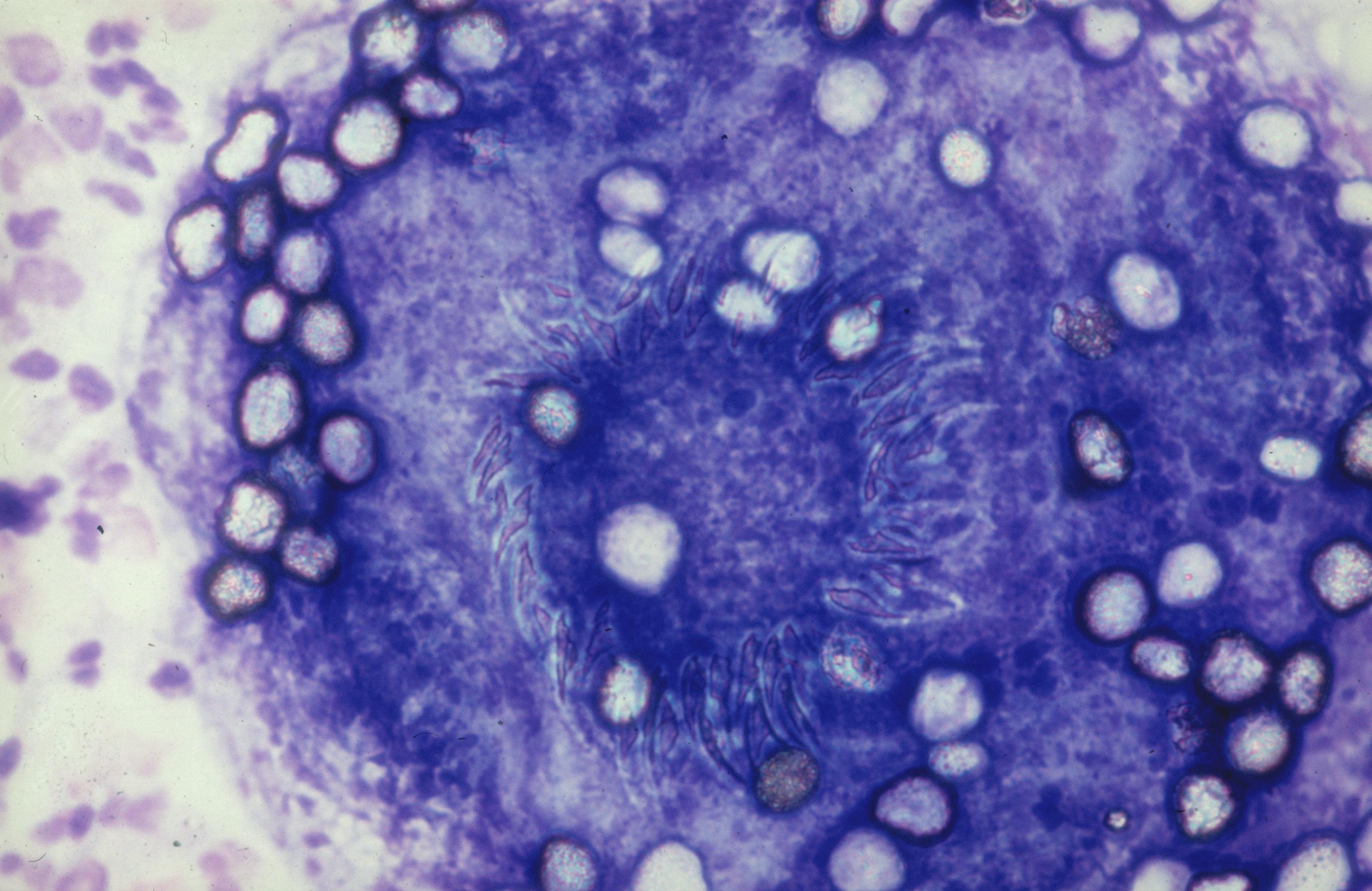
 Lung, biopsy, higher magnification of…
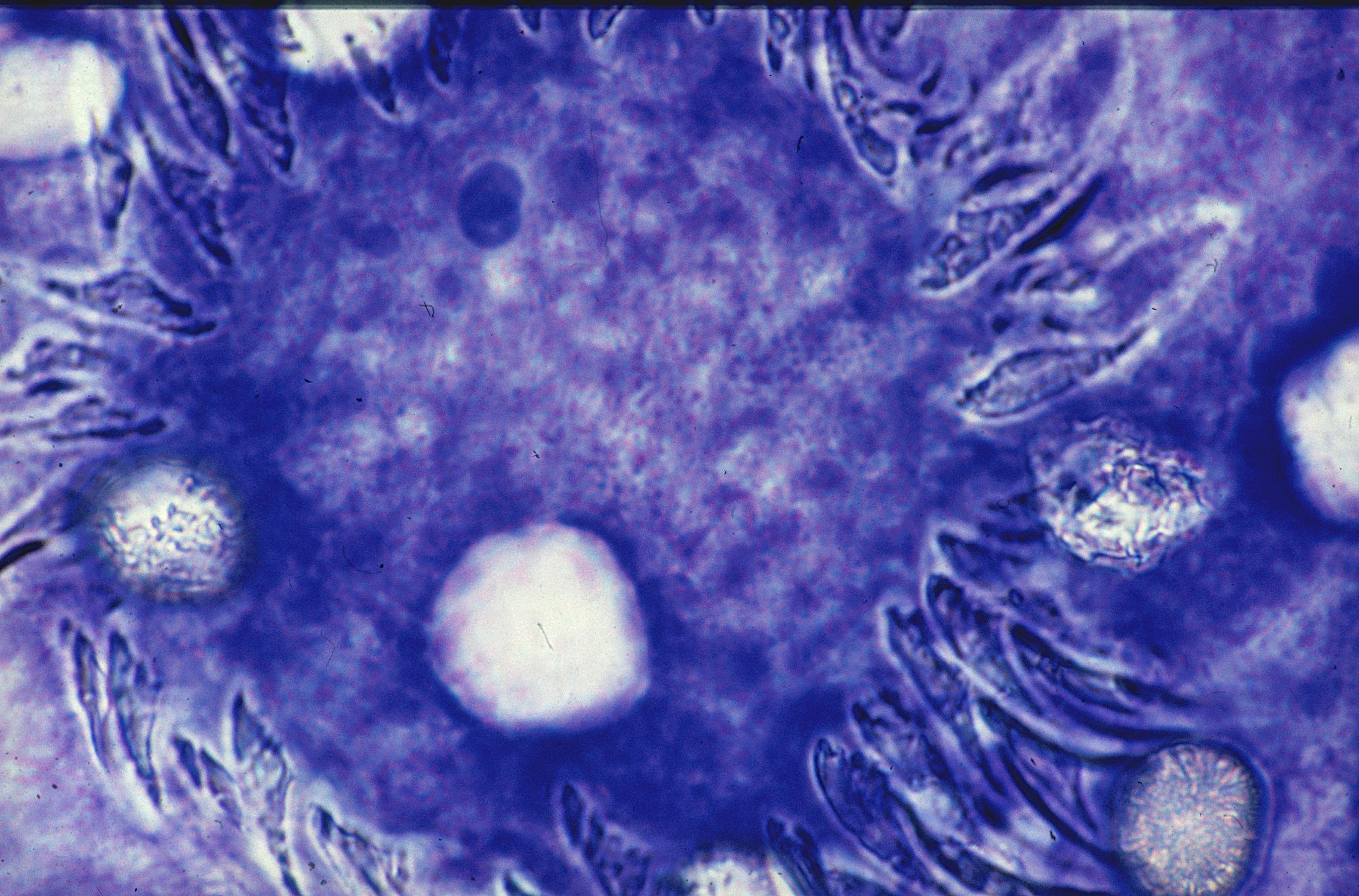
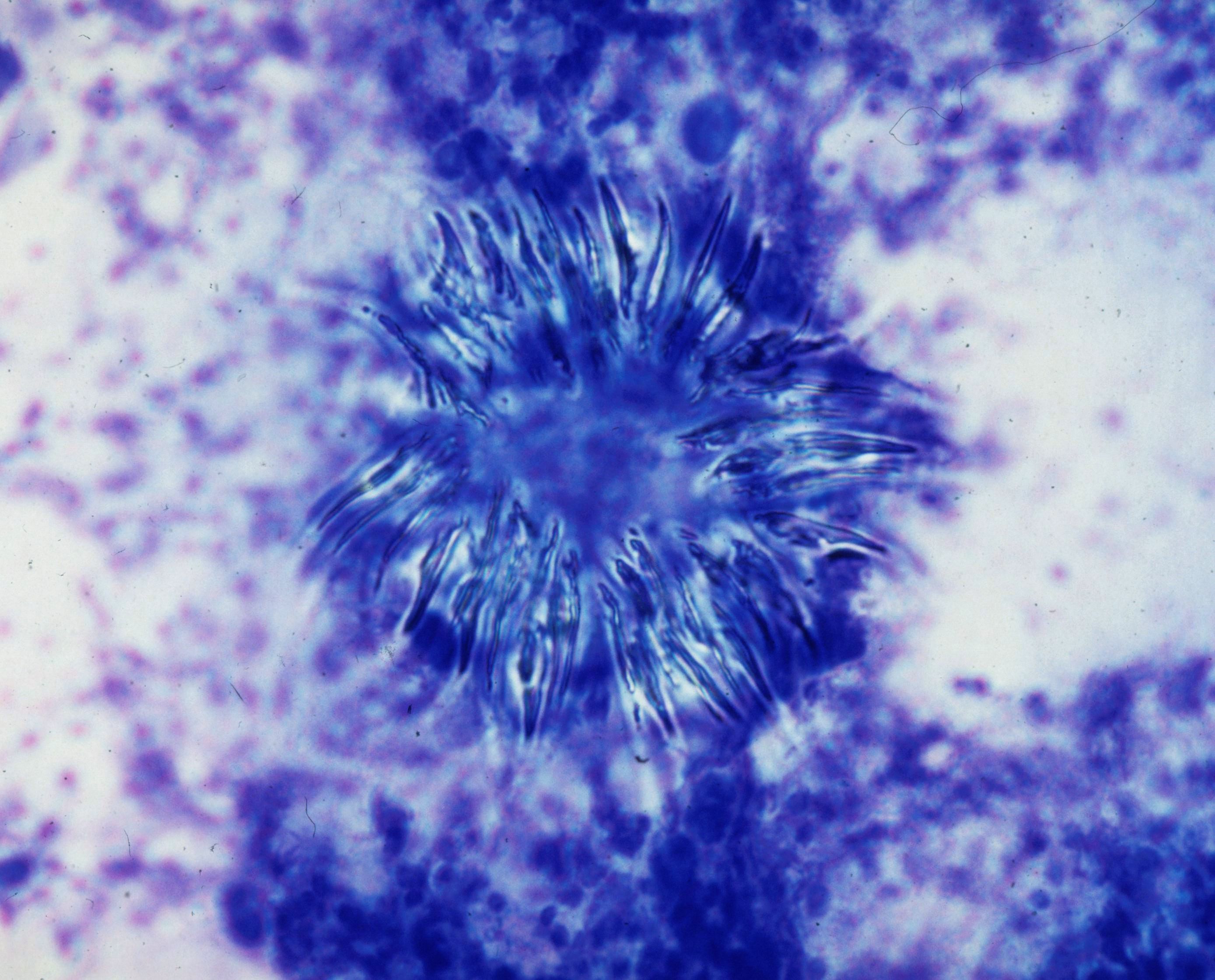 Lung, biopsy, hooklets (MGG)
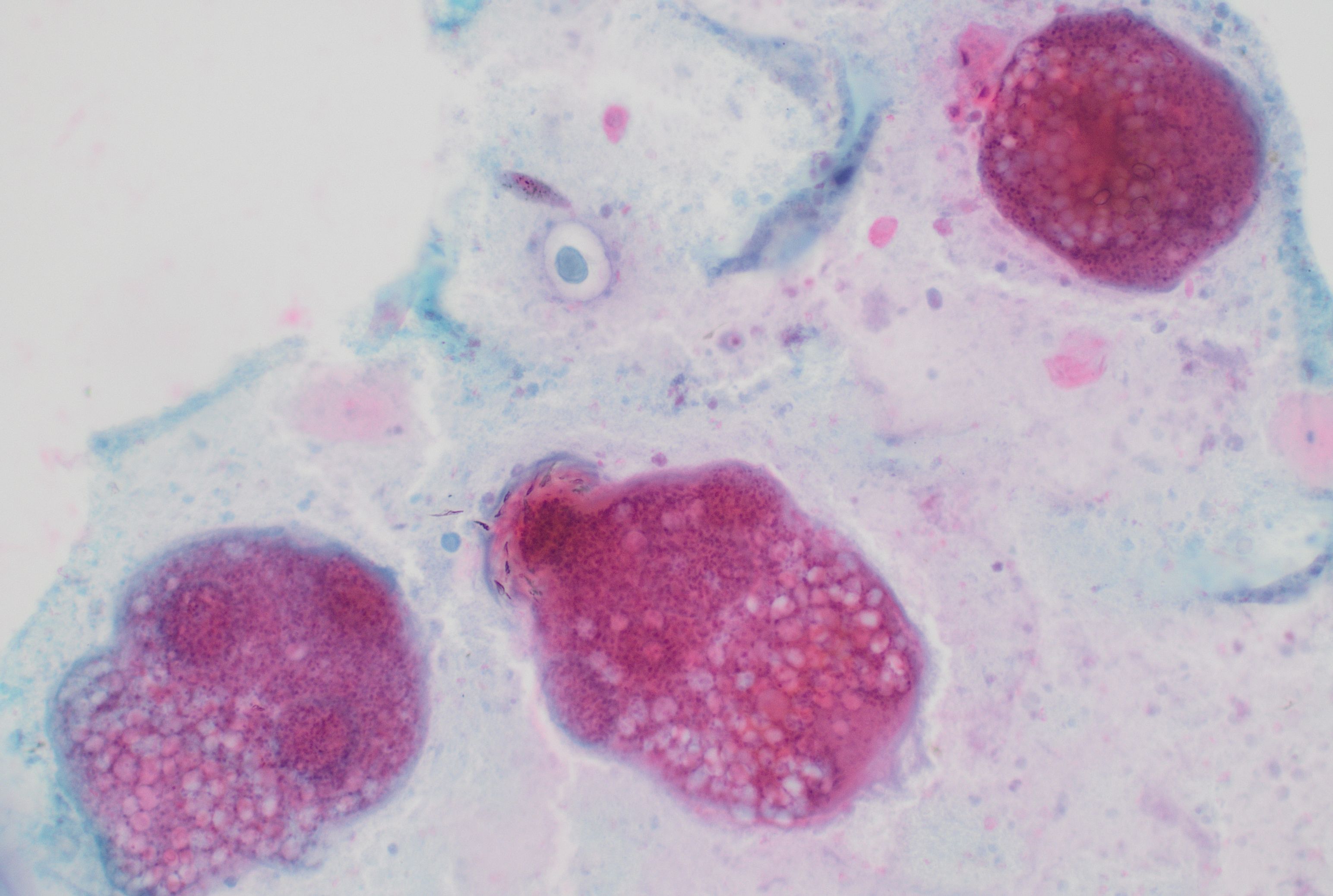 Neck, FNA, protoscolices with hooklets and…
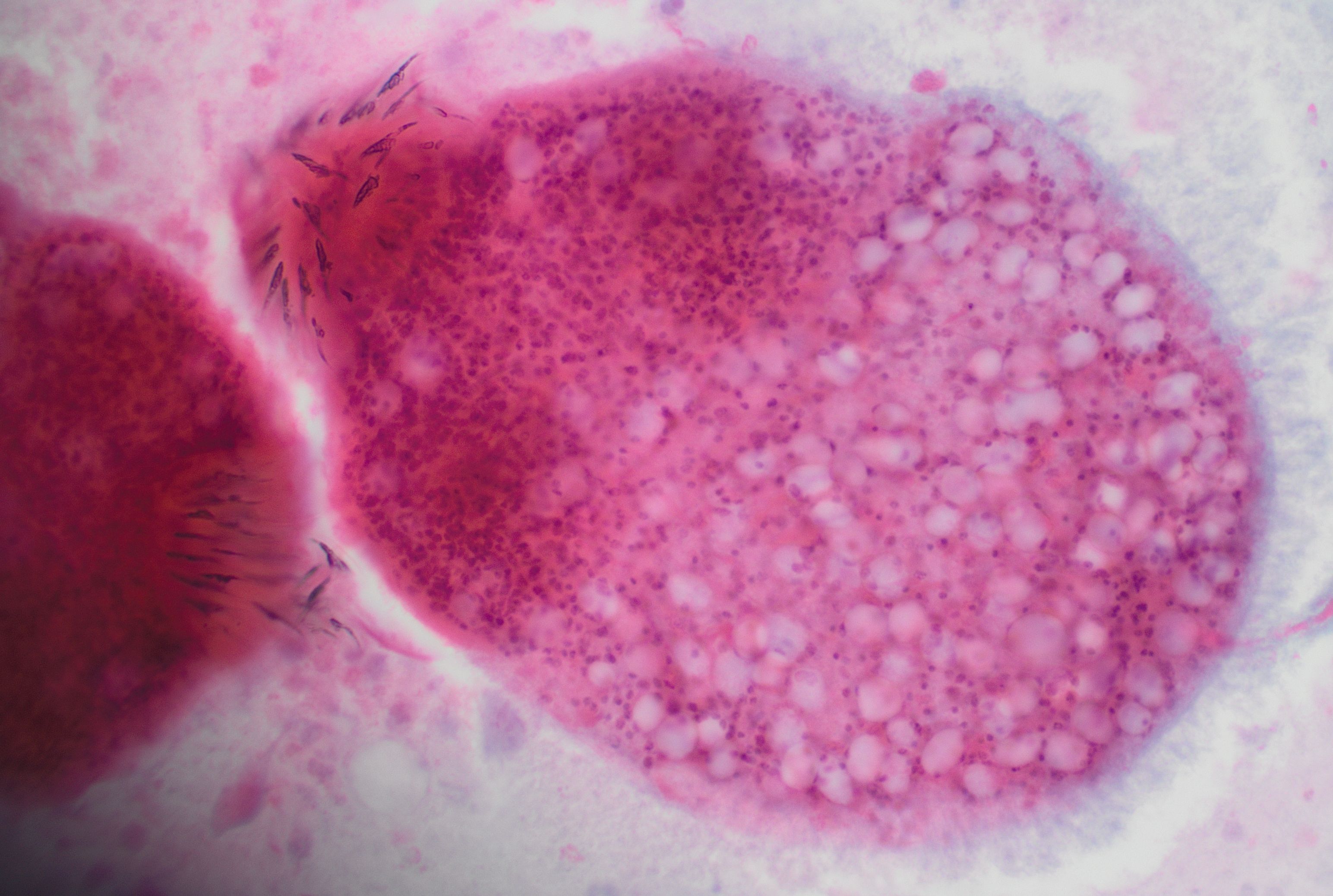 Neck, FNA, protoscolices with hooklets and…
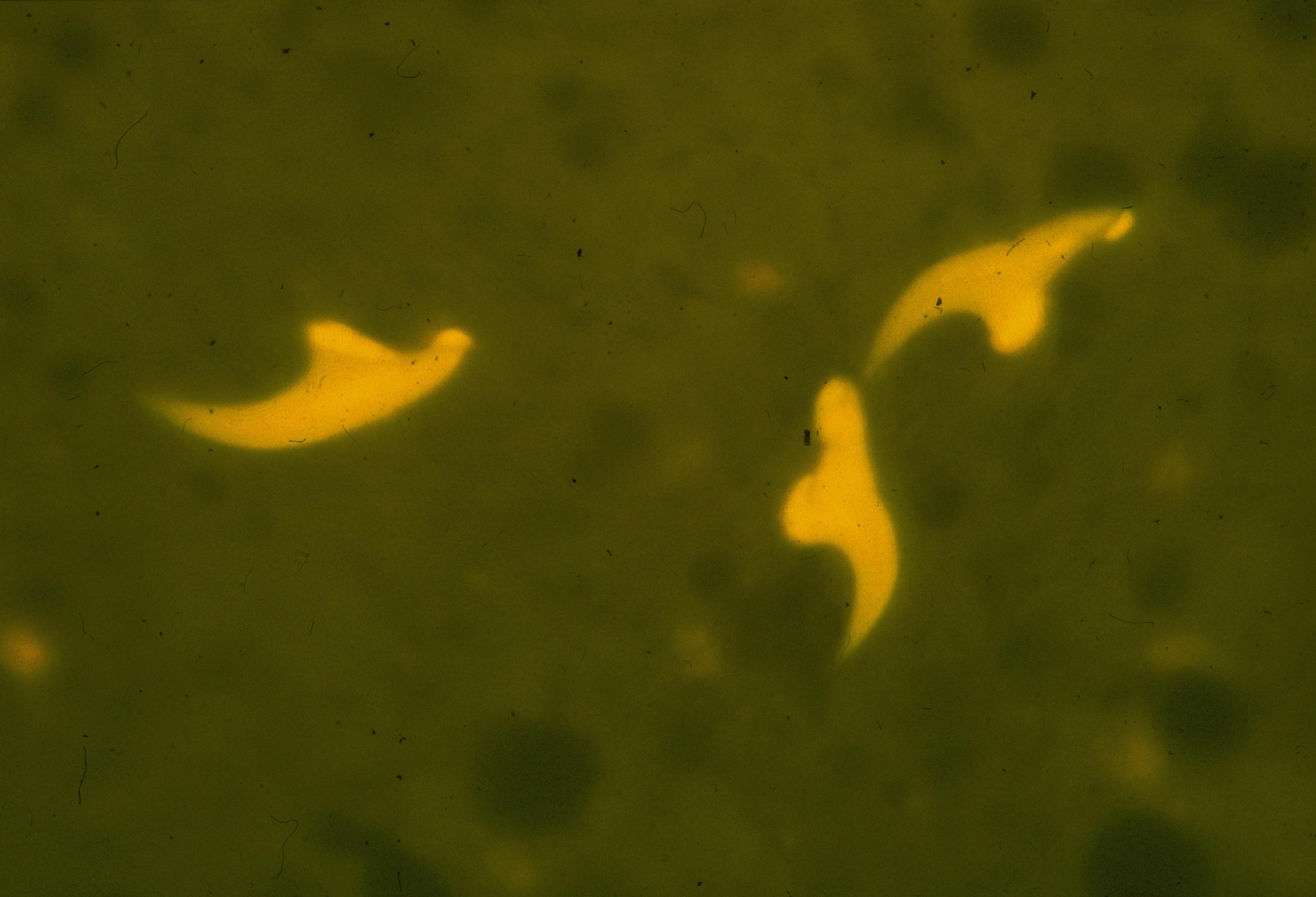 Lung, biopsy, hooklets (Auramine)
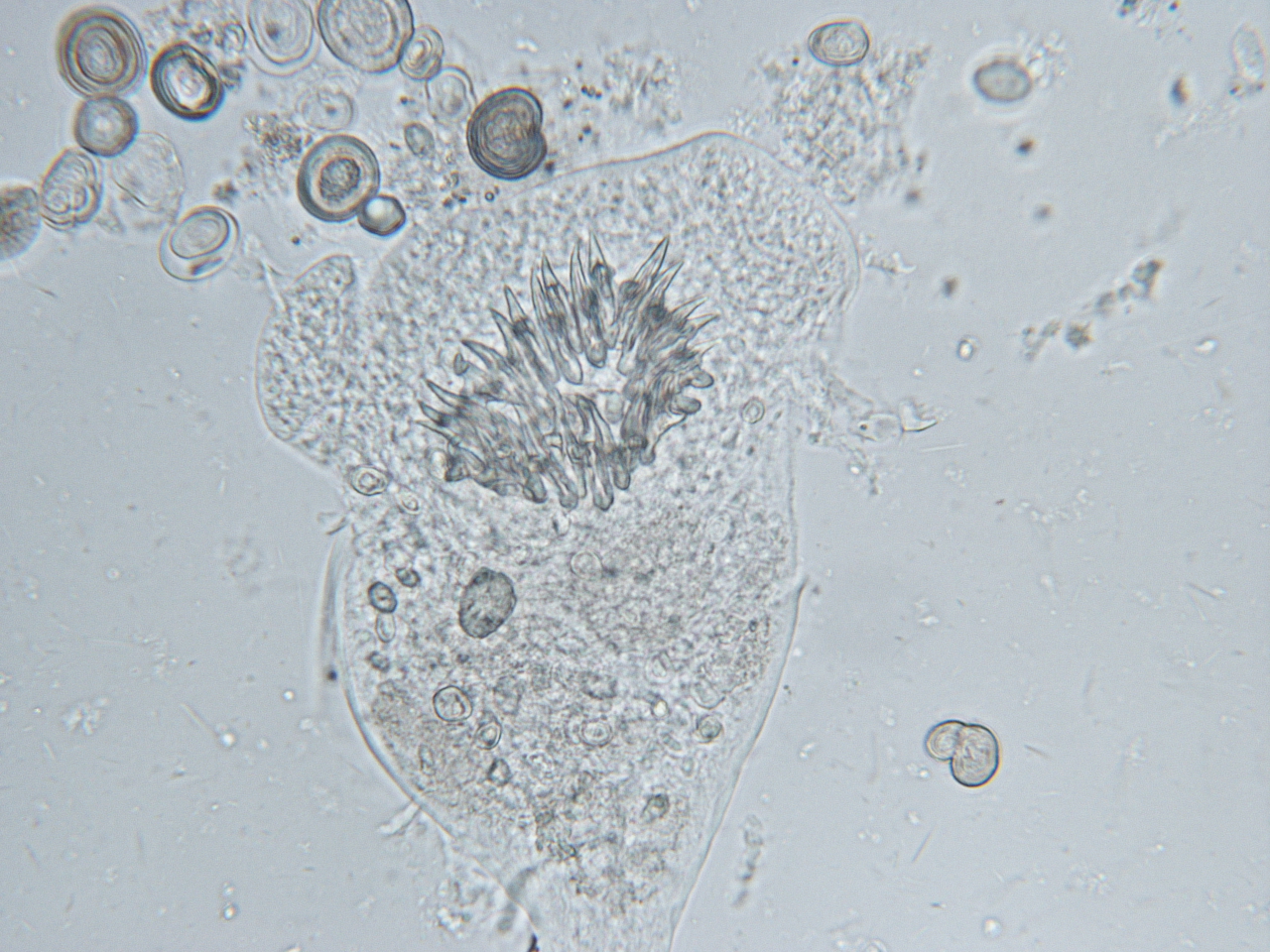 Lung, biopsy, protoscolex with calcareous…
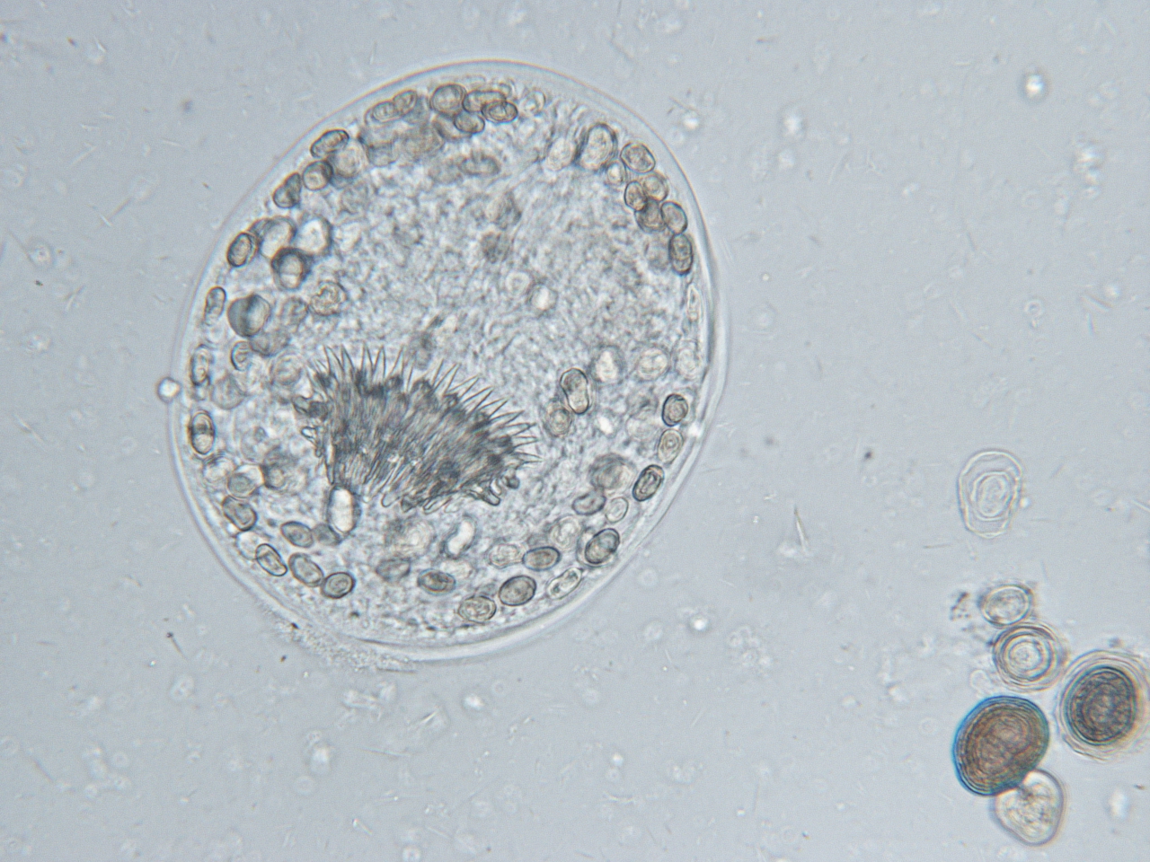 Lung, biopsy, protoscolex with calcareous…
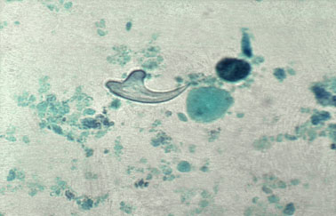 Lung, biopsy, hooklet (PAP; courtesy of…
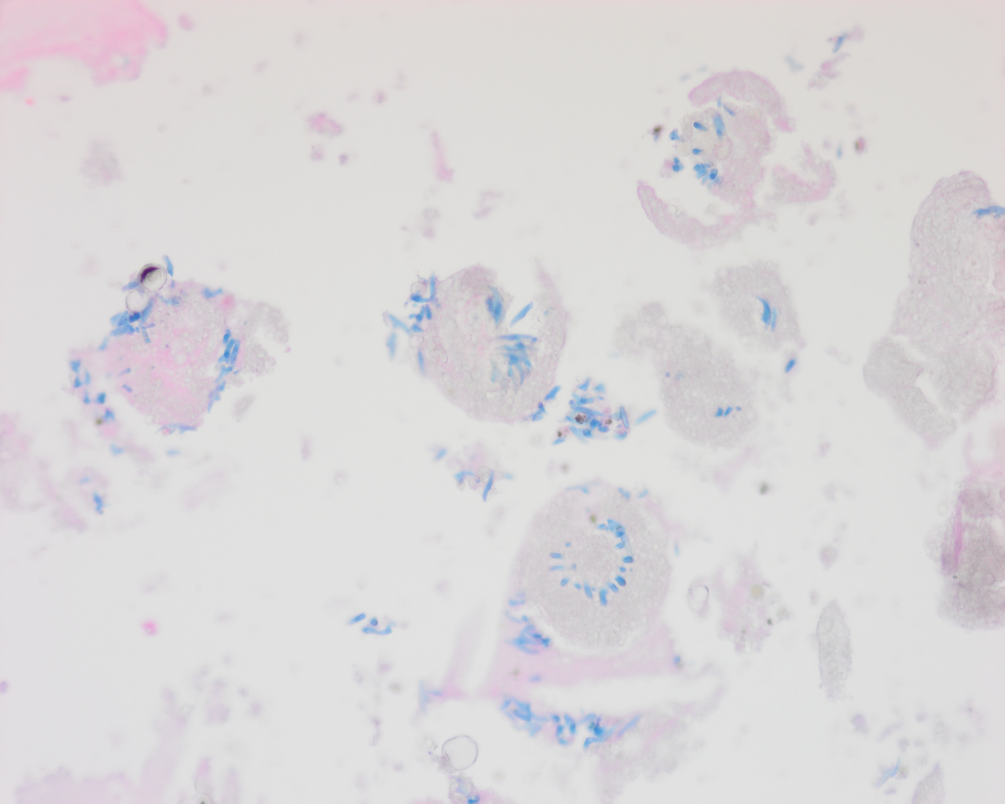 Liver, echinococcus cyst with non-viable…
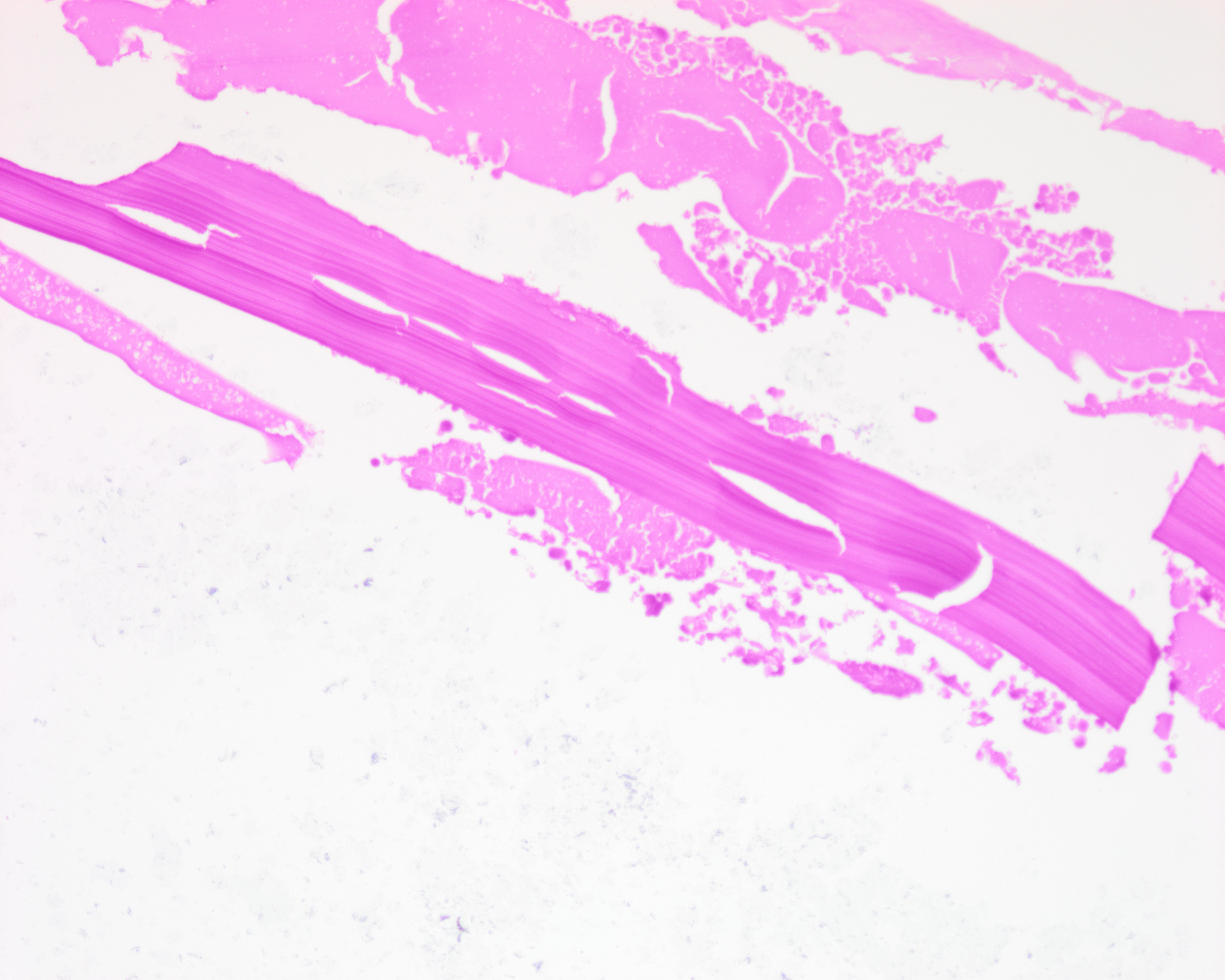 Liver, echinococcus cyst, remnants of a…
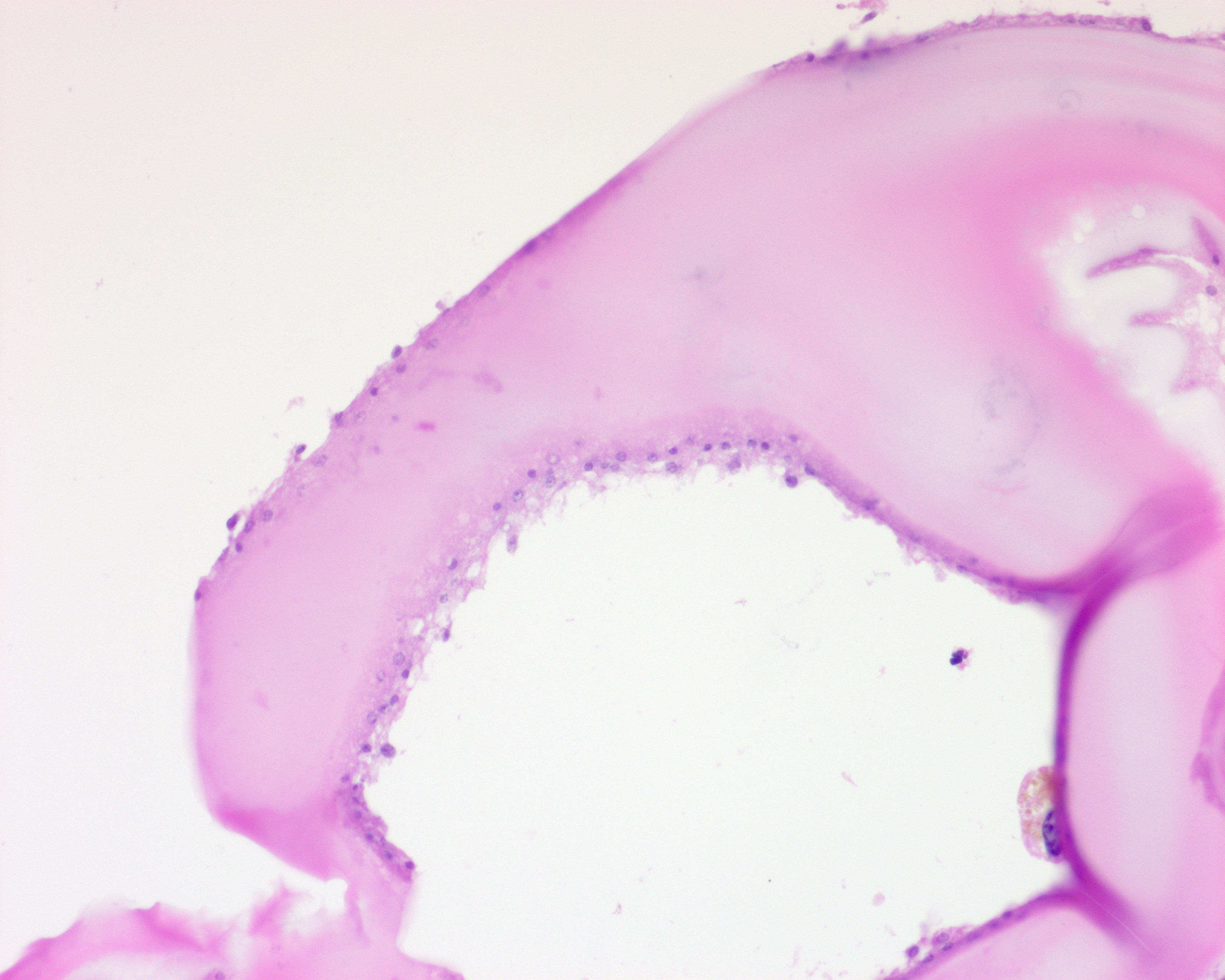 Liver, echinococcus cyst, laminated membrane…
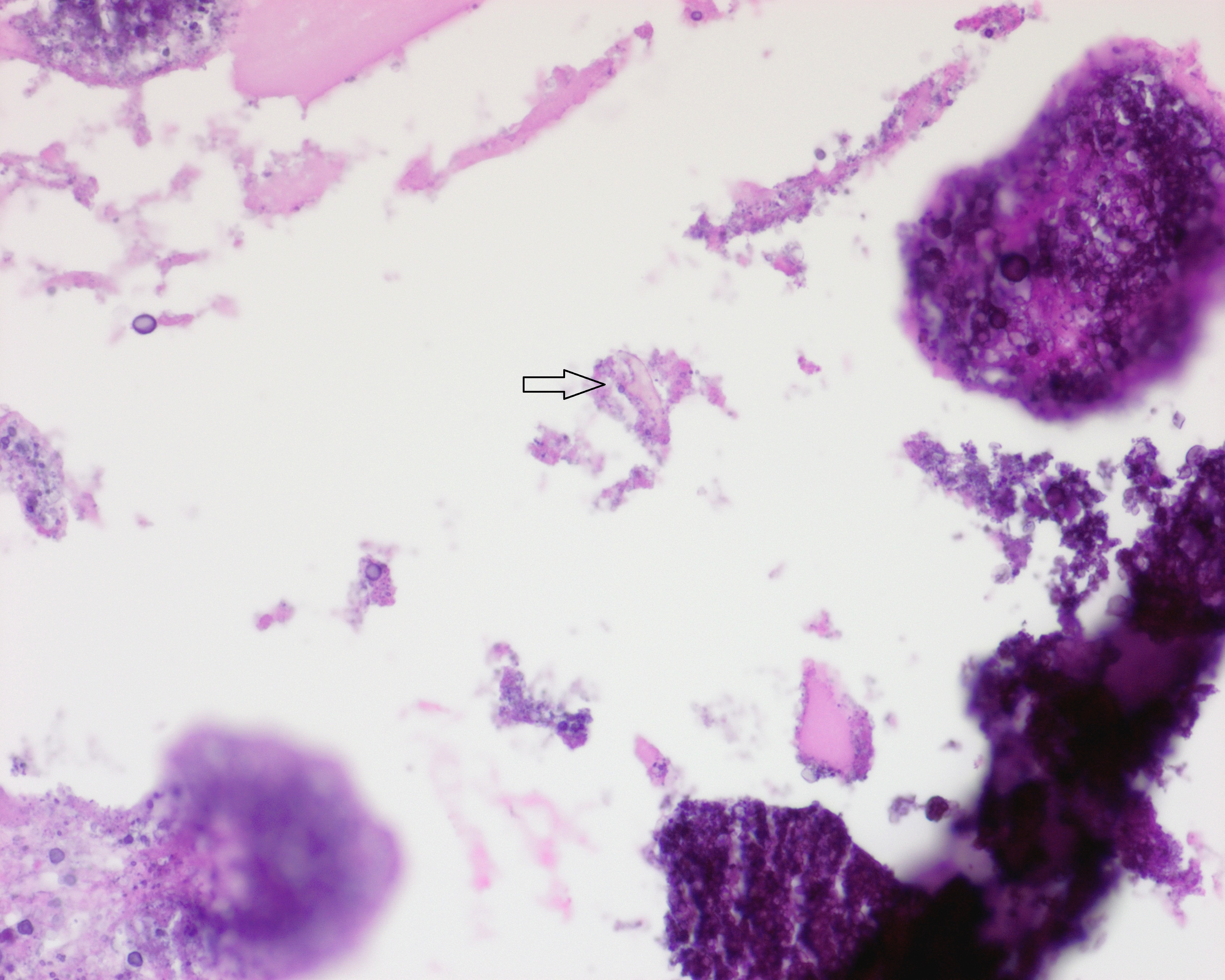 Liver, non-viable echinococcus cyst, single…
Morphology
Cytology:
In aspirations of cysts, only protoscolices (see histology) and/or hooklets can be identified. Hooklets can be highlighted with MGG, PAP, and Auramine stain.
Histology:
Viable cyst components:
- Fibrotic pericyst (host tissue reaction)
- Laminated membrane
- Germinal layer with viable brood capsules containing intact protoscolices (larval stage)
Alveolar echinococcosis forms multiple irregular cystic spaces with collapsed and folded membranes and germinal layers; protoscolices are rarely produced.
Ruptured, traumatized, or dead cysts may contain neutrophilic or granulomatous inflammation and varying amounts of necrotic debris, calcifications, and degenerating helminth fragments and free hooklets.
Protoscolices are small spherical larval forms with invaginated heads and rostral hooks, and calcareous corpuscules.
Histologically, hooklets can be highlighted with Giemsa (blue) and Ziehl-Neelsen (red) stains, or with polarised light.
The lamellar structure of the laminated membrane can be highlighted with GMS and PAS stain.
Differential Diagnosis:
- Cysticercosis
- Coenurosis
- Lipid pseudomembranes of membranous lipodystrophy (no laminar layers of the membrane)
- Noninfectious cystic lesions
Clinical notes
Four species are pathogenic to humans and have different clinical presentations:
E. granulosus (cystic echinococcosis)
E. multilocularis (alveolar echinococcosis)
E. oligarthus & E. vogeli (polycystic neotropic echinococcosis)
E. granulosus and E. multilocularis show a worldwide distribution, whereas E. oligarthus and E. vogeli are only found in Central and South America.
Infections with Echinococcus spp. most commonly affect the liver and, secondly, the lung, but may involve any region of the body.
The cysts are slow-growing masses that often do not show any symptoms for many years.
Possible symptoms depend on the location and size of the cysts.
Rupture of the cyst, spontaneous or due to a mechanical cause, can lead to immediate anaphylaxis and dissemination to other organs.
Unilocular hydatid cyst contains fluid, whereas the fluid in alveolar hydatid cyst has a more gelatinous consistency.
Echinococcosis, particularly alveolar echinococcosis, can mimic an invasive neoplasm of the liver.
Because of the risk of peritoneal implants or dissemination, biopsy is not recommended; however, it may be useful for confirming the diagnosis in seronegative patients.
Ancillary Testing
ELISA; indirect hemagglutination test; indirect fluorescent antibody tests - negative test results do not rule out infection, cross reactions with other cestode antigens; immunoblot to confirm positive test result; PCRAdditional Information
Echinococcus spp. are cyclophyllidean cestodes (tapeworms).
Hydatid cyst, or hydatid disease, is a synonym for echinococcosis.
Humans become infected by ingesting eggs found in the feces of definitive hosts (wild and domestic canids). The eggs hatch inside the small intestine. The released oncospheres penetrate the intestinal wall and migrate through the circulatory system to various organs, where they develop into a hydatid cyst.
Humans are aberrant intermediate hosts and do not transmit infection.
Adult tapeworms are never found in humans.
Cervical hydatid cysts are very uncommon, even in endemic areas, representing fewer than 1% of all cases. They often present as benign-appearing neck masses. Neurological symptoms caused by compression of adjacent neural structures are particularly rare (1).
References
- Rezaei, J., Gandomkar, H., Chenarani, E. et al. Hydatid cyst of the neck presenting with left upper limb paresthesia in a 29-year-old man: a case report. J Med Case Reports 19, 445 (2025). https://doi.org/10.1186/s13256-025-05523-5