
- EN
- DE
Fungi
Differential Diagnosis of Candida Species:
Aspergillus species and "Aspergillus-like" fungi

- Organ System
- Respiratory Tract, Soft Tissue & Bone
- Diagnostic Method
- Cytology, Histology

- Geographical Region
- Dry Areas,
- Islands,
- Tropical and Subtropical Region,
- Polar and Subpolar Zone,
- Temperate Zone
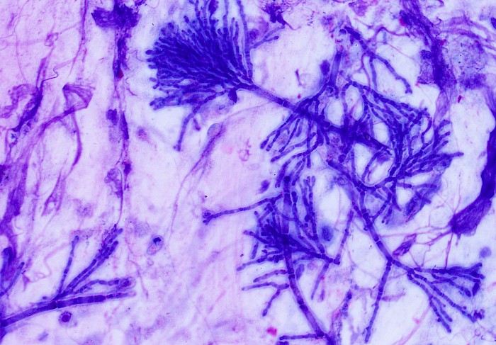
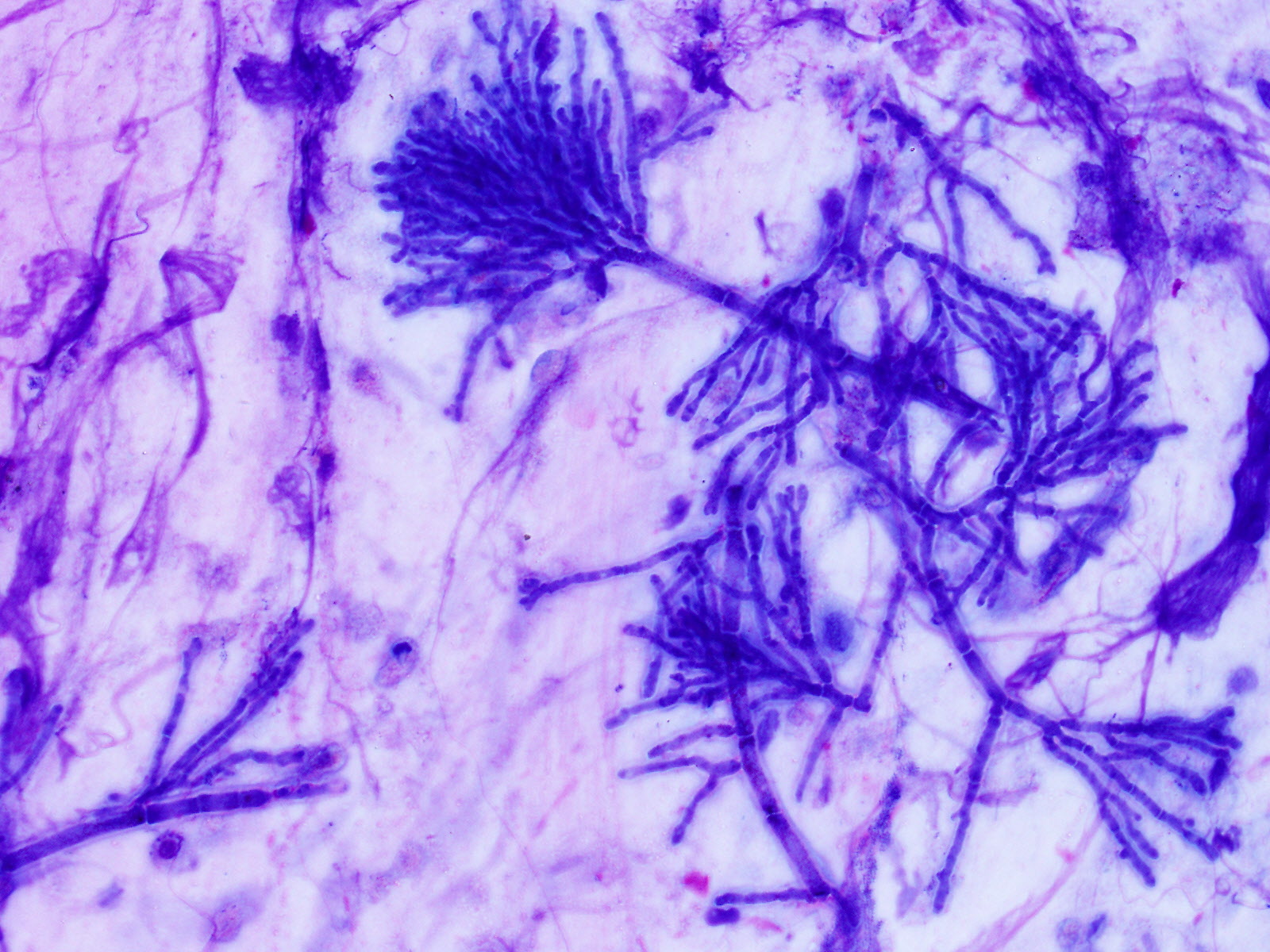 Sputum, septate hyphae with acute-angle…
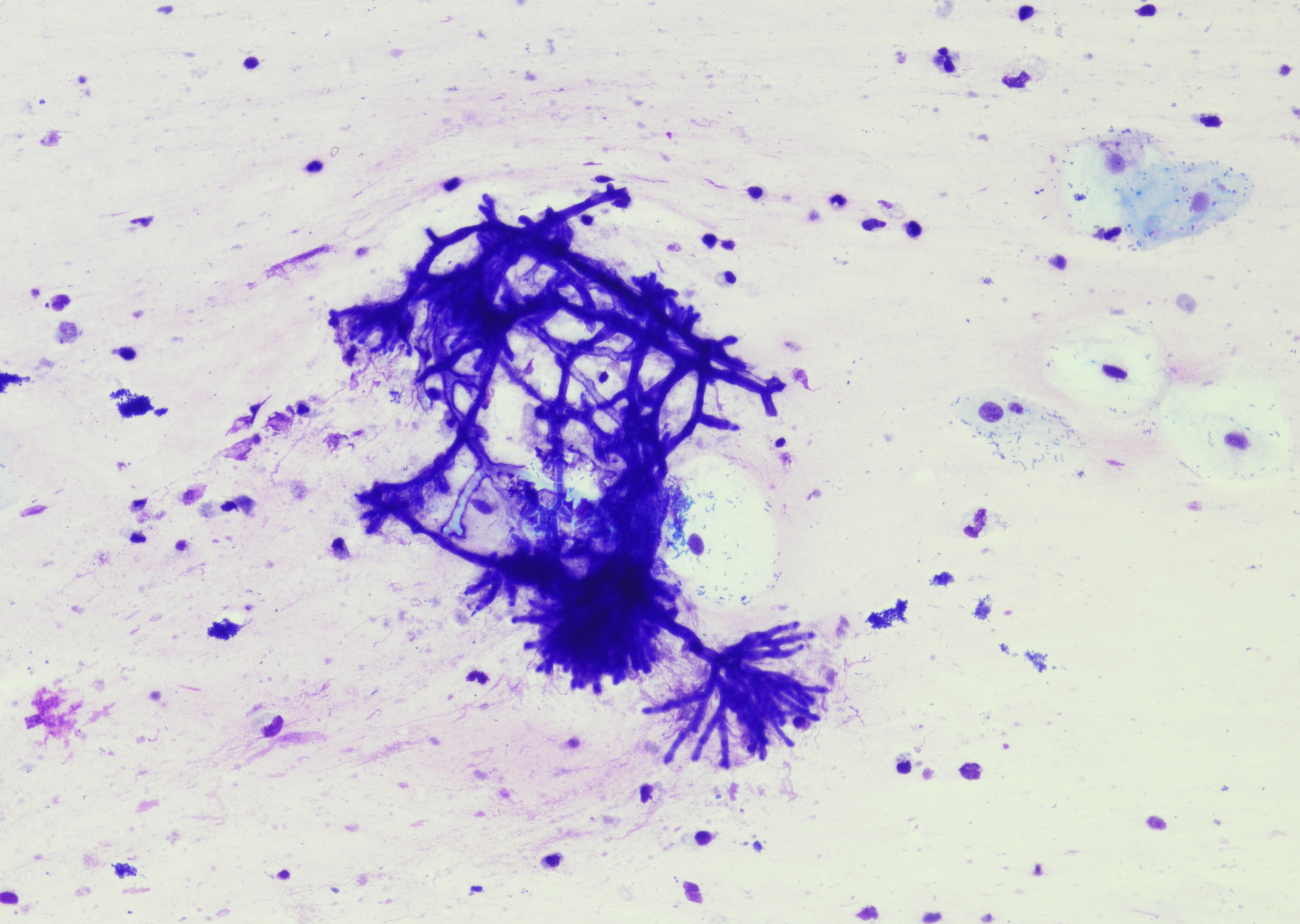 Sputum, narrow hyphae with acute-angle…
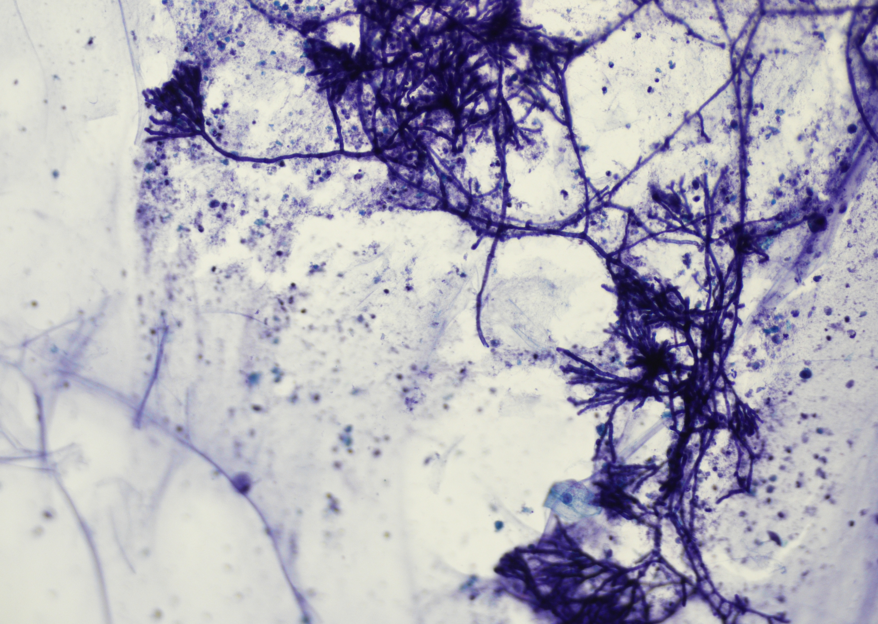 Sputum, narrow hyphae with acute-angle…
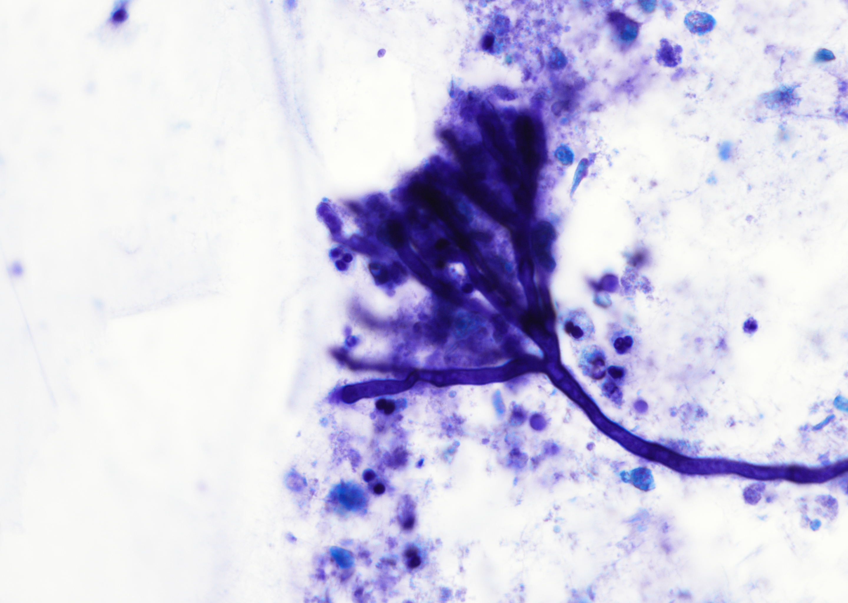 Sputum, hyphea with acute-angle branching at…
 BAL, cystic fibrosis patient with allergic…
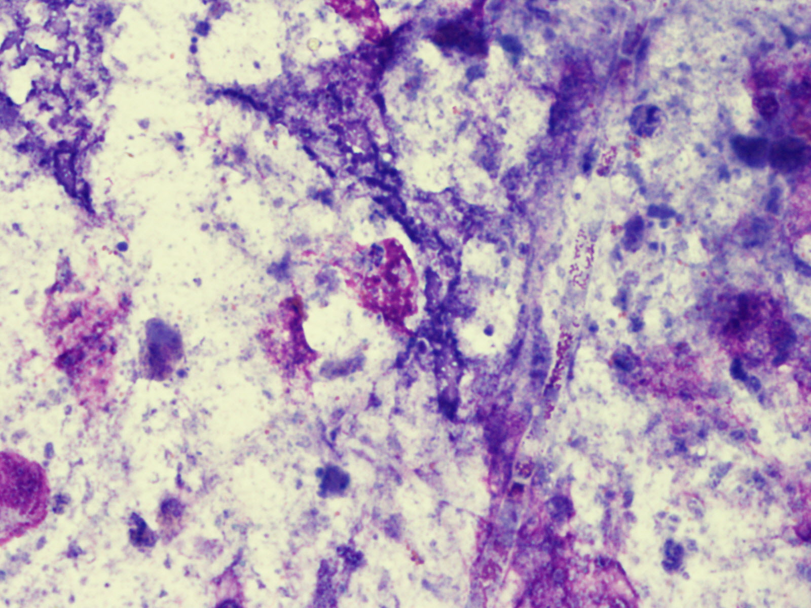 BAL, cystic fibrosis patient with allergic…
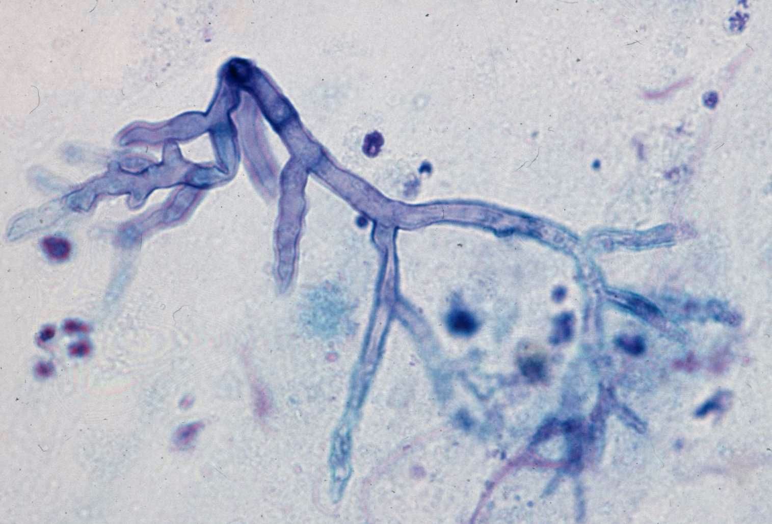 Lung, cytobrush, hollow-appearing…
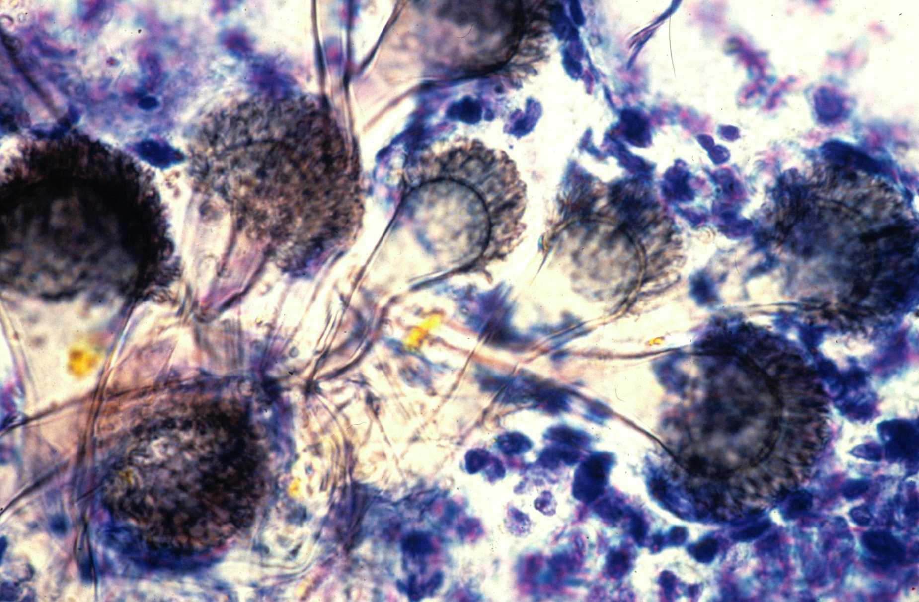 Lung, cytobrush, conidiophores (MGG, x600)
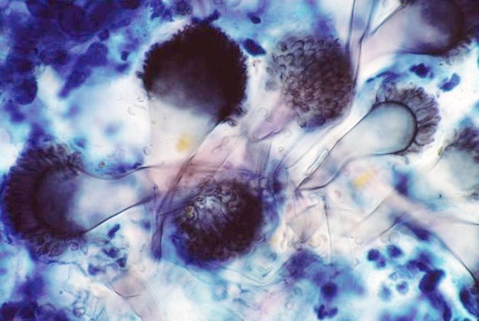 Lung, cytobrush, conidiophores (PAP, x600)
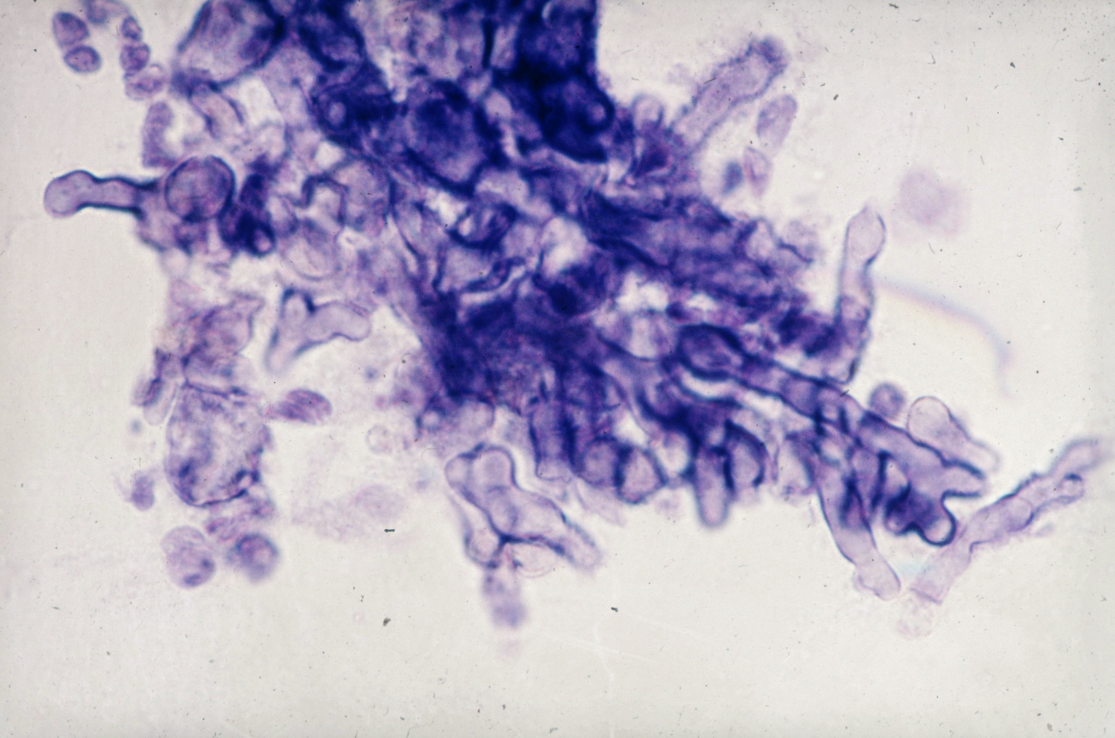 Lung, cytobrush, typically clustered…
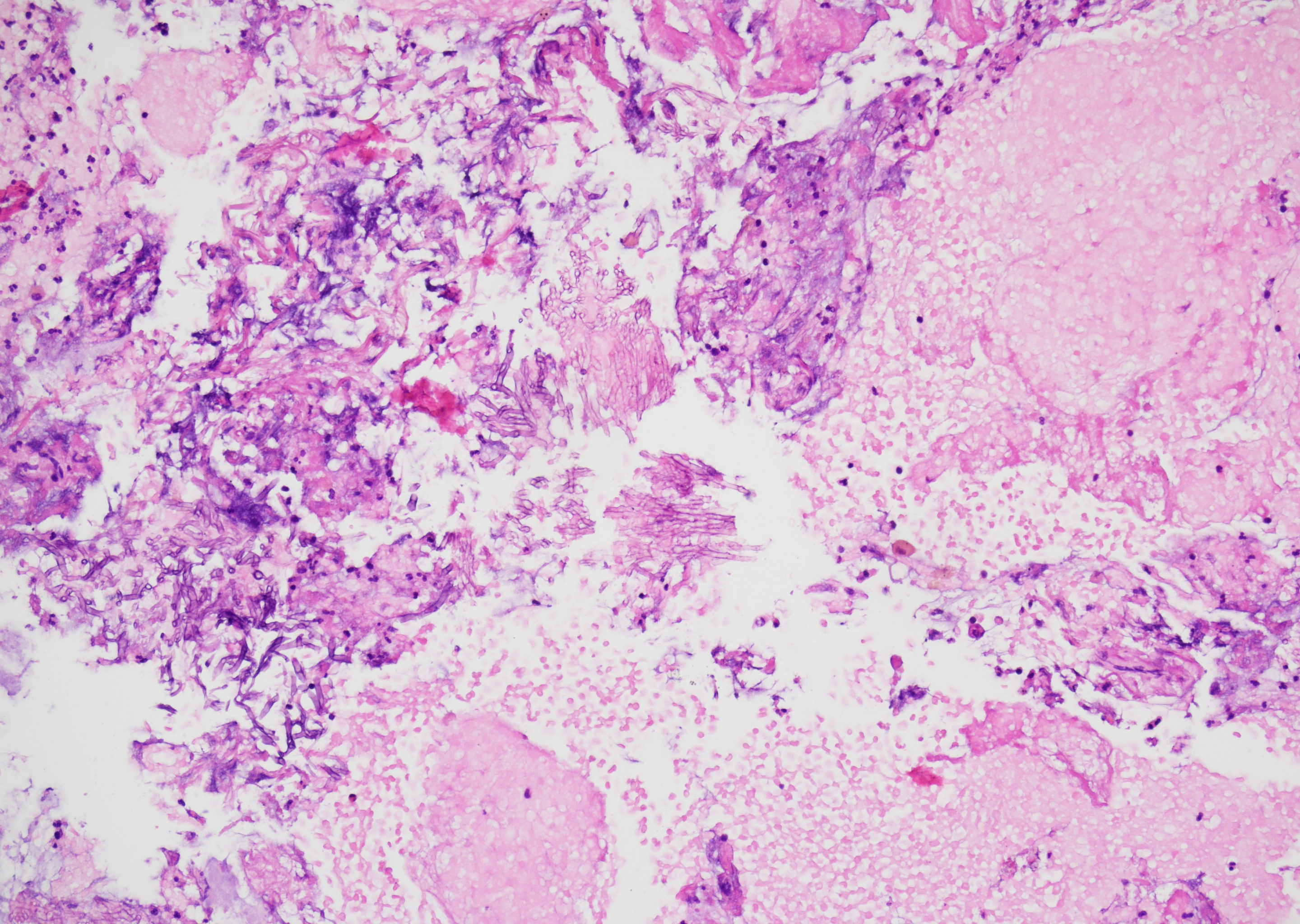 Lung FNA with cell block, demonstrating…
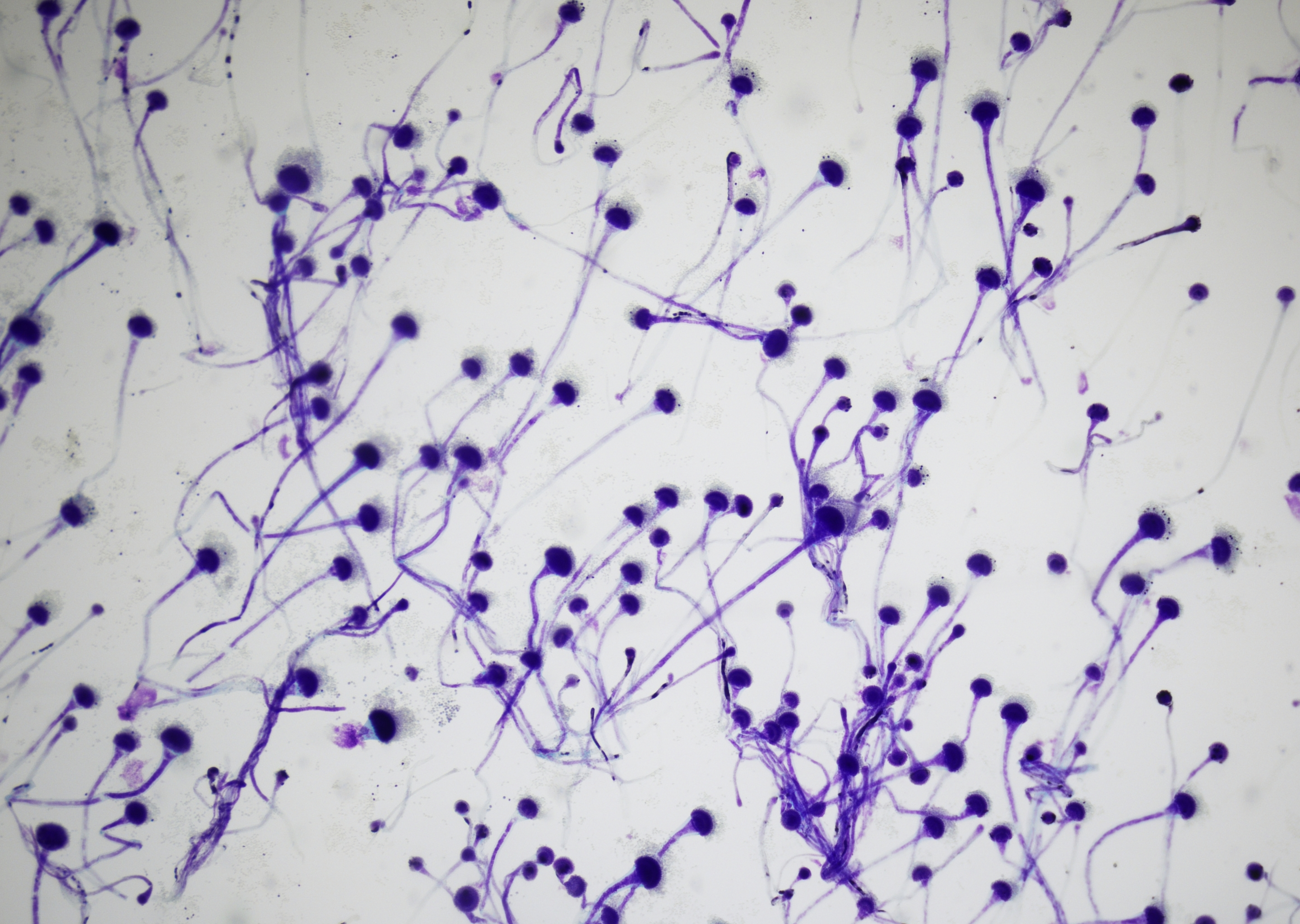 Sputum, fungal culture, tape mount…
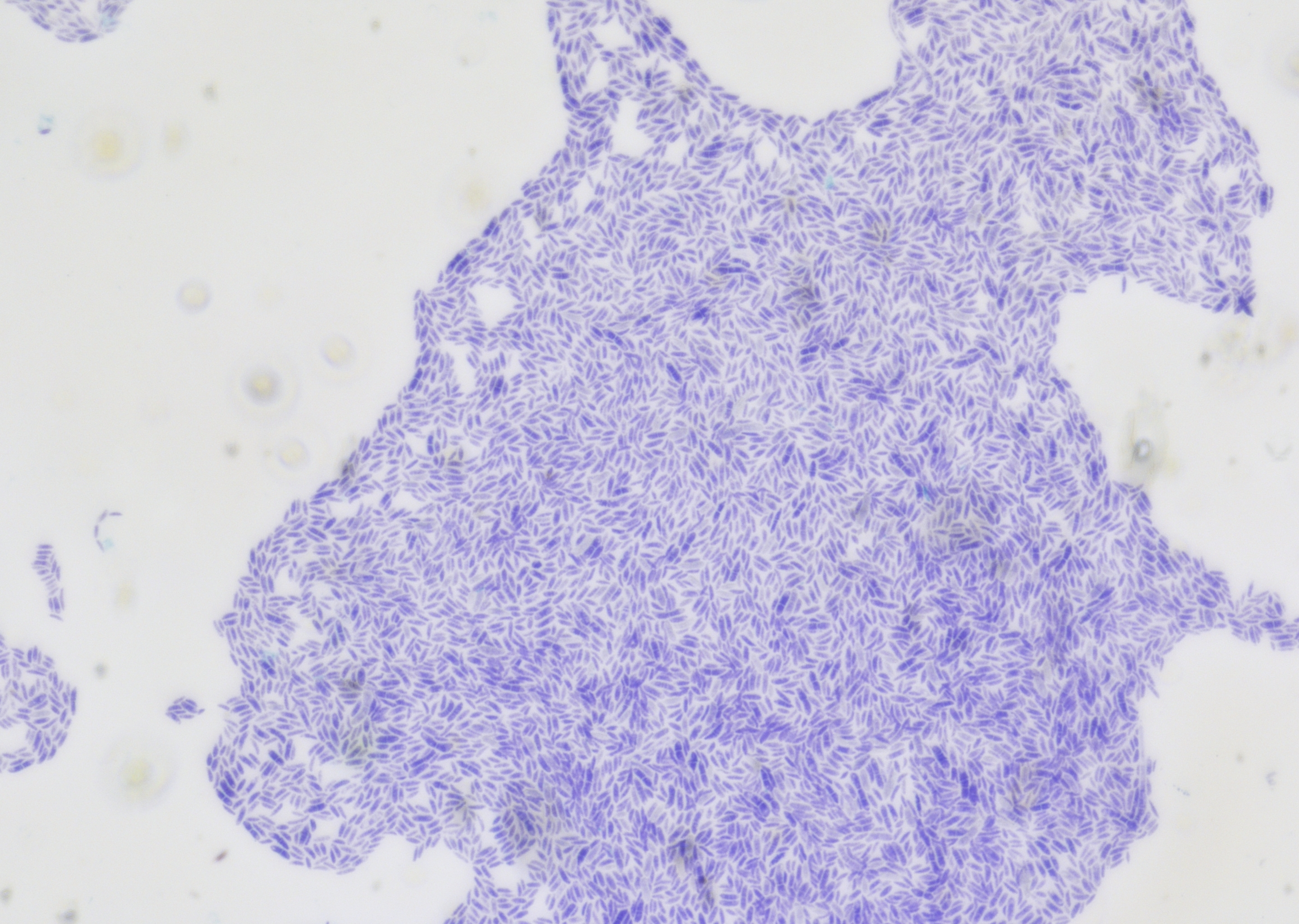 Sputum, fungal culture, tape mount with…
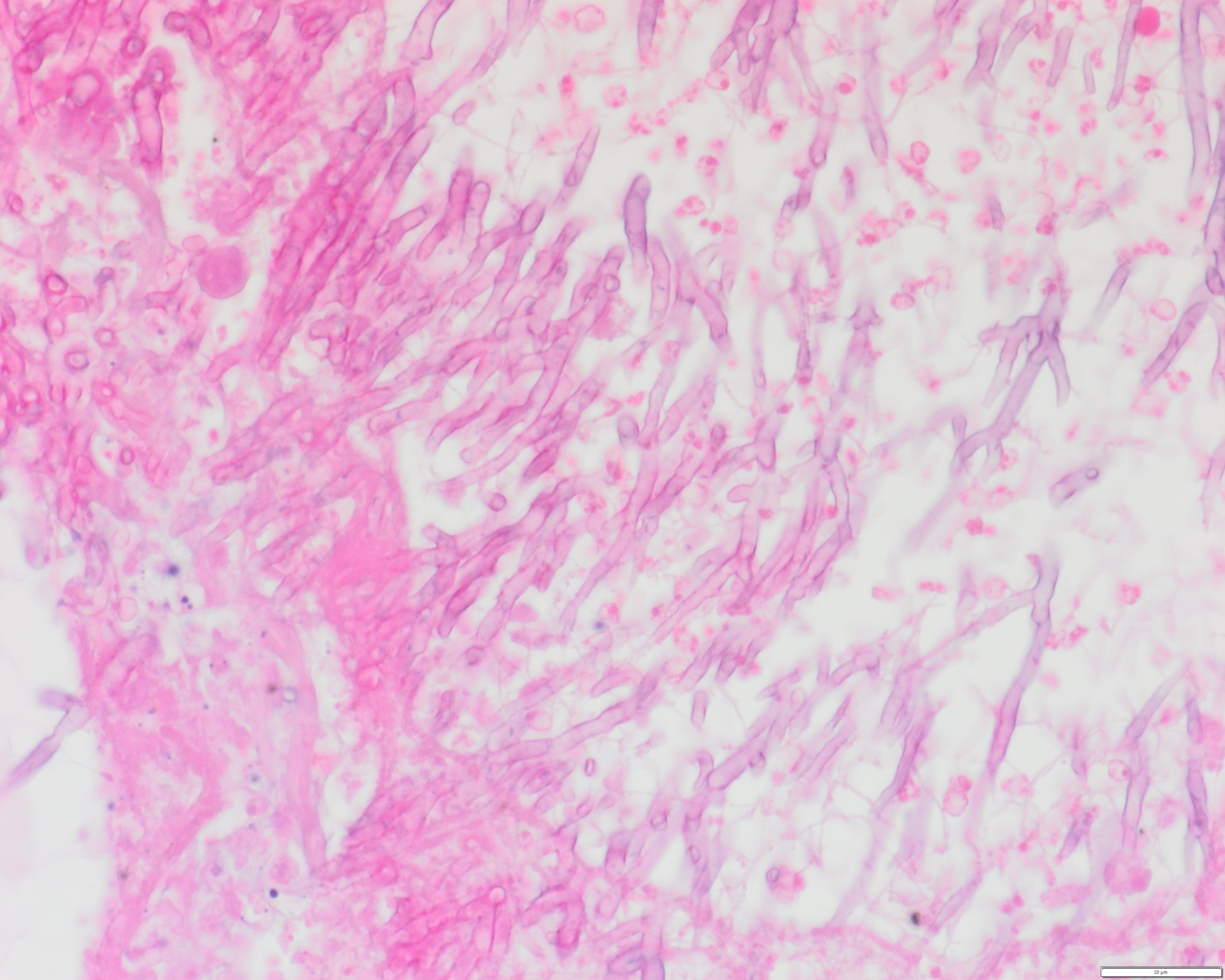 Lung section, autopsy case…
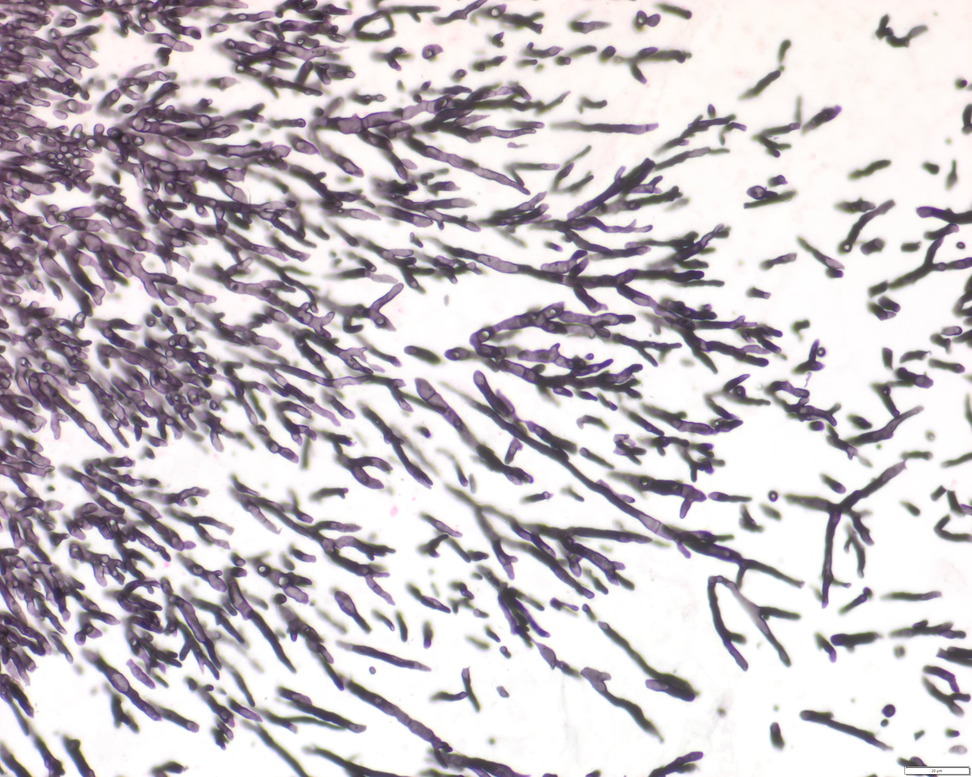 Lung section, autopsy case…
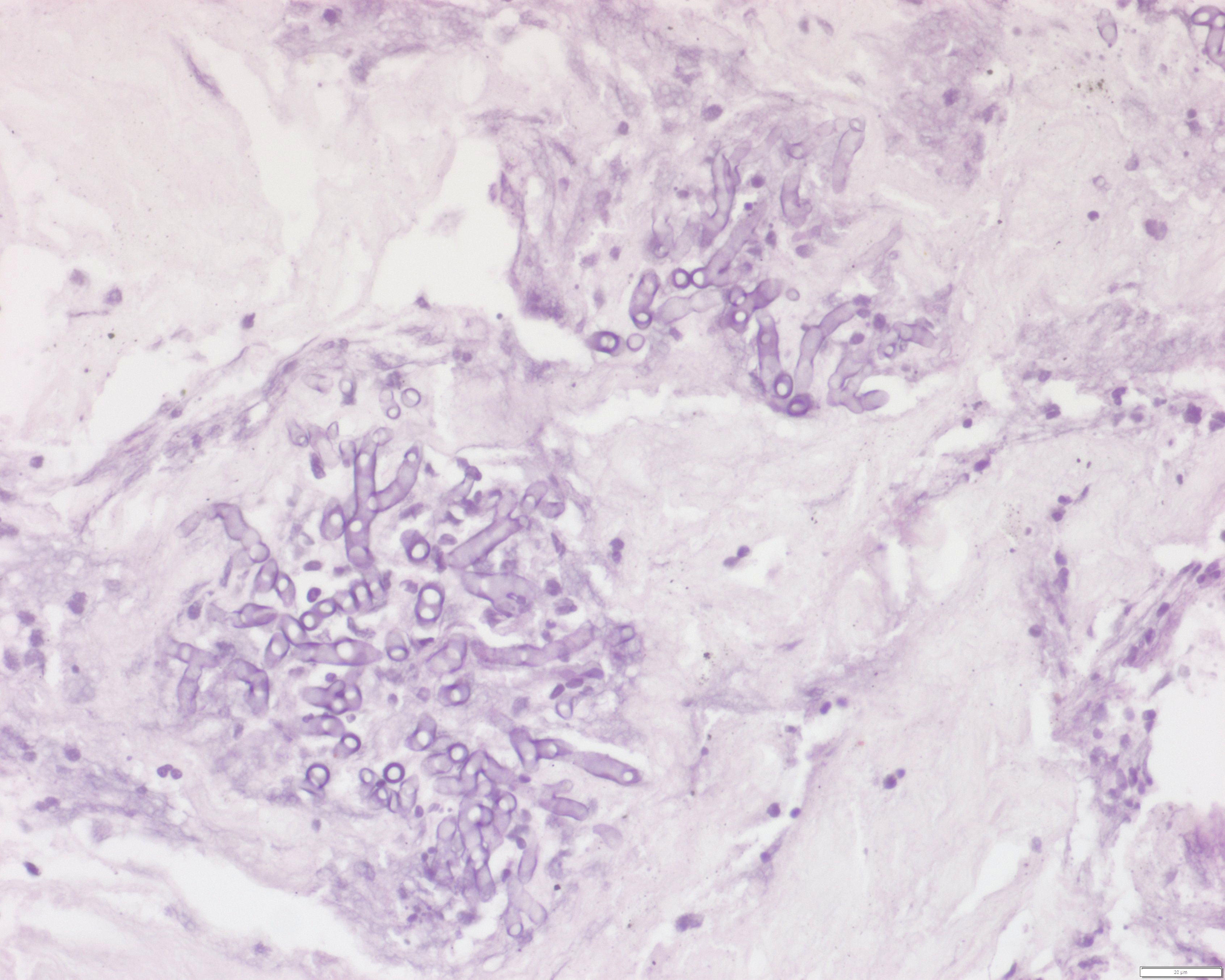 Lung biopsy of suspect nodule, invasive…
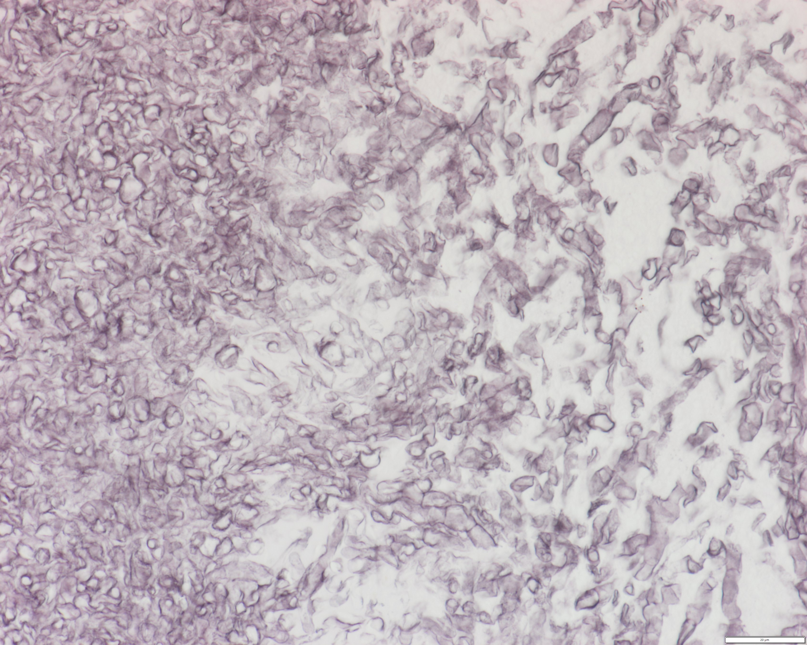 Paranasal sinus, FESS, non-invasive fungal…
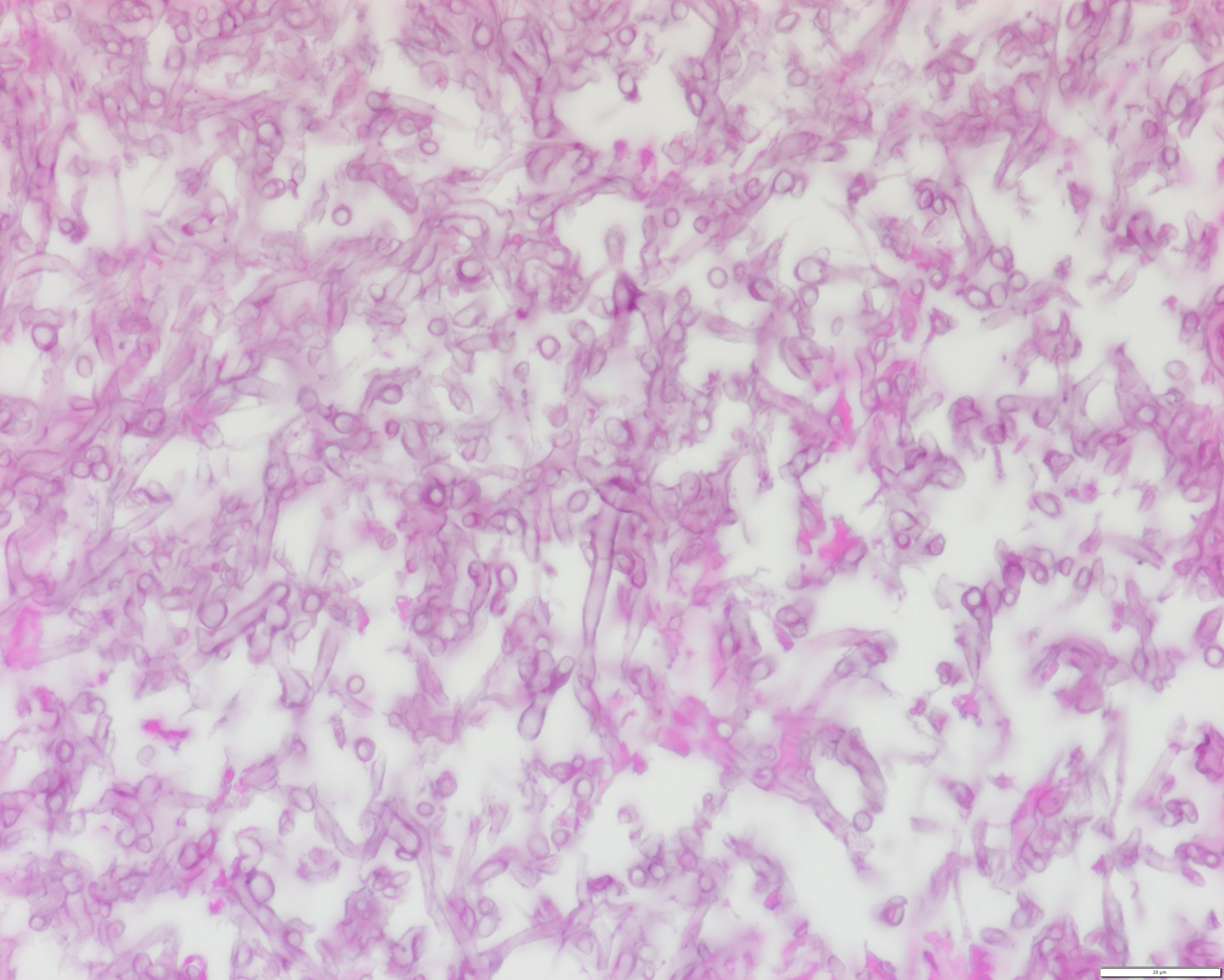 Paranasal sinus, FESS, non-invasive fungal…
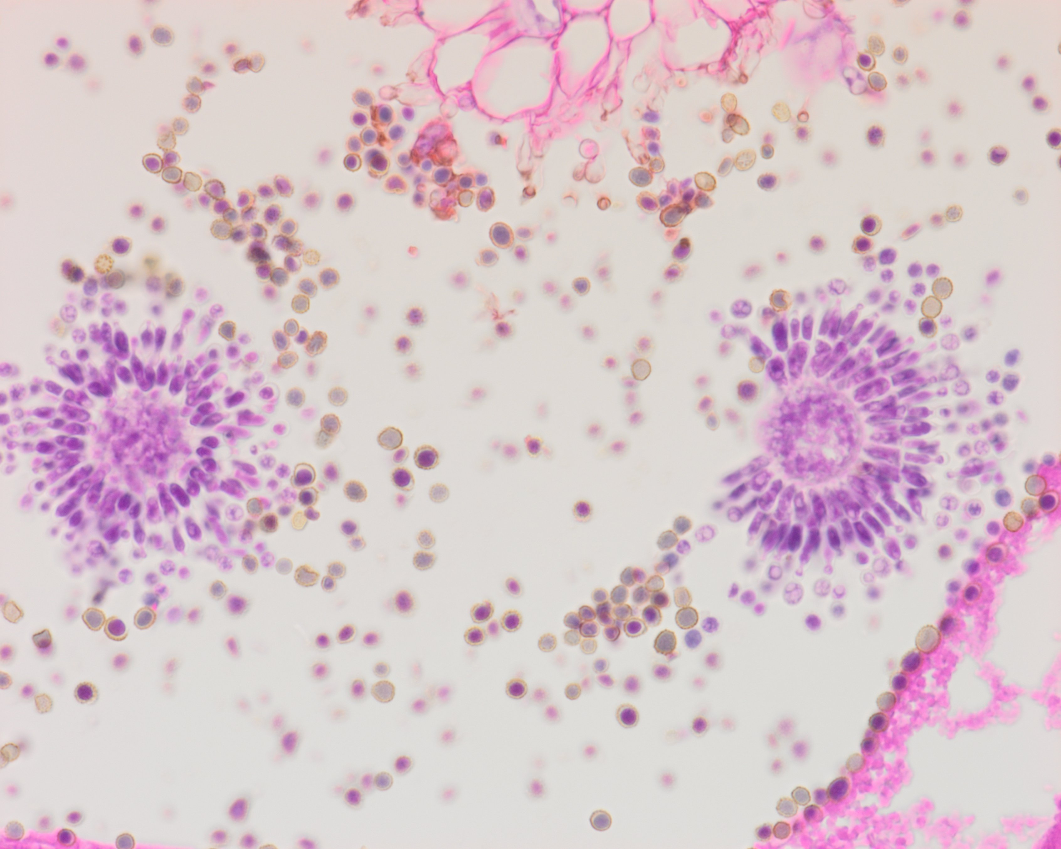 Paranasal sinus, FESS, Aspergillus spp. with…
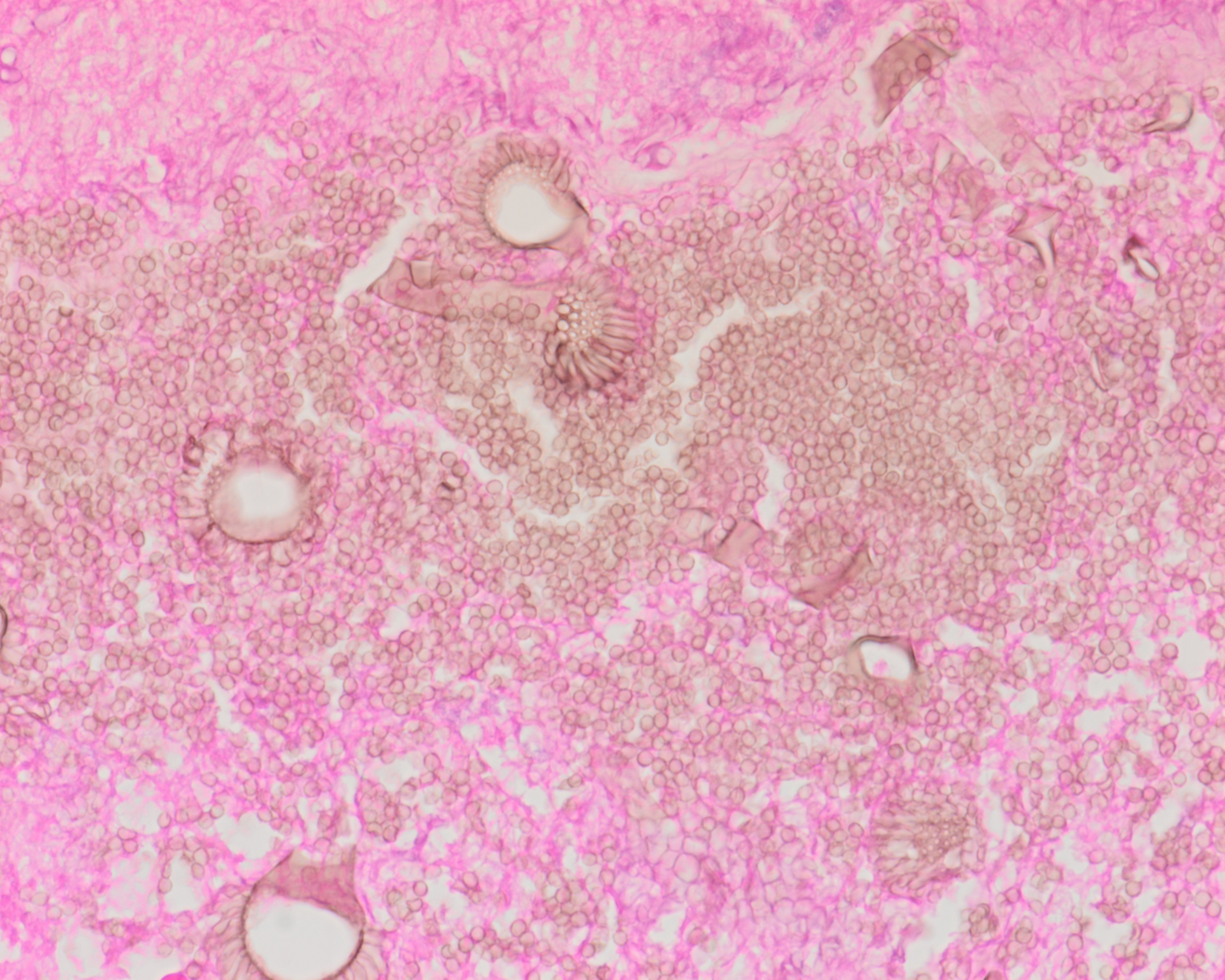 Paranasal sinus, FESS, Aspergillus spp. with…
Morphology
Cytology & Histology:
Hyaline (non-pigmented) molds with multiple true septations and dichotomous branching.
Common features of Aspergillus spp.:
- Septate hyaline hyphae with parallel side walls and dichotomous, acute-angle branching at 45°
- The diameter of hyphae is around 3-6 µm
- The diameter of conidia is around 2-3 µm
- Dark brown or black pigmented conidia can be present in infections caused by A. niger, A. fumigatus, and A. carbonarius
- The fruiting heads (conidiophores) can be observed in oxygenated areas, such as paranasal sinuses or cavitary lung lesions
- Necroinflammatory background
Identification of curved or banana-shaped fusiform conidia (15-30 µm) may suggest infection by Fusarium spp..
In general, other genera in this group of hyaline molds can show similar or identical morphological characteristics; therefore, they can't be truly distinguished morphologically alone. Sometimes, if conidia or conidiophores are visible, their morphology can provide clues, but identification through culture, FFPE tissue PCR, or MALDI-TOF is necessary.
Histology in allergic fungal rhinosinusitis usually shows prominent lamellar mucin, abundant eosinophils, desquamated epithelial cells, and may reveal Charcot-Leyden crystals, with usually scant fungal hyphae.
Special stains to highlight fungal hyphae: PAS, GMS
Differential Diagnosis:
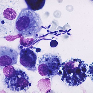
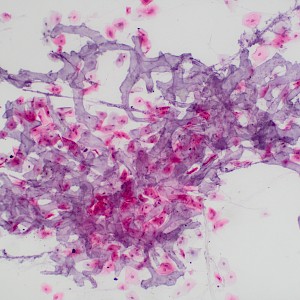
Infection with hyaline pauciseptate molds, such as mucormycosis, or with Candida spp.
Clinical notes
The main source of infection is inhalation of conidia, especially in susceptible individuals who are either allergic or immunosuppressed and are unable to clear inhaled conidia.
Involvement or infection usually occurs in sites that are directly exposed to air (lung and nasal cavities, air sinuses) or the surrounding environment (ear, eye, skin), with the lung as the most commonly involved site.
Prolonged neutropenia is a significant risk factor for invasive fungal disease. In critically ill and immunocompromised patients, invasive aspergillosis shows a high mortality rate.
Infections with hyaline septate molds are predominantly caused by Aspergillus spp., with Fusarium and Scedosporium spp. representing the second and third most common causes of human disease.
Clinical manifestations of disease include:
- Asymptomatic colonization, formation of a fungal ball
- Allergic disease
- Localized infection
- Locally invasive or destructive infection
- Disseminated infection
Invasive pulmonary aspergillosis and acute invasive fungal rhinosinusitis are often associated with angioinvasion, secondary coagulative necrosis, and infarction.
Saprophytic lung infection arises in preexisting lung cavities and is most often seen as a fungal ball. A fungal ball can also sometimes be seen in oxygenated areas, such as the sinonasal cavities. In immunocompromised individuals, it may lead to invasive infection.
Allergic disease represents an allergic reaction to fungal hyphae and/or conidia. It is usually seen in individuals with a history of atopy and asthma, and may lead to allergic bronchopulmonary aspergillosis, hypersensitivity pneumonitis (extrinsic allergic alveolitis), or allergic sinusitis.
Infections by Fusarium spp. include superficial infections such as keratitis, especially following ocular trauma or in contact lens users, onychomycosis, skin infections, and invasive disease, such as fungemia or deep tissue infections, in immunocompromised patients.
Ancillary Testing
PCR (blood, BAL, FFPE tissue), culture isolation, Galactomannan antigen test (serum, BAL), β-D-glucan assay (serum), MALDI-TOFAdditional Information
Hyaline septate molds are found ubiquitously in the environment.
Genera of hyaline septate molds include Aspergillus, Fusarium, Scedosporium, Paecilomyces, and Penicillium spp..
The most common involved humanpathogenic species of Aspergillus spp.:
- A. flavus
- A. fumigatus
- A. niger
- A. terreus
Differential Diagnosis
References
Taccone, F. S., Van den Abeele, A. M., Bulpa, P., et al. (2015). Epidemiology of invasive aspergillosis in critically ill patients: clinical presentation, underlying conditions, and outcomes. Critical care (London, England), 19(1), 7. https://doi.org/10.1186/s13054-014-0722-7