- EN
- DE

Parasites
Leishmania donovanii Complex

- Organ System
- Respiratory Tract, Blood & Bone Marrow, Lymphatic System, Skin and Mucosa
- Diagnostic Method
- Cytology, Histology

- Geographical Region
- Dry Areas,
- Tropical and Subtropical Region,
- Temperate Zone
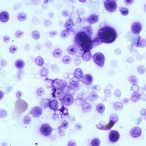
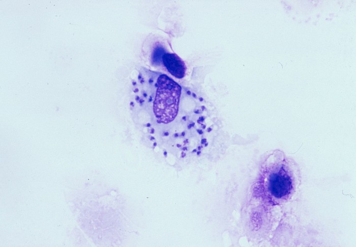
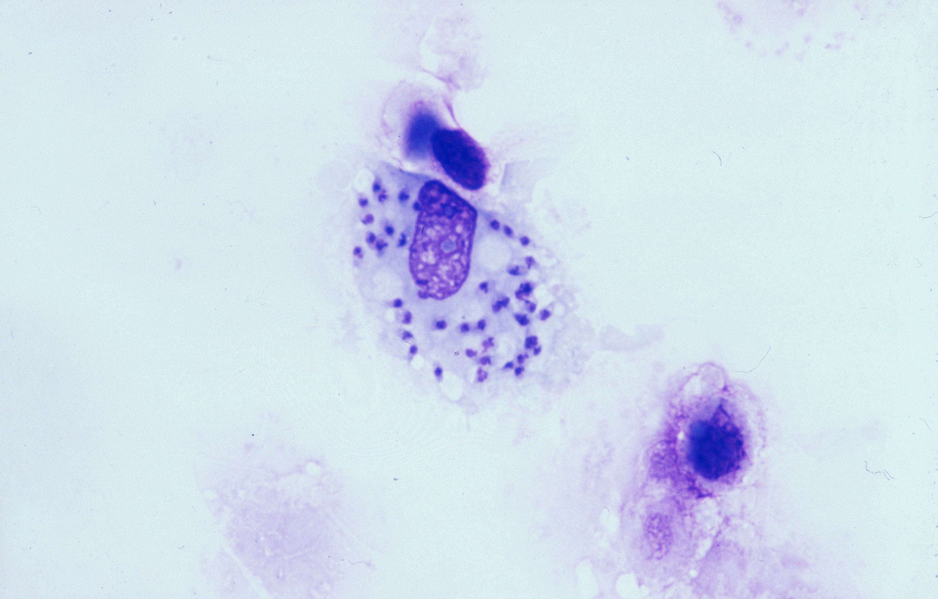 BAL, intracellular 2-4 µm large, round-oval…
 BAL, intracellular 2-4 µm large, round-oval…
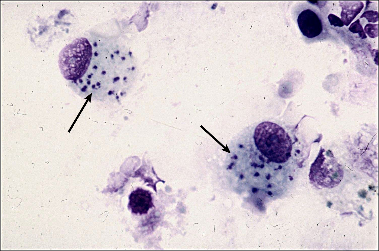 BAL, intracellular 2-4 µm large, round-oval…
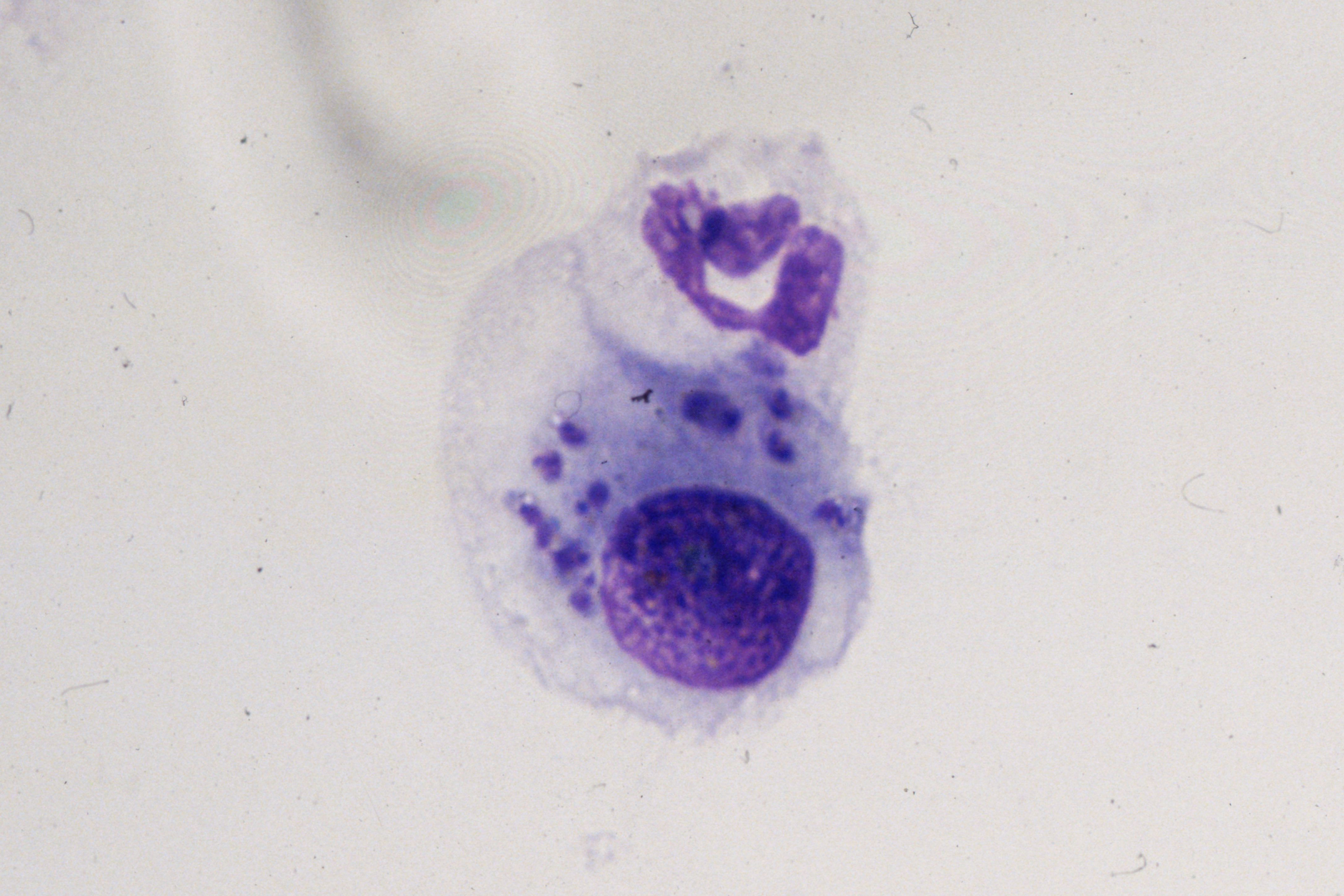 BAL, intracellular 2-4 µm large, round-oval…
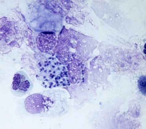 Lung, cytobrush, intracellular 2-4 µm large…
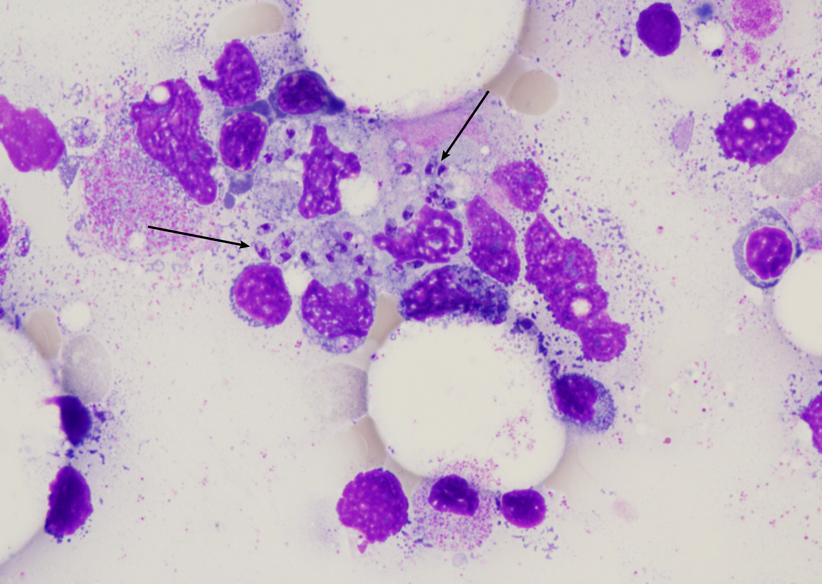 Bone marrow aspirate with plenty of…
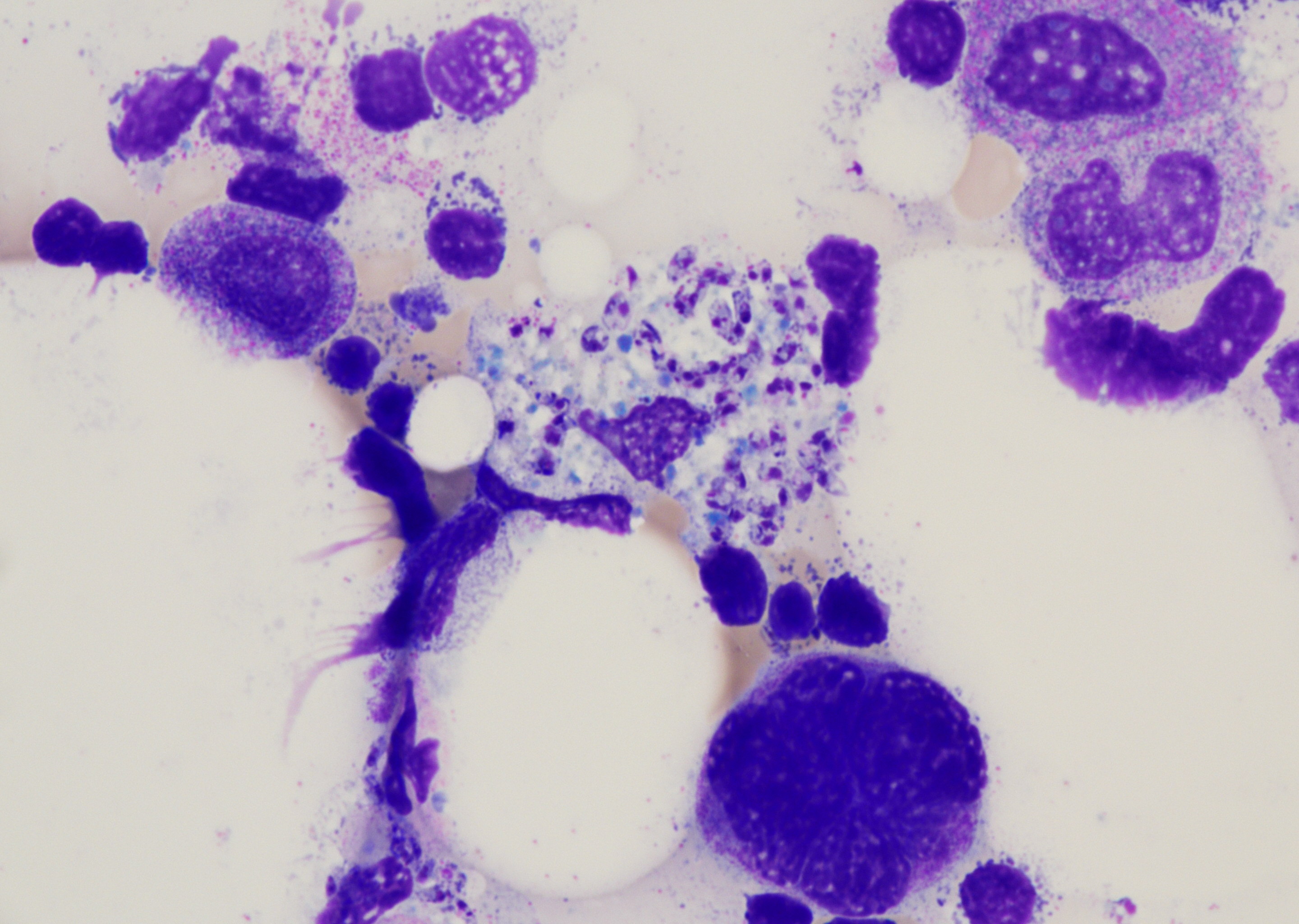 Bone marrow aspirate with plenty of…
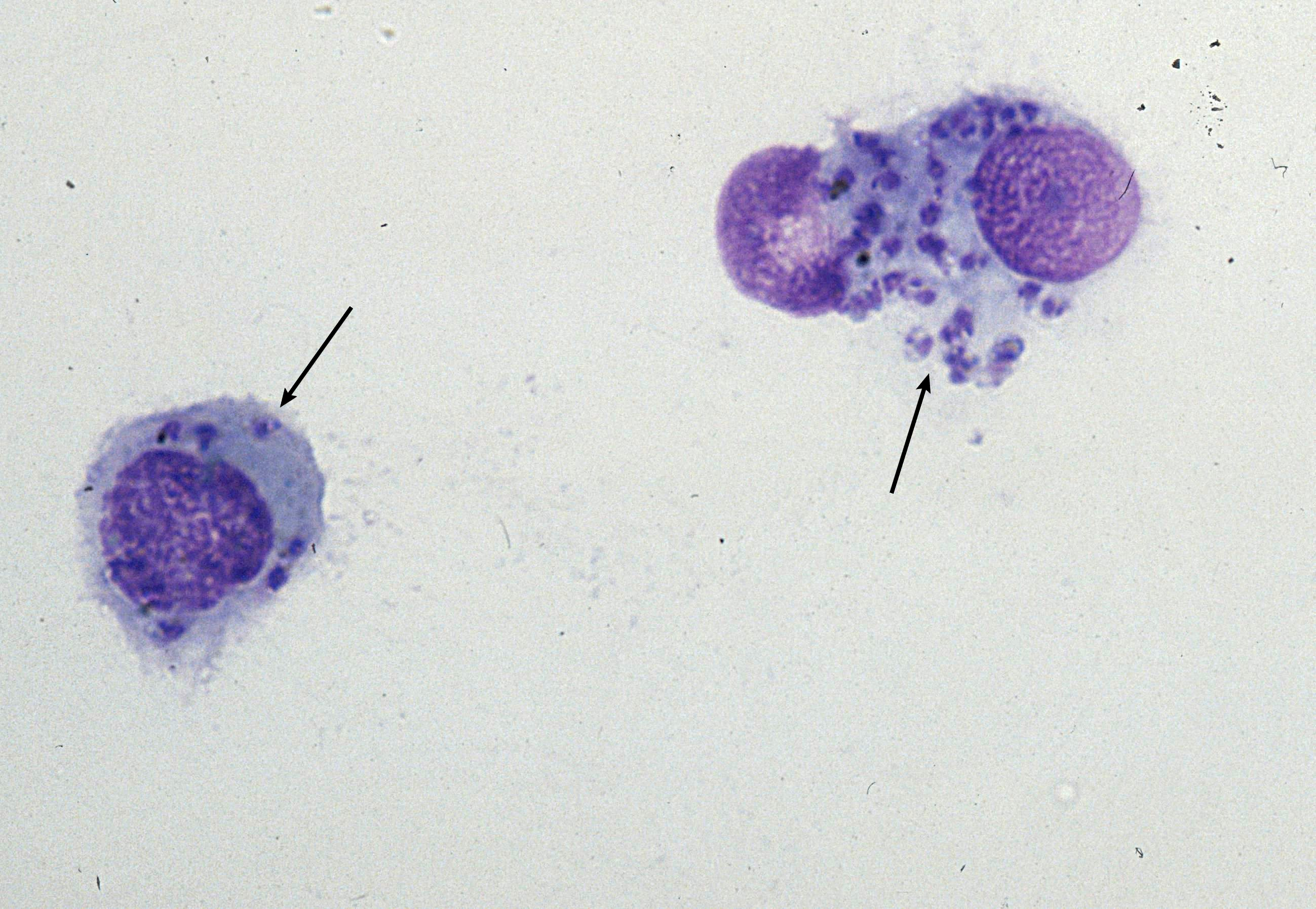
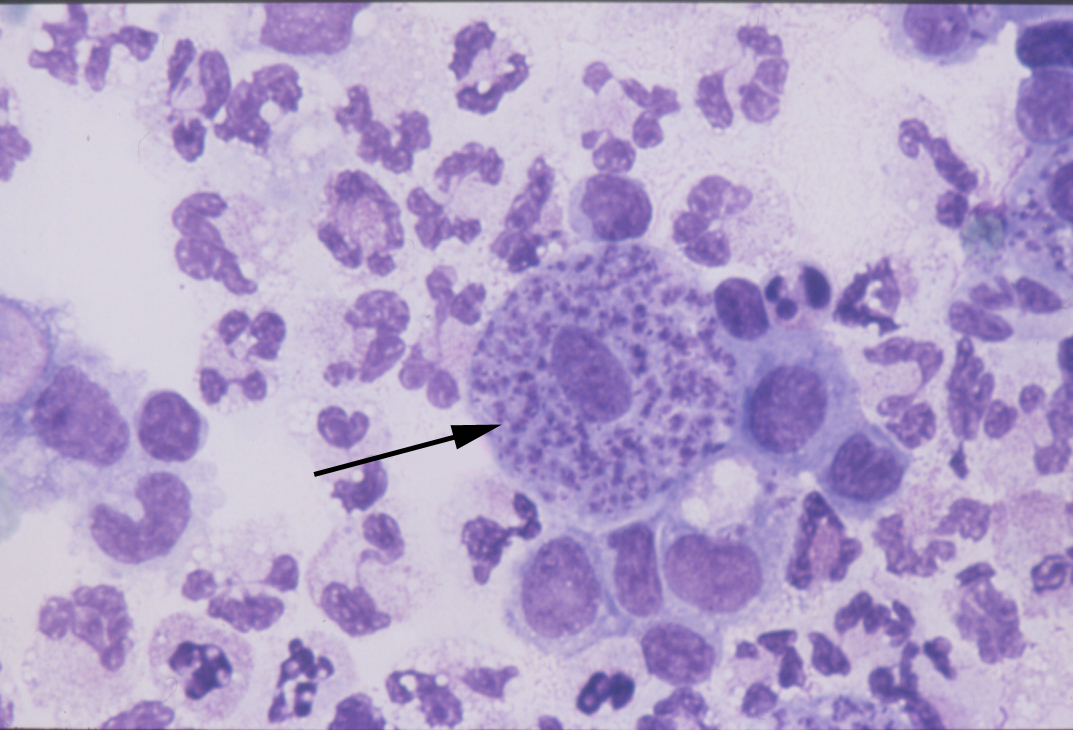 Lung, cell with amastigotes (arrow)…
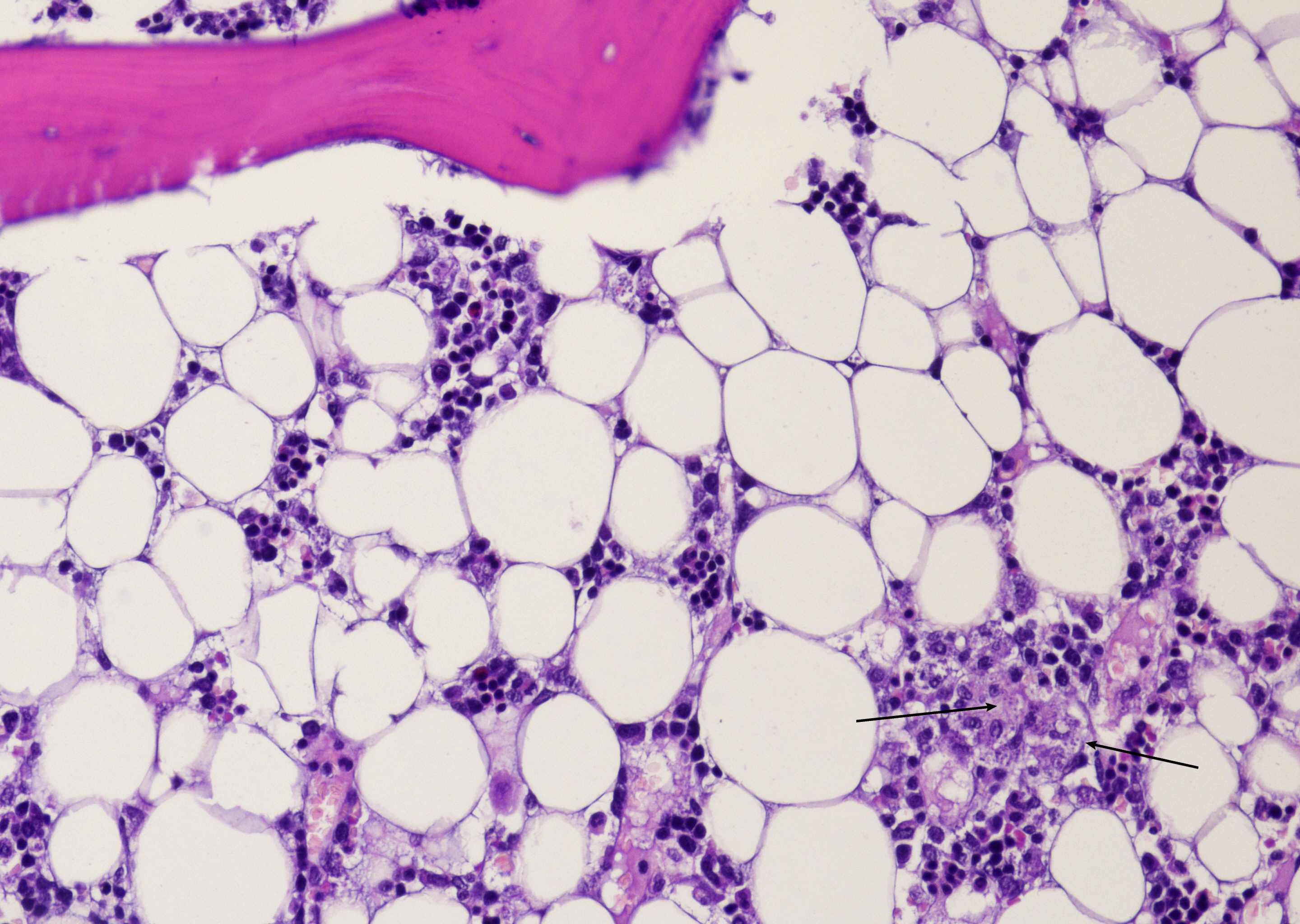 Bone marrow with plenty of amastigotes…
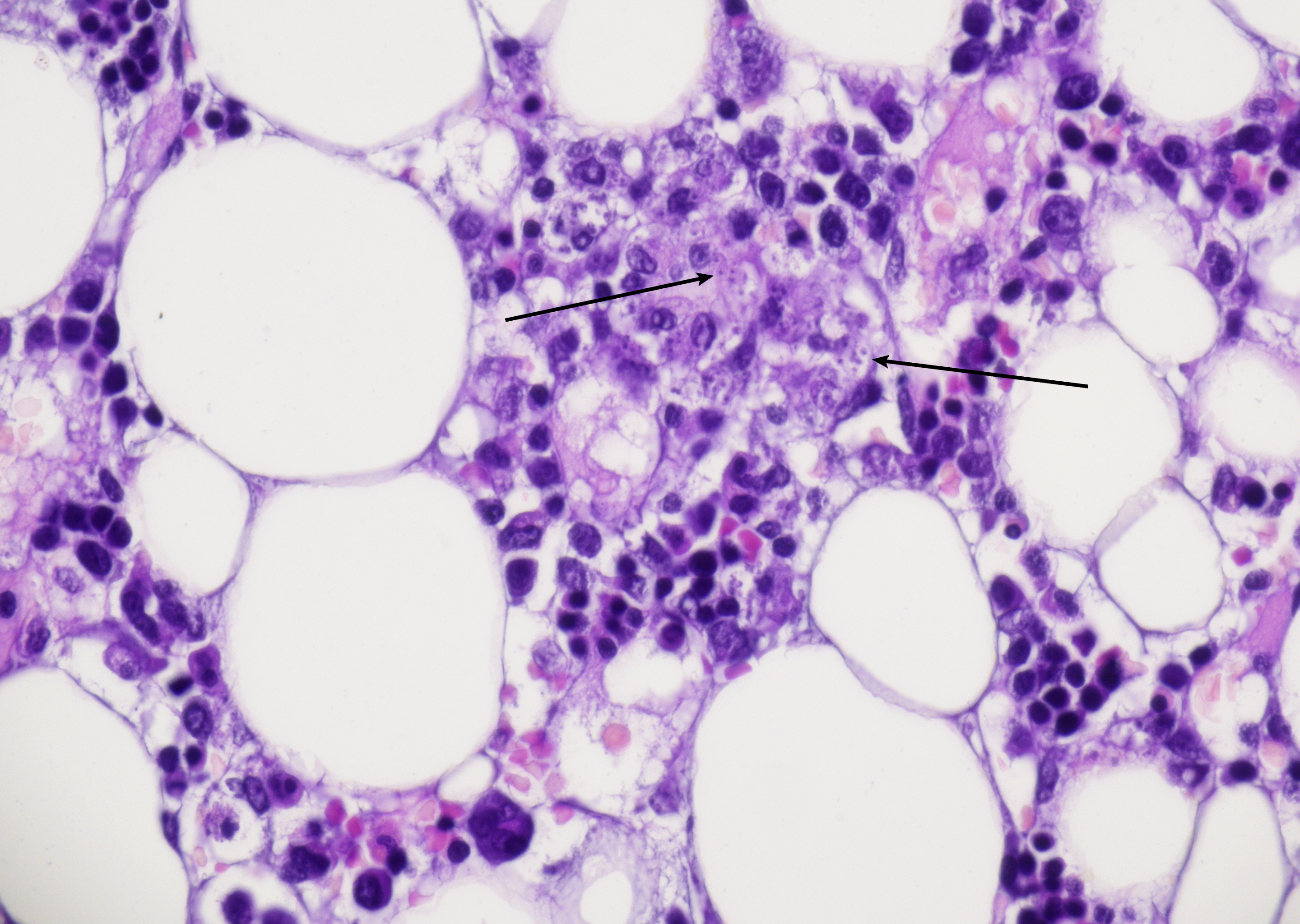 Bone marrow with plenty of amastigotes…
Morphology
Cytology:
- Background can show acute or granulomatous inflammation, with or without plasma and giant cells
- Amastigotes are the intracellular pathogenic form of parasites, 2-4 µm in size, round-to-oval with a thin cell membrane, a large nucleus, and a rod-shaped kinetoplast, lacking an external flagellum
- Amastigotes can be arranged in a ring-like pattern along the inner margin of the cytoplasm of histiocytes, often described as the "marquee sign"
- Bone marrow or spleen aspirate with macrophages densely packed with amastigotes (Leishman-Donovan bodies), with a dot-dash-like pattern; numerous free amastigotes in the background
Histology:
- Bone marrow core biopsy shows hypercellularity with marked macrophage expansion and numerous intramacrophage amastigotes on Giemsa or HE
- Liver with sinusoidal and portal macrophage infiltration (Kupffer cell hyperplasia)
- Spleen with white-pulp depletion and diffuse macrophage/plasma cell infiltrates
- Lymph nodes with paracortical histiocytosis; Amastigotes intramacrophagic; granulomas may be present; necrosis is uncommon
- Amastigotes are GMS-negative.
Differential Diagnosis:
- Trypanosoma cruzi - both amastigotes are indistinguishable; confirmation by serology or PCR is necessary
- Histoplasmosis - amastigotes are GMS-negative
- Malakoplakia
Clinical notes
Patients show 5 main symptoms: Hepatosplenomegaly, pancytopenia, lymphadenopathy, fever, and, in the advanced stages, cachexia - fatal if untreated.
Chronic cough indicates the rare involvement of the lungs. Imaging studies reveal, in addition to massive hepatosplenomegaly, diffuse interstitial consolidations in the lungs.
Ancillary Testing
PCR/RT-PCR on bone marrow, blood (EDTA), or tissue; Antigen/Serology: rK39 rapid test (good rule-in in endemic settings), rK28/ELISA or DAT (direct agglutination test) as alternatives; Montenegro skin test (Leishmanin test)Additional Information
Leishmania spp. are hemoflagellates and have two morphological stages: amastigotes (human tissue) and promastigotes (within vector).
Infection with certain Leishmania spp. can cause cutaneous, mucocutaneous, and visceral leishmaniasis.
The subspecies within the L. donovani complex cause visceral disease (syn. kala-azar): L. donovani, L. chagasi, and L. infantum.
Visceral leishmaniasis occurs endemically in tropical and subtropical regions, and rarely in the Mediterranean area. It predominantly affects children and young adults.
The pathogens, transmitted percutaneously by sandflies, multiply within macrophages. Through the bloodstream, the pathogens spread to the liver, spleen, bone marrow, and lymph nodes. In severe infections, all organs may be affected.
The incubation period is 3 to 8 months.