
- EN
- DE

Bacteria
Differential Diagnosis of Nocardia Species:
Actinomyces Species

- Organ System
- Respiratory Tract, Genitourinary Tract, Soft Tissue & Bone
- Diagnostic Method
- Cytology

- Geographical Region
- Dry Areas,
- Islands,
- Tropical and Subtropical Region,
- Polar and Subpolar Zone,
- Temperate Zone
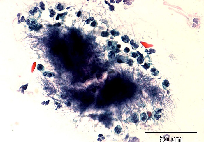
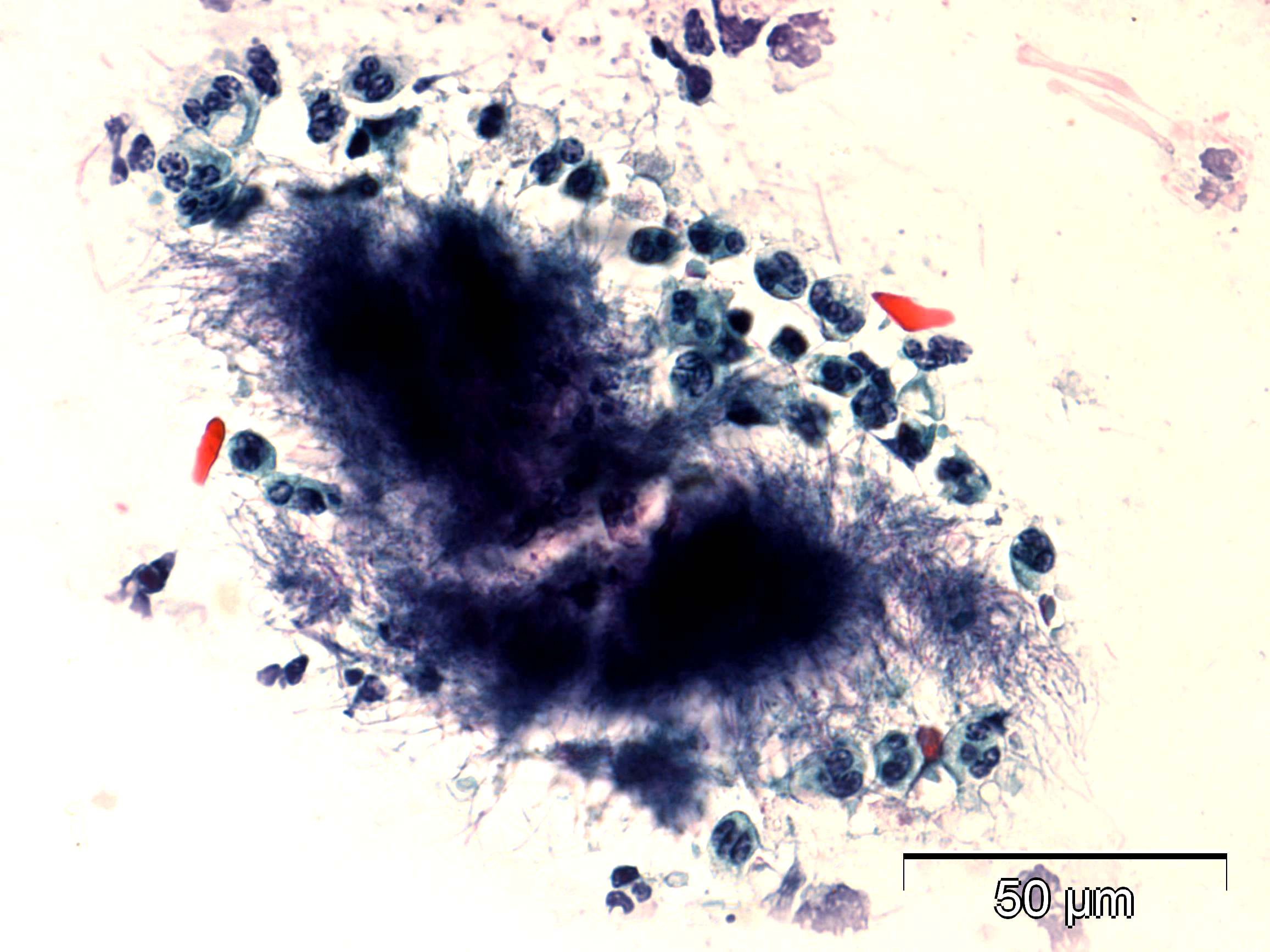 Cervical smear, basophilic actinomycetic…
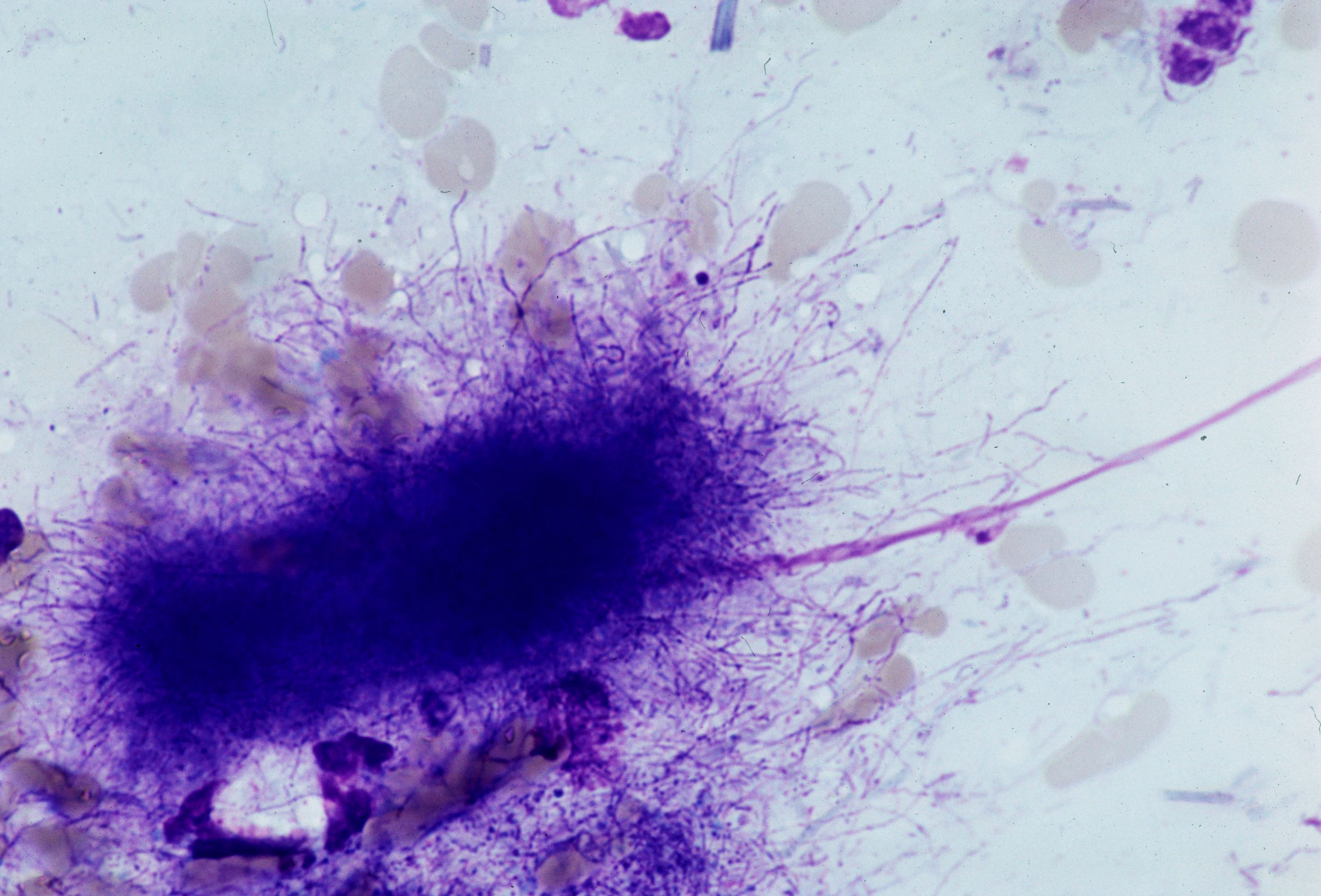 Lung, cytobrusch, actinomycetic filamentous…
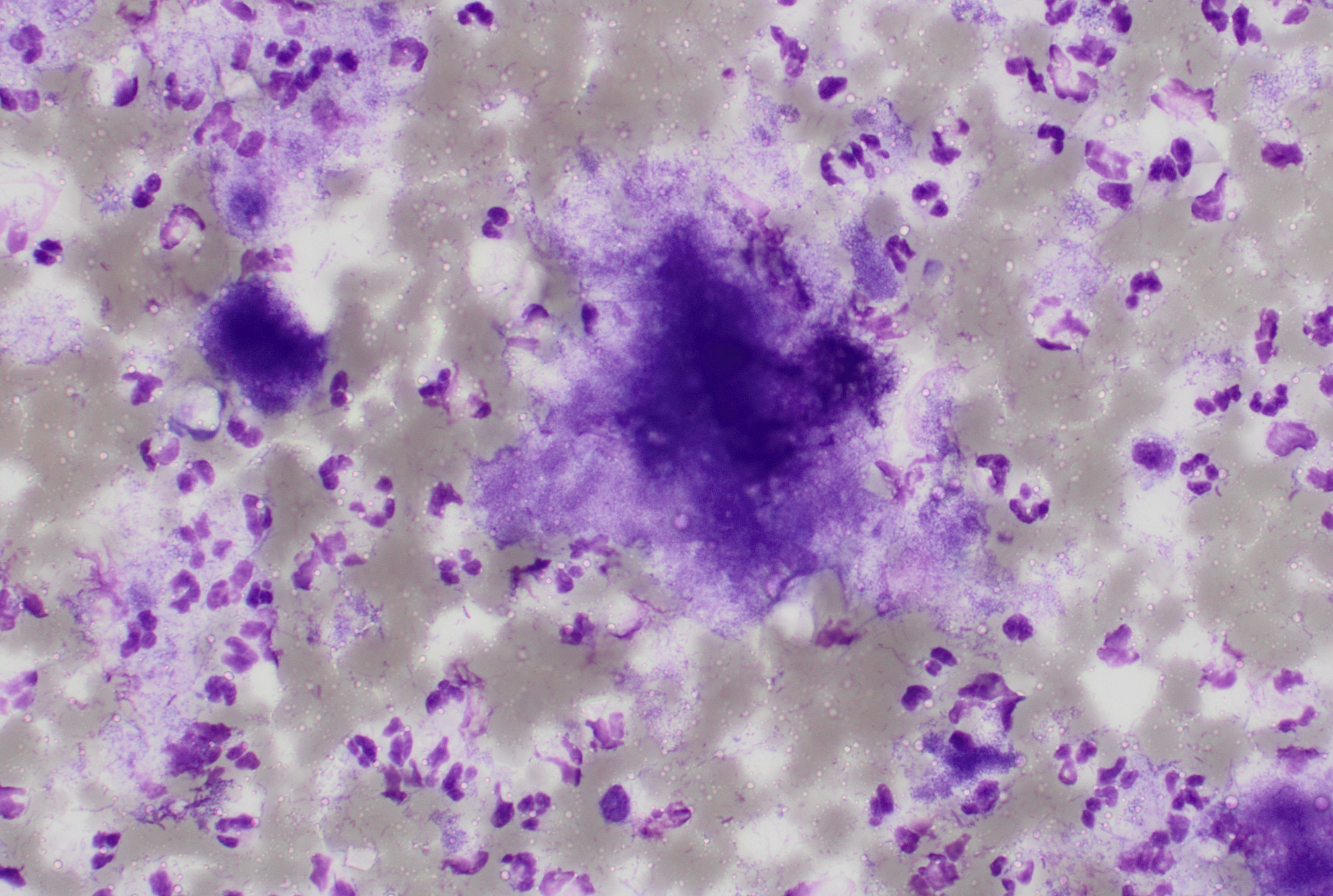 Jaw, FNA, granular bacterial clumps of…
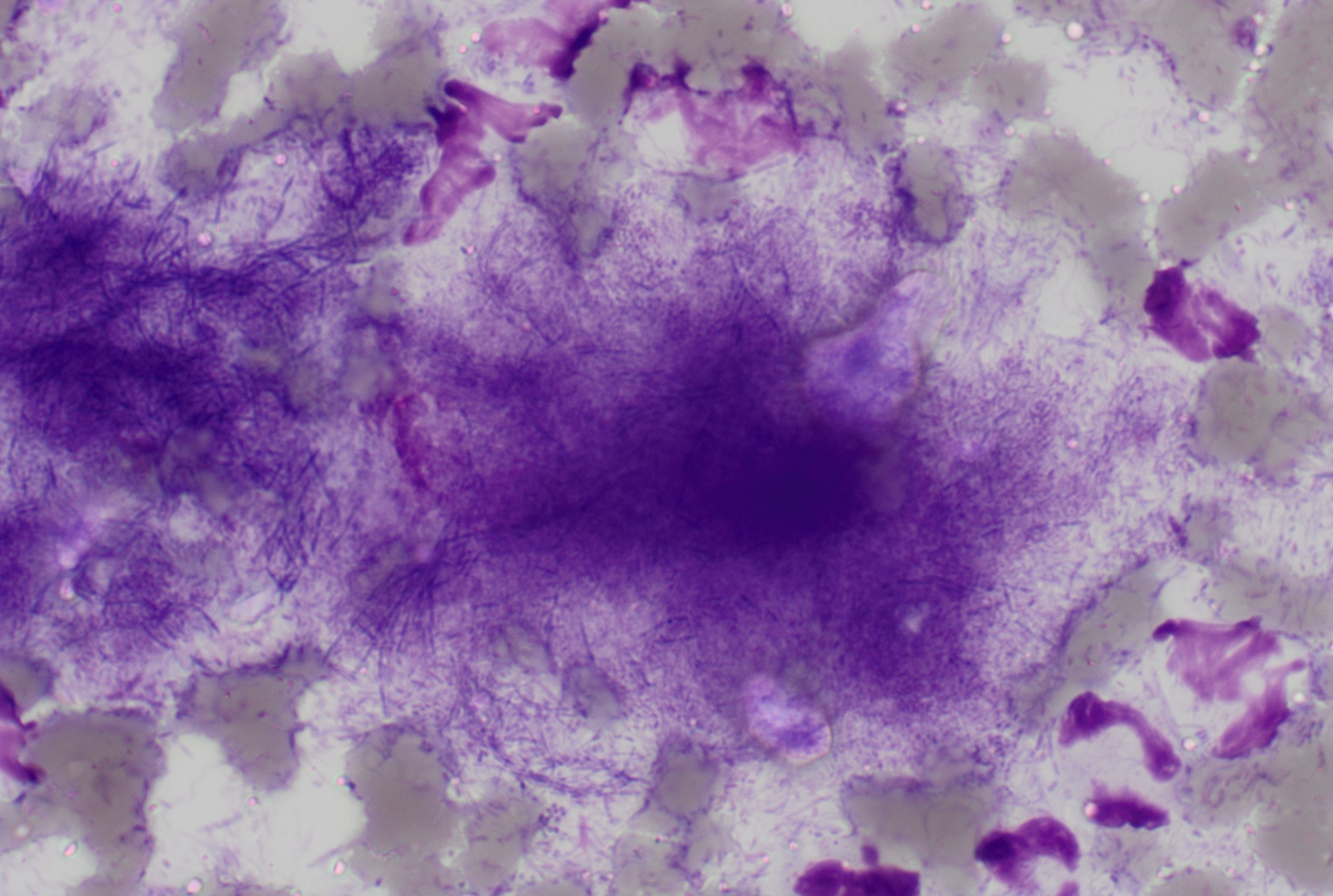 Jaw mass, FNA, granular clumps of…
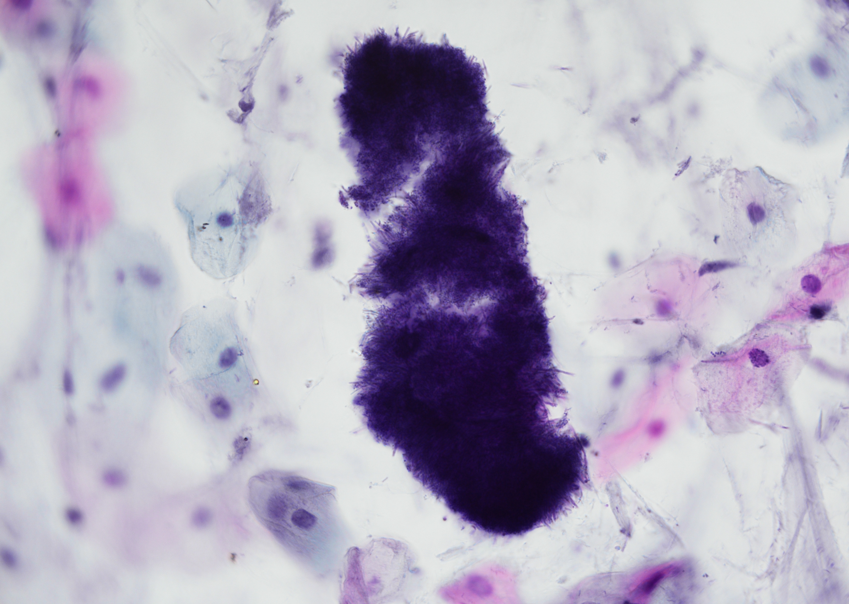 Sputum, squamous epithelial cells and…
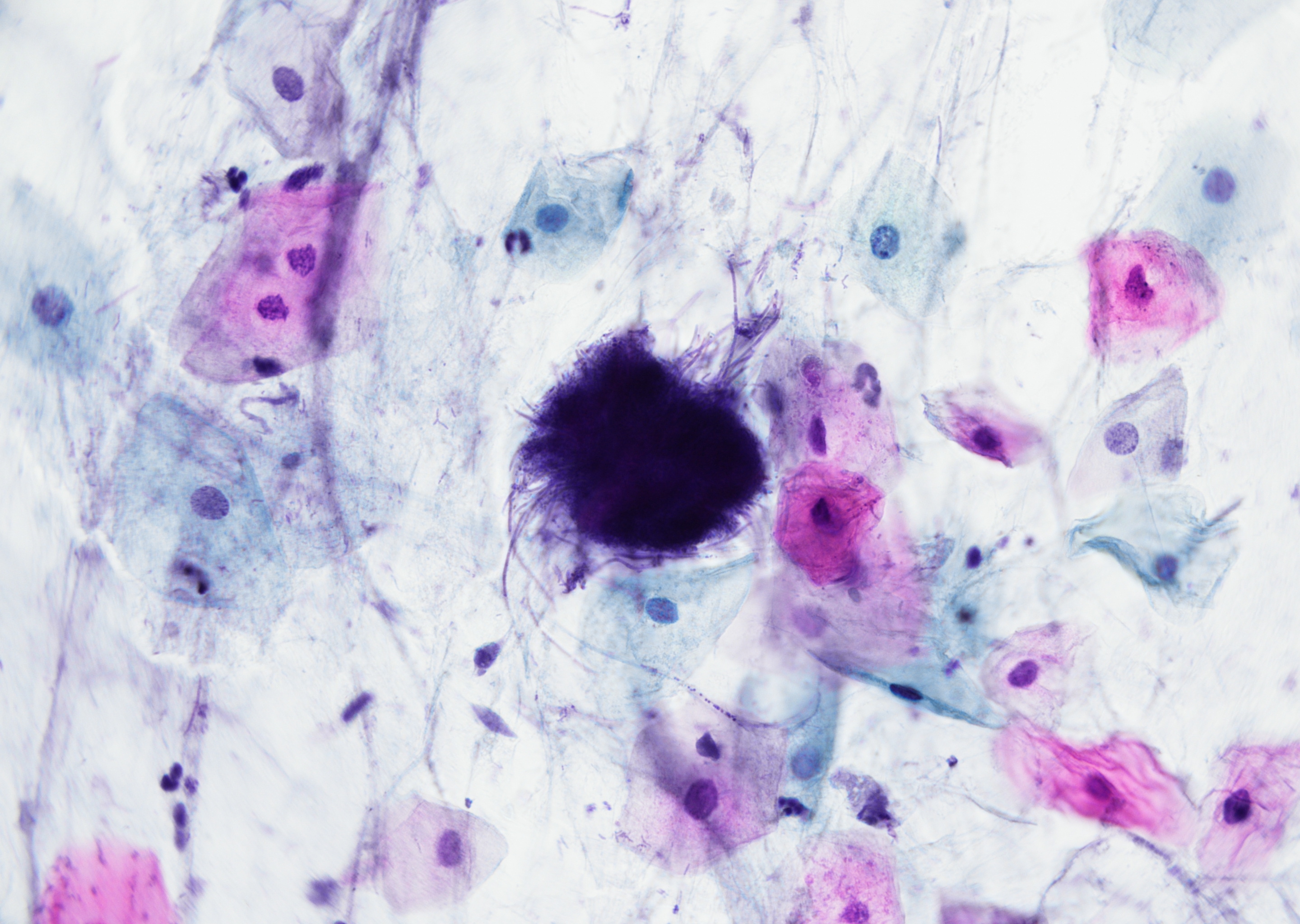 Sputum, squamous epithelial cells…
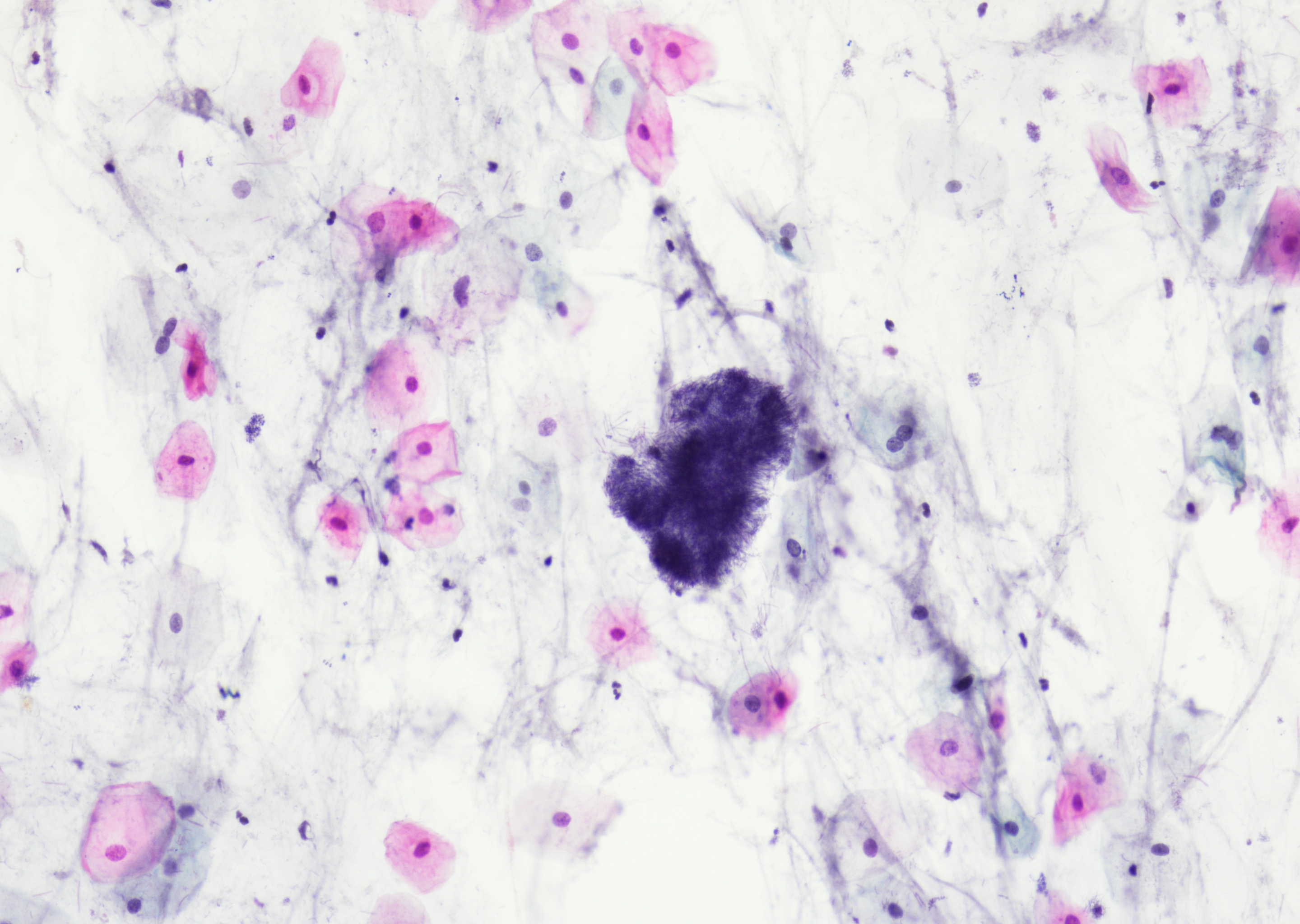 Sputum, filamentous, branching colonies…
Morphology
Cytology:
- Basophilic filamentous, branching gram-positive colonies with an eosinophilic splendore-hoeppli rim (“sulfur granules”)
- Central dense core with radiating fibres that branch at acute angles
- Central dense core stains dark purple or black on Papanicoloau stain
- Background often neutrophil-rich +- multinucleated giant cells
- AFS (ZN) negative
Histology:
- Compact bacterial colonies with eosinophilic club-like material
- AFS (ZN) negative
- Highlighted by Gram stain, PAS, and GMS/Grocott
- Surrounding fibrosis
- Mixed suppurative inflammation
Differential diagnosis:
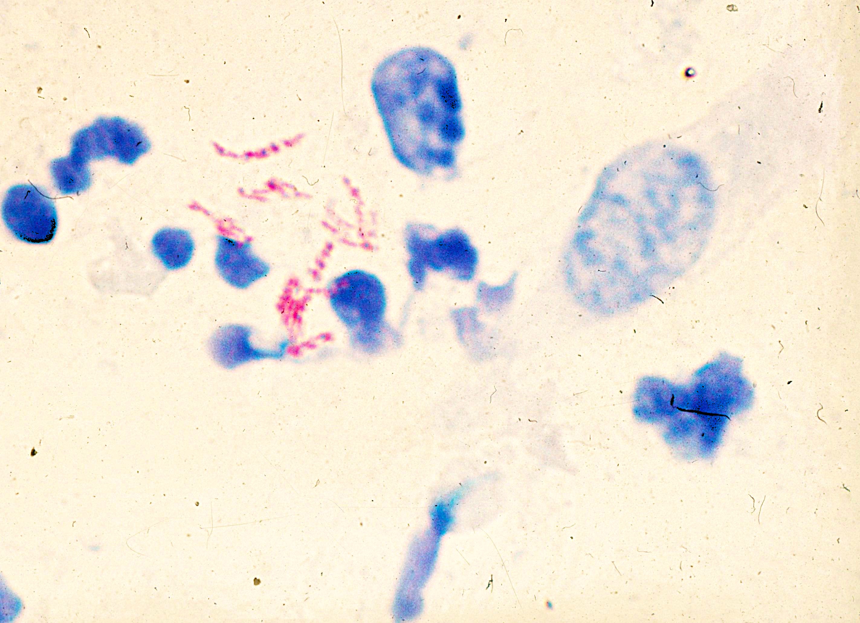
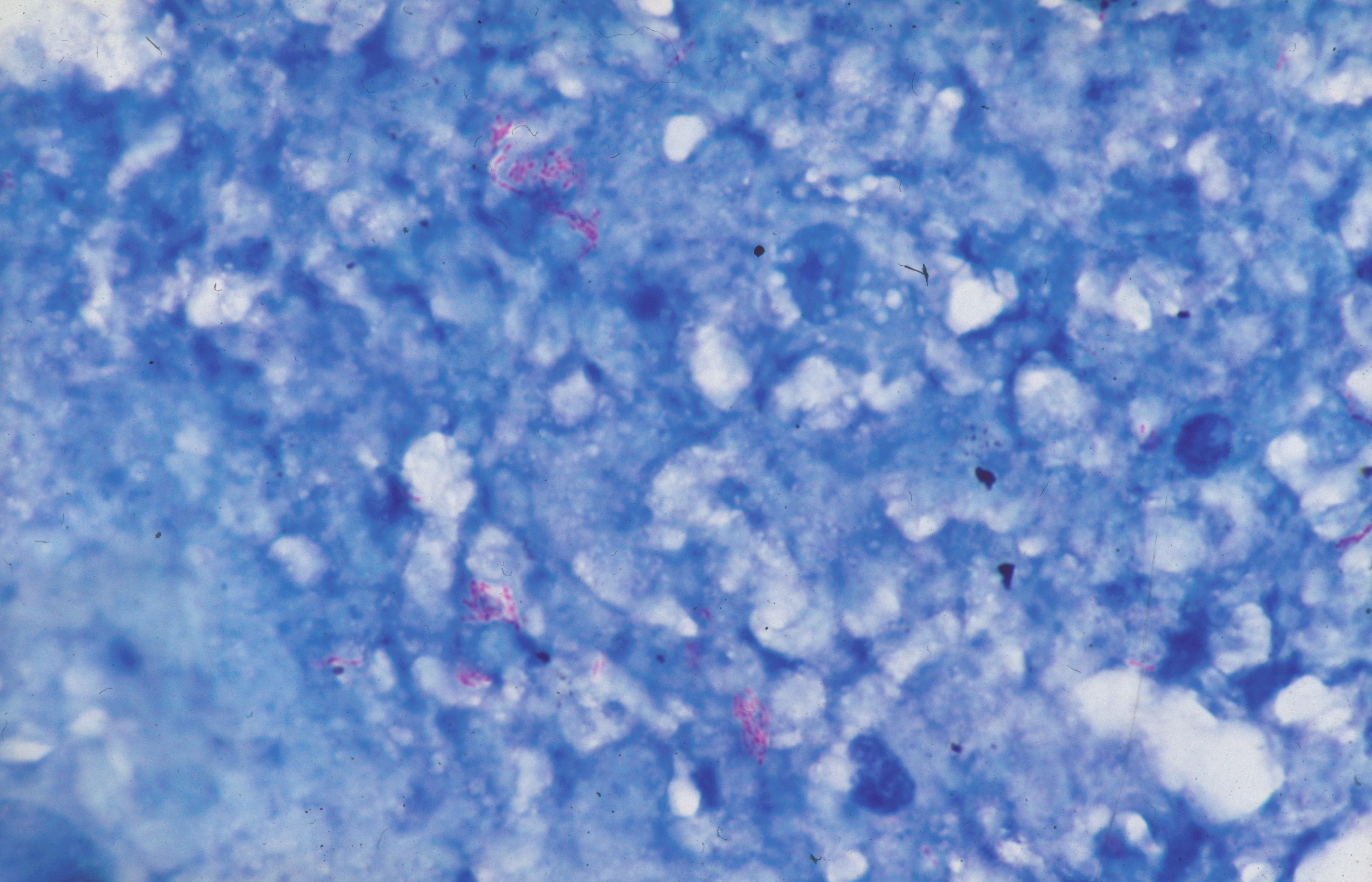
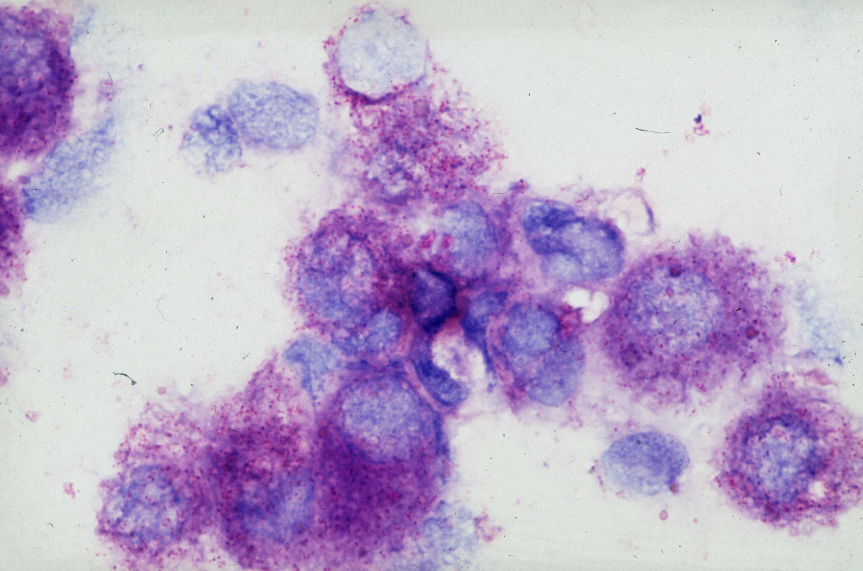
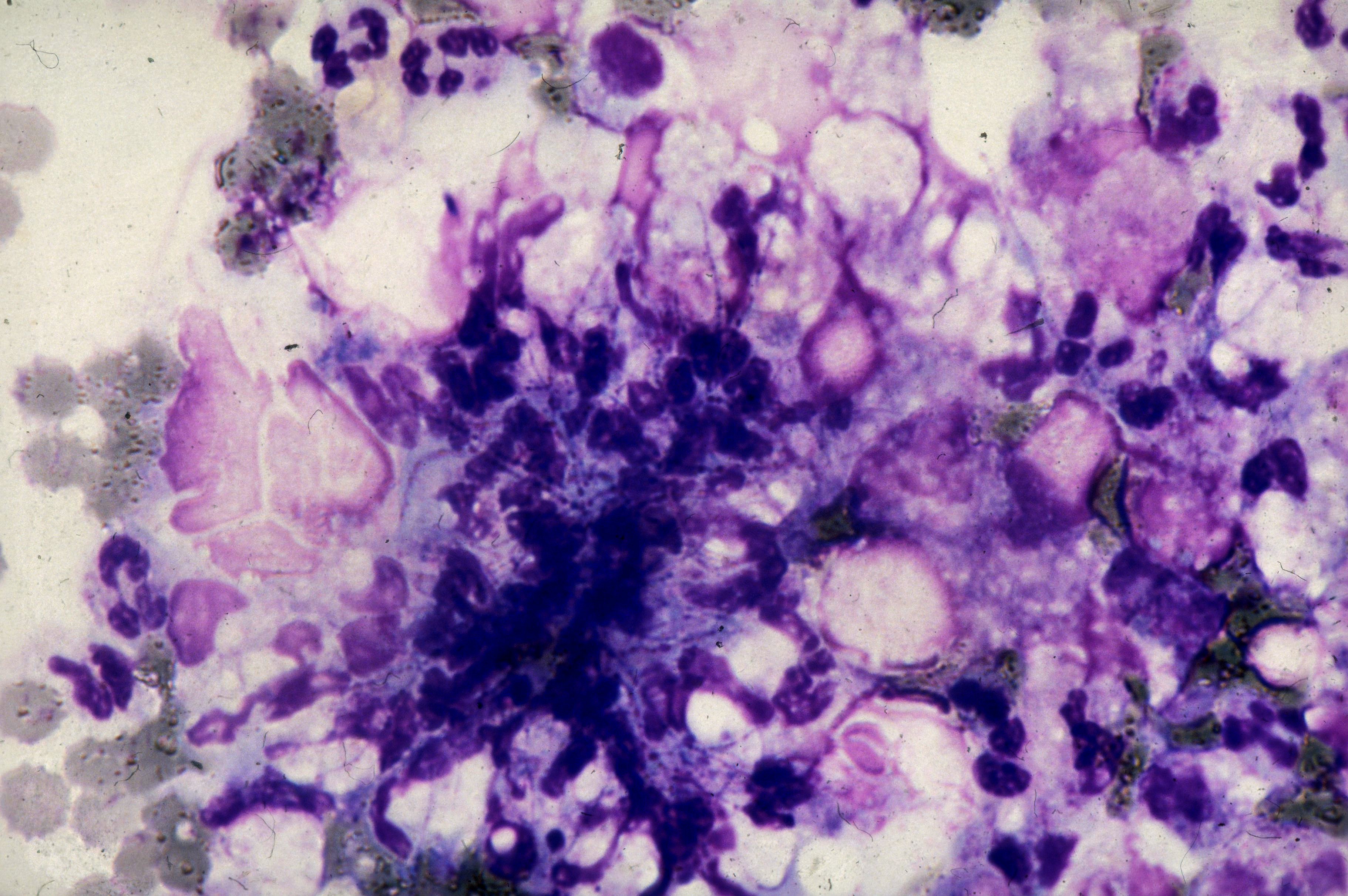
- Nocardia spp. - partially acid-fast aerobes; Actinomyces are non-acid-fast anaerobes
- Pseudoactinomycotic radiate granules - particularly in IUD-associated specimens: broad irregular eosinophilic clubs without a central filamentous bacterial core; Gram and GMS stains are negative or non-specific. (1)
Clinical notes
Chronic, indurated masses with draining sinus tracts (“lumpy jaw”); thoracic disease can mimic malignancy; abdominopelvic forms after perforation or IUD. Generally good but slow response to prolonged high-dose penicillin therapy; additional drainage or surgical debridement may be required. (2)
Asymptomatic individuals with Actinomyces-like organisms on cervical cytology are more likely to be colonised than infected; neither antibiotic treatment nor IUD removal is required. (FSRH Guideline: Intrauterine Contraception. March 2023)
Ancillary Testing
Anaerobic culture with prolonged incubation; MALDI-TOF; 16S rRNA sequencing if needed. Consider broad PCR from tissue in culture-negative cases. Colonies: small, rough, heaped “molar-tooth” colonies on anaerobic blood agar, firmly adherent, white to gray-yellow and often chalky, with spidery/rhizoid edges after prolonged anaerobic incubation.Additional Information
Detection in non-sterile mucosal samples may represent colonisation (oral/GI/genital tract commensal); tissue invasion, sulfur granules and associated suppurative inflammation support true infection.
Differential Diagnosis
References
- Pritt B, Mount SL, Cooper K, Blaszyk H. Pseudoactinomycotic radiate granules of the gynaecological tract: review of a diagnostic pitfall. J Clin Pathol. 2006 Jan;59(1):17-20. doi: 10.1136/jcp.2005.028977. PMID: 16394276; PMCID: PMC1860258.
- Valour F, Sénéchal A, Dupieux C, Karsenty J, Lustig S, Breton P, Gleizal A, Boussel L, Laurent F, Braun E, Chidiac C, Ader F, Ferry T. Actinomycosis: etiology, clinical features, diagnosis, treatment, and management. Infect Drug Resist. 2014 Jul 5;7:183-97. doi: 10.2147/IDR.S39601. PMID: 25045274; PMCID: PMC4094581.